Материал: BASIC CLINICAL SYNDROMES IN INTERNAL DESEASES CLINIC
Atrial flutter (irregular and regular forms). Symptoms
1) the number of pulses does not exceed 250-300 per min .;
2) pulses through the atrioventricular node are conducted more rhythmically than with atrial fibrillation.
3) The clinically irregular form does not differ from atrial fibrillation. With a regular form, the pulse and heart rate are rhythmic.
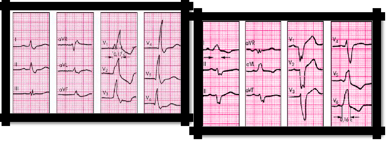
4) ECG: Irregular shape - the same signs as with atrial fibrillation, but the f waves are of high amplitude - ―saw teeth‖.
5) The regular shape is distinguished by the same R-R intervals.
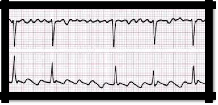
Pic 3.12 Atrial fibrillation (upper) and atrial flutter (lower)
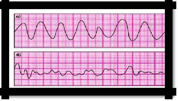
Pic 3.13 ventricular flutter and fibrillation
Ventricular flutter and fibrillation. Symptoms
faint;
pallor of the skin;
pulse and blood pressure are not determined;
39
ECG: individual disordered deformed complexes, on which it is difficult to distinguish individual teeth.
IV. Conductivity disorders - blockade:
sinoauricular;
atrial;
atrioventricular I, II, III degree;
g) intraventricular.
a) sinoauricular block. Symptoms
with auscultation of the heart and palpation of the pulse, periodic loss of heart beat and pulse beat is detected. On the ECG: against the background of the correct sinus rhythm, periodically, cardiac complex falls out (the P wave and QRS complex are not recorded), the duration of the diastole doubles.
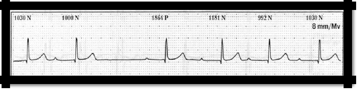
Pic 3.14 Sinoauricular block
b) Atrial block. Symptoms
It is detected only electrocardiographically. ECG: there is a change in the P waves, they are deformed, their duration exceeds normal conductivity - up to 0.1 s.
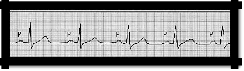
Pic 3.15 Atrial block
c) Atrioventricular. Symptoms
I degree - detected only electrocardiographically by lengthening the P-Q interval of more than 0.2 s;
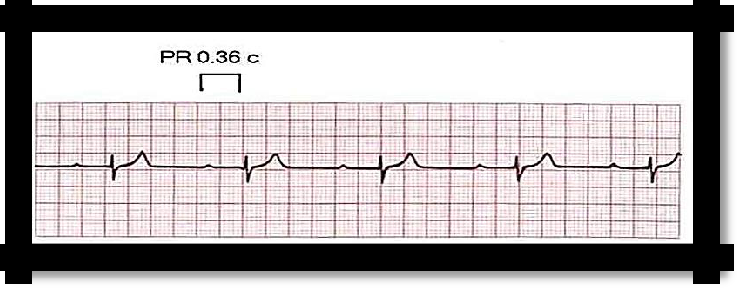
Pic 3.16 I degree
40
degree: type 1 or Mobitz I in case of violation (deceleration) of atrioventricular conduction up to the complete blocking of the pulse.
1) a feeling of "freezing" or a stop in the work of the heart;
2) slight dizziness;
3) with auscultation and palpation of the pulse - periodic loss of heart beat and pulse beat;
4) ECG: gradual lengthening of the P-Q interval until the ventricular QRS complex falls out (Samoilov-Wenckebach periods)
.
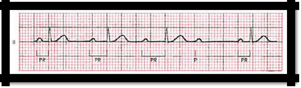
Pic 3.17 II degree: type 1 or Mobitz I
Type 2 or Mobitz II is characterized by a constant duration of the P-Q interval with periodic loss of the QRST complex.
"Fading", "interruptions", "stop" in the work of the heart;
dizziness, darkening in the eyes, short-term loss of consciousness;
heart contractions and pulse are rare, arrhythmic.
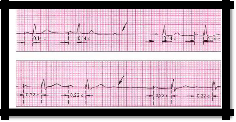
Pic 3.18 Type 2 or Mobitz II
AV blocks of the II degree 2: 1 are also secreted when every second ventricular contraction falls out, while the pulse remains rhythmic.
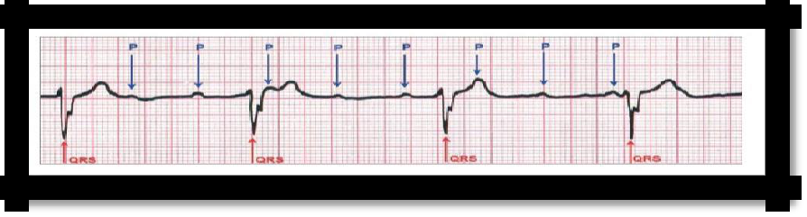
degree of blockade (complete transverse blockade of the heart). The atrium contracts under the guidance of the sinus node, and the ventricles - atrioventricular, independent of each other.
1) a rare rhythmic pulse, large in size, heart rate - 30-45 in 1 min. ;
2) increased heart size due to diastolic overflow with its blood;
3) heart sounds are muffled, but loud I tone can be periodically determined (―cannon tone‖ of Strazhesko);
4) Morgagni-Edems-Stokes syndrome, caused by a violation of the regional blood circulation of various organs, primarily Ts.N.S. as a result of the loss of not one, but several contractions of the heart in a row:
41
sudden loss of consciousness, the patient falls; with a duration of fainting of 15-20 seconds. general muscle cramps (epileptiform) occur;
deep breathing, pallor of the skin;
ECG: P wave without communication with QRST, correct P wave alternation, correct Q-T alternation (R-R are equal to each other, but R-R is longer than R-P).
Pic 3.18 III degree of blockade
d) Intraventricular block. Symptoms:
With auscultation of the heart, splitting or bifurcation of tones due to asynchronism in the activity of the ventricles.
Allocate: Complete and incomplete blockade of the right and left legs, left anterior and left posterior legs of the bundle of His.
CAUSES, Congenital conduction disorders and acquired due to heart disease.
Signs of a full leg block:
expansion of the QRS complex for more than 0.11 seconds, discordance of the T wave and QRS complex
With blockade of the right leg in leads V1, V2, R is high (R, RSr types) and the T wave is negative.
With blockade of the left leg, QS prevails in leads V1, V2 and the T wave is positive
Pic 3.19 Right bundle branch block Pic 3.20 Left bundle branch block
Generalized heart rhythm and conduction disorder syndrome
42
REASONS: any diseases and dysfunction of the heart; reflex effects on cardiac conduction and automatism.
GENERAL SYMPTOMS:
complaints of palpitations, interruptions in the work of the heart, a feeling of "freezing", "stopping" of the heart, sometimes followed by a strong blow (with extrasystole);
irregular heartbeat, tachycardia, bradycardia;
pulse deficiency (most characteristic for atrial fibrillation);
irregularity of tones during auscultation, polyphony of I tone at the apex (atrial fibrillation), ―cannon‖ tone of Strazhesko (complete atrioventricular block), premature contraction of the heart with a characteristic loud I tone, periodic loss of tones (with blockade);
heart rhythm disturbance, especially paroxysmal, can provoke acute coronary, cardiac or vascular insufficiency, or aggravate chronic forms thereof;
the type of arrhythmia or blockade can be finally verified using an ECG.
13. Syndromes of valvular heart disease.
Valvular heart defects are acquired, less often congenital morphological changes in the valvular apparatus, leading to a violation of its function and hemodynamics. Two periods in the clinical course of heart defects:
compensation period (without the development of heart failure)
decompensation period (with the development of heart failure)
Mitral valve insufficiency
With mitral valve insufficiency, the function of the valve apparatus is impaired, which is characterized by incomplete closure of the valves of the bicuspid valve during systole and the absence of a period of closed valves.
REASONS: rheumatic endocarditis (in 75% of cases), infectious endocarditis, atherosclerosis, trauma, systemic diseases of the connective tissue, myocardial infarction with rupture of the papillary muscles, congenital splitting of the mitral valve cusps, relative mitral valve insufficiency (with aortic defects, arterial hypertension, myocarditis myocardial dystrophy, cardiosclerosis, subaortic muscle stenosis), mitral valve prolapse.
Complaints: There are no complaints at the compensation stage. In the decompensated phase - shortness of breath, palpitations, interruptions and pain in the heart, with congestion in the lungs - coughing, hemoptysis.
Palpation - apical impulse is displaced to the left, spilled;
Percussion - an increase in the boundaries of relative dullness to the left, and in the case of a sharp increase in the left atrium - and up. During decompensation (Kitaev’s reflex) - expansion to the right;
During auscultation, weakening of the I tone, systolic murmur at the apex, decreasing a different timbre, is brought to the left axillary region or to the second and third intercostal space to the left of the sternum, takes more than ½ systole, amplifies in the exhalation phase and on the left side, a moderately pronounced accent II tone on the pulmonary artery (Kitaev's reflex);
ECG: signs of left atrial hypertrophy (―P mitrale‖) and left ventricle, during decompensation (Kitaev’s reflex) - right ventricular hypertrophy;
FCG: confirms auscultation data;
X-ray: an increase in the left departments of the heart (ventricle and atrium), the heart acquires a mitral configuration, the displacement of the contrasted esophagus along an arc of large radius (in the left lateral projection). During decompensation (Kitaev’s reflex) - expansion to the right;
43
Echocardiography: expansion of the cavities of the left ventricle and left atrium, multidirectional movement of the cusps of the mitral valve, their thickening and lack of closure in systole, pulmonary hypertension;
Doppler echocardiography: a turbulent flow of blood into the left atrium according to the degree of regurgitation.
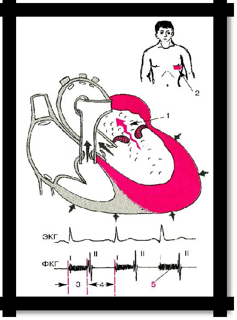
Pic 3.21 Mitral insufficiency (hemodynamics)
Mitral stenosis
- narrowing of the left atrioventricular foramen.
REASONS: most often - acute rheumatic fever (rheumatic endocarditis); rarely - systemic diseases of the connective tissue (rheumatoid arthritis, systemic lupus erythematosus), mitral valve calcification.
Complaints: There are no complaints during the compensation period.
During decompensation: shortness of breath, hemoptysis, palpitations, interruptions and pain in the heart, with severe decompensation - swelling on the legs, pain in the right hypochondrium, an increase in the abdomen;
Ortner's symptom - a sharp increase in the left atrium can cause a violation of swallowing (compression of the esophagus) and hoarseness in connection with compression of the recurrent nerve.
Examination: acrocyanosis, cyanotic blush in the form of a ―butterfly‖ (―mitral‖ face), poor physical development, infantilism, ―heart hump‖.
Palpation: positive cardiac impulse, diastolic trembling ("cat purr") at the apex.
Percussion: the boundaries of relative and absolute dullness are shifted up and to the right (due to hypertrophy of the left atrium and right ventricle);
Auscultation: Amplification of the I tone at the apex (―clapping‖ I tone), ―mitral click‖ or tone of opening the mitral valve. The combination of ―clapping‖ I tone, II
44
tone and tone of opening of the mitral valve creates a peculiar melody of a three-membered rhythm - ―quail rhythm‖. Based on the heart, emphasis and bifurcation of the II tone on the pulmonary artery. At the apex, diastolic noise is heard locally, without irradiation, sometimes with a presystolic amplification. On the pulmonary artery, you can listen to diastolic murmur (Graham-Steel noise);
The pulse is small, often the pulse is different - on the left radial artery is weaker than on the right (Popov's symptom), arrhythmia is possible (usually, atrial fibrillation);
HELL - a downward trend;
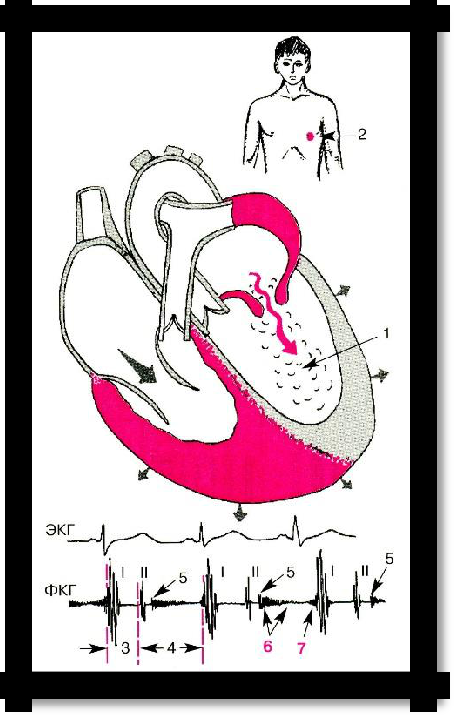
Pic 3.22 Mitral stenosis, hemodynamics
ECG: signs of left ventricular hypertrophy (―P mitrale‖) and right ventricle, atrial fibrillation (a specific symptom for mitral stenosis);
FCG: confirms the signs of auscultation;
X-ray - ―mitral configuration‖ of the heart: flatness of the waist, an increase in the left atrium, determined by the deviation of the esophagus contrasted with barium in the left lateral projection (along an arc of small radius), hypertrophy of the right ventricle, often the bulge of the pulmonary artery arch;
Echocardiography: unidirectional movement of the anterior and posterior cusps of the mitral valve forward, the rate of early diastolic closure of the anterior cusp and the amplitude of its movement is reduced, thickening of the valve, expansion of the cavity of the right ventricle and left atrium, reduction of the diameter of the mitral orifice.
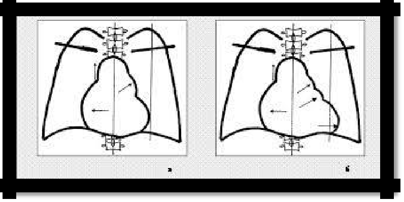
Pic 3.23 Mitral configuration of the heart:
mitral stenosis (a), mitral insufficiency (b)
45
Stenosis of the aortic orifice
REASONS: rheumatic endocarditis, infectious endocarditis, aortic atherosclerosis, congenital
aortic stenosis, syphilis.
SIGNS:
1. Complaints: there are no complaints in the compensation stage.