Материал: Атлас Ханц фениш

|
382 |
Sense organs |
|
|
|||
|
|
1 |
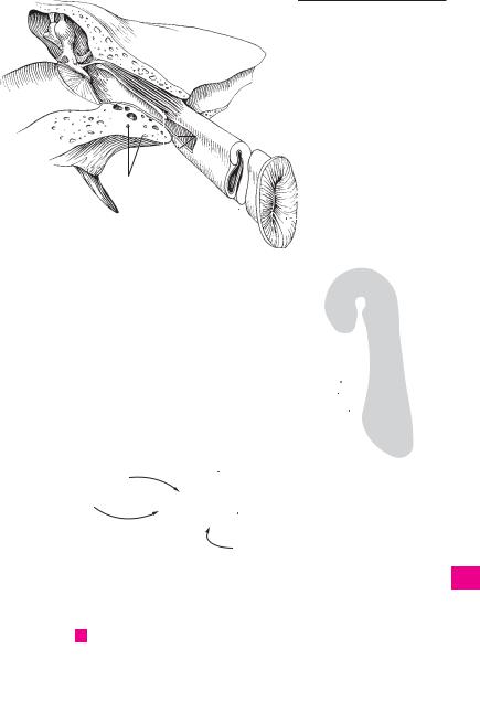
Auditory ossicles (malleus, incus and stapes). |
19 |
Incudomallear joint. Articulatio incudomal- |
||
1 |
|||||||
|
|
Ossicula auditoria (auditus). They operate col- |
|
learis. Joint between the incus and malleus. It |
|||
|
|
|
lectively as a bent lever system transferring |
|
occasionally exhibits an articular cavity. A |
||
2 |
|
|
sound waves from the tympanic membrane to |
20 |
Incudostapedial articulation. Articulatio in- |
||
|
|
|
the inner ear. |
|
cudostapedialis. Union between the lenticular |
||
|
|
|
|
|
|
||
3 |
|
2 Stapes (stirrup). A stirrup-shaped auditory os- |
|
process of the long crus of the incus and the |
|||
|
|
sicle. Its base is inserted into the fenestra vesti- |
|
stapes. A |
|||
|
|
|
|
||||
|
|
|
buli. A B |
|
21 |
Tympanostapedial syndesmosis. Syndesmo- |
|
4 |
|
|
|
||||
|
|
|
|
||||
3 |
Head of stapes. Caput stapedis. It lies opposite |
|
sis tympanostapedialis. Connective tissue at- |
||||
|
|
|
the base of the stapes and articulates with the |
|
taching the base of the stapes to the oval win- |
||
5 |
|
|
lenticular process of the incus. A B |
|
dow. It is broader anteriorly than posteriorly. B |
||
|
4 |
Anterior |
crus (limb.). Crus anterius. The vir- |
22 |
Ligaments of auditory ossicles. Ligg. os- |
||
|
|||||||
6 |
|
|
tually straight anterior limb of the stapes. A B |
|
siculorum auditoriorum. |
||
|
|
23 |
Anterior ligament of malleus. Lig. mallei an- |
||||
|
5 |
Posterior crus (limb.). Crus posterius. The |
|||||
|
|||||||
|
|
terius. Ligament that arises from the anterior |
|||||
7 |
|
|
more curved posterior limb of the stapes. A B |
|
|||
|
|
|
process of the malleus, lies in the anterior mal- |
||||
6 |
Base (footplate) of stapes. Basis stapedis. The |
|
|||||
|
|
lear fold and extends as far as the petrotym- |
|||||
8 |
|
|
plate of bone inserts into the fenestra vestibuli |
|
panic fissure. D |
||
|
|
(ovalis). A B |
|
||||
|
|
|
24 |
Superior ligament of malleus. Lig. mallei su- |
|||
|
|
7 Incus (anvil). The central ossicle situated be- |
|||||
9 |
|
|
perius. It passes from the head of the malleus to |
||||
|
|
tween the head of the malleus and the head of |
|
the roof of the epitympanic recess. C D |
|||
|
|
|
the stapes. A D |
25 |
Lateral ligament of malleus. Lig. mallei |
||
10 |
|
|
|||||
8 |
Body of incus. Corpus incudis. It articulates |
||||||
|
laterale. It unites the neck of the malleus with |
||||||
|
|
|
with the malleus by means of a saddle-shaped |
|
the upper margin of the tympanic notch. C |
||
|
|
|
|
||||
11 |
|
|
joint. A |
|
26 |
Superior ligament of incus. Lig. incudis su- |
|
9 |
Long crus (limb.). Crus longum. Long process |
||||||
|
|
perius. Courses approximately parallel to the |
|||||
|
|
||||||
12 |
|
|
which projects almost vertically downward be- |
|
superior ligament of the malleus and connects |
||
|
|
hind the manubrium of the malleus with the |
|
the body of the incus with the roof of the |
|||
|
|
|
|
||||
|
|
|
lenticular process at its tip. A |
|
epitympanic recess. C D |
||
13 |
|
|
|
||||
10 |
Lenticular process. Processus lenticularis. Small |
27 |
Posterior ligament of incus. Lig. incudis post- |
||||
|
|
|
bony projection on the tip of the long crus. It ar- |
|
erius. It passes from the short limb of the incus |
||
14 |
|
|
ticulates with the stapes. A |
|
to the lateral wall of the tympanic cavity. C D |
||
|
11 |
Short crus (limb). Crus breve. Small posteri- |
28 |
Stapedial membrane. Membrana stapedialis. |
|||
|
|||||||
15 |
|
|
orly directed process attached to the incudal |
|
Thin membrane between the limbs and base of |
||
|
|
fossa by a ligament. A |
|
the stapes. B |
|||
|
|
|
|
||||
|
12 |
Malleus [[hammer]]. It lies between the tym- |
29 |
Anular ligament of stapes. Lig. anulare stape- |
|||
16 |
|||||||
|
|
panic membrane and the incus. A C |
|
diale. Ligament situated between the base of |
|||
|
13 |
Handle |
(manubrium) of malleus. Manu- |
|
the stapes and the margin of the oval window. It |
||
17 |
|
is broader anteriorly than posteriorly. B |
|||||
|
|
brium mallei. Its outer surface is fused to the |
|
||||
|
|
|
|
||||
|
|
|
tympanic membrane as far as the lateral |
|
|
||
18 |
|
|
process. A |
|
|
||
|
14 Head of malleus. Caput mallei. It has a convex |
|
|
||||
|
|
|
|||||
19 |
|
|
articular surface for accomodation of the body |
|
|
||
|
|
of the incus. A |
|
|
|||
|
|
|
|
|
|||
|
15 Neck of malleus. Collum mallei. Structure con- |
|
|
||||
20 |
|
|
|||||
|
|
necting the head and manubrium of the mal- |
|
|
|||
|
|
|
leus. A |
|
|
|
|
21 |
|
|
|
|
|
||
16 |
Lateral process of malleus. Processus later- |
|
|
||||
|
|
|
alis. Short process that projects from the end of |
|
|
||
22 |
|
|
the manubrium and produces the mallear |
|
|
||
|
|
|
prominence. A |
|
|
||
|
17 |
Anterior process of malleus. Processus ante- |
|
|
|||
23 |
|
|
|||||
|
|
|
rior. Long, very thin process. It extends into the |
|
|
||
24 |
|
|
petrotympanic fissure in the newborn, but |
|
|
||
|
|
diminishes in the adult. A |
|
|
|||
18 Articulations of auditory ossicles. Articula-
25tiones ossiculorum auditoriorum. They are not true joints but syndesmoses.

|
384 |
Sense organs |
|
|
|
|
|||
|
|
1 |
Muscles of auditory ossicles. Musculi os- |
13 |
AUDITORY |
(PHARYNGOTYMPANIC, EUSTA- |
|||
1 |
|||||||||
|
|
siculorum auditoriorum. The following two |
|
CHIAN) TUBE. Tuba auditoria (auditiva). Narrow |
|||||
|
|
|
muscles are attached to the ossicles. |
|
4 cm long, partly cartilaginous, partly bony tube |
||||
2 |
2 |
M. tensor tympani. Muscle located in the sem- |
|
between the middle ear and the nasopharynx |
|||||
|
|
|
icanal for the tensor tympani muscle above the |
|
for aeration of the tympanic cavity. A C |
||||
|
|
|
|
||||||
|
|
|
auditory tube. Its tendon bends laterally around |
14 |
Tympanic ostium of auditory tube. Ostium |
||||
3 |
|
|
|||||||
|
|
the cochleariform process almost at a right |
|
tympanicum tubae auditoriae. Opening of the |
|||||
|
|
|
angle and inserts at the base of the manubrium |
|
auditory tube on the anterior wall of the tym- |
||||
4 |
|
|
of the malleus. I: Mandibular nerve. A |
|
panic cavity. It usually lies slightly above the |
||||
|
3 |
M. stapedius. Muscle that originates in a bony |
|
floor of the tympanic cavity. A |
|||||
|
|
||||||||
5 |
|
|
canal on the posterior wall of the tympanic cav- |
15 |
Osseous part of auditory tube. Pars ossea |
||||
|
|
ity. Its tendon passes through an opening at the |
|
tubae auditoriae. Laterally and posterosuperi- |
|||||
|
|
|
apex of the pyramid before inserting at the head |
|
orly situated portion that involves about 1/3 of |
||||
6 |
|
|
of the stapes. By tilting the stapes, it exerts a |
|
the tube’s length. It lies below the semicanal for |
||||
|
|
|
dampening effect on sound vibrations reaching |
|
the tensor tympani muscle and emerges be- |
||||
|
|
|
the inner ear. I: Nerve to stapedius from the fa- |
|
tween the carotid canal and the foramen spino- |
||||
7 |
|
|
|
||||||
|
|
cial nerve. B |
|
|
|
sum. A |
|
||
|
4 |
Mucous membrane of tympanic cavity. Tunica |
16 |
Isthmus [tubae auditoriae]. Narrow portion of |
|||||
8 |
|
|
mucosa cavitatis tympanicae. It consists of |
|
the tube between the cartilaginous and bony |
||||
|
|
|
simple squamous and/or cuboidal epithelium |
|
parts of the auditory tube. A |
||||
|
|
|
and a delicate vascular-rich lamina propria. |
17 |
Air cells. Cellulae pneumaticae. Small fossae in |
||||
9 |
|
|
|||||||
5 |
Posterior mallear fold. Plica mallearis poste- |
|
the wall of the osseous part of the auditory tube. |
||||||
|
|
|
rior. Fold extending from the base of the handle |
|
A |
|
|||
10 |
|
|
of the malleus to the posterior, upper part of the |
18 |
Cartilaginous part of auditory tube. Pars car- |
||||
|
|
|
tympanic ring. It contains the posterior portion |
|
tilaginea [tubae auditoriae]. Located anterome- |
||||
|
|
|
|
||||||
|
|
|
of the chorda tympani. D |
|
dially and has a length of about 2.5 cm. A |
||||
11 |
|
|
|
||||||
6 |
Anterior mallear fold. Plica mallearis anterior. |
19 |
Cartilage of auditory tube. Cartilago tubae |
||||||
|
|||||||||
|
|
|
Fold extending from the base of the manubrium |
|
auditoriae. Hook-shaped in cross section. It be- |
||||
|
|
|
|
||||||
12 |
|
|
to the anterior, upper part of the tympanic ring. |
|
comes lower lateroposteriorly and contains |
||||
|
|
|
It contains the anterior segment of the chorda |
|
elastic cartilage only in the angle between the |
||||
|
|
|
|
||||||
|
|
|
tympani, the anterior process of the malleus |
|
two cartilaginous laminae. A |
||||
13 |
|
|
|
||||||
|
|
and the anterior ligament of the malleus. D |
20 |
Medial cartilaginous lamina. Lamina (car- |
|||||
|
|
7 Fold of chorda tympani. Plica chordae tym- |
|
tilaginis) medialis. Broad plate of cartilage. C |
|||||
14 |
|
|
|||||||
|
|
pani. Fold created by the chorda tympani on the |
21 |
Lateral cartilaginous lamina. Lamina (car- |
|||||
|
|
|
neck of the malleus between the two above- |
|
tilaginis) |
lateralis. Narrow anterolaterally |
|||
|
|
|
|
||||||
15 |
|
|
mentioned folds. D |
|
|
directed plate of cartilage. C |
|||
|
7 a Recess of |
tympanic |
membrane. Recessus |
|
|||||
|
22 |
Membranous lamina. Lamina membranacea. |
|||||||
|
|
||||||||
|
|
|
membranae tympanicae. Mucosal pouch in the |
||||||
|
|
|
|
Membranous portion of the wall of the car- |
|||||
16 |
|
|
|
||||||
|
|
tympanic cavity. |
|
|
|||||
|
|
|
|
tilaginous part of the auditory tube. A C |
|||||
|
8 |
Anterior recess of tympanic membrane. Re- |
23 |
Tunica mucosa. Mucous membrane of auditory |
|||||
17 |
|
|
cessus [membranae tympani] anterior. Mucosal |
|
tube lined by a simple ciliated epithelium. C |
||||
|
|
|
pouch between the anterior mallear fold and |
24 |
Glands of auditory tube. Glandulae tubariae. |
||||
|
|
|
the tympanic membrane. D |
||||||
18 |
|
|
|
Mucous glands especially in the cartilaginous |
|||||
9 |
Superior recess of tympanic membrane |
|
|||||||
|
part of the tube. C |
||||||||
|
|
|
(Prussak’s |
pouch). |
Recessus [membranae |
25 Pharyngeal opening of auditory tube. Ostium |
|||
19 |
|
|
tympani] superior. Recess bounded laterally by |
||||||
|
|
|
pharyngeum tubae auditoriae. Funnel-shaped |
||||||
|
|
|
the flaccid part of the tympanic membrane, me- |
|
to slit-like opening above the levator eminence |
||||
|
|
|
dially by the head and neck of the malleus and |
|
|||||
20 |
|
|
|
at the level of the inferior nasal meatus 1 cm |
|||||
|
|
the body of the incus. D |
|
||||||
|
|
|
lateral and anterior to the posterior wall of the |
||||||
|
10 |
Posterior recess of tympanic membrane. Re- |
|
||||||
|
|
pharynx. A |
|
||||||
21cessus [membranae tympani] posterior. Mucosal pouch between the posterior mallear fold and tympanic membrane. D
2211 Incudal fold. Plica incudialis. Mucosal fold extending from the roof of the epitympanic recess
23to the head of the incus or from the short limb of the incus to the posterior wall of the tympanic cavity. D
2412 Stapedial fold. Plica stpedialis. Mucosal fold extending from the posterior wall of the tympanic
25cavity to the stapes. It covers the stapedius muscle and the stapes. B

|
386 |
Sense organs |
|
|
||
|
|
1 |
EXTERNAL EAR. Auris externa. The part of the |
20 |
Cymba conchalis. Upper, slit-like part of the |
|
1 |
||||||
|
|
ear consisting of the auricle (pinna) and exter- |
|
concha between the crura of helix and anthelix. |
||
|
|
|
nal acoustic meatus. |
|
A |
|
2 |
|
|
|
|||
2 |
External acoustic (auditory) meatus (canal). |
21 |
Cavity of concha. Cavitas (cavum) conchalis. |
|||
|
|
|
Meatus acusticus externus. Flat, partly car- |
|
Main part of the concha located below the crus |
|
|
|
|
|
|||
3 |
|
|
tilaginous, partly bony, S-shaped canal about |
|
of the helix and behind the tragus. A |
|
|
|
2.4 cm long with a diameter of about 6 mm. C |
22 |
Antitragus. Small tubercle present on the infe- |
||
|
|
|
||||
|
3 |
External opening of acoustic canal. Porus |
|
rior continuation of the anthelix and separated |
||
4 |
|
|||||
|
|
acusticus extrnus. C |
|
from the tragus by the intertragic incisure. A D |
||
|
|
3 a Tympanic notch. Incisura tympanica. Defect |
23 |
Tragus. Flat projection in front of the external |
||
|
|
|||||
5 |
|
|
between the greater and lesser tympanic |
|
opening of the acoustic canal. A |
|
|
|
|
spines. In the newborn, it is the superior gap be- |
24 |
Anterior notch. Incisura anterior (auris). It lies |
|
|
|
|
||||
|
|
|
tween the still free ends of the tympanic ring. |
|
between the tragus (supratragic tubercle) and |
|
6 |
|
|
|
|||
|
|
See p. 16.8 |
|
the crus of the helix. A |
||
|
4 |
Cartilaginous part of external acoustic mea- |
25 |
Intertragic incisure. Incisura intertragica. |
||
7 |
||||||
|
|
tus. Meatus acusticus externus cartilagineus. |
|
Notch between tragus and antitragus. A D |
||
|
|
|
Lateral, cartilaginous third of the external |
26 |
Auricular [[Darwin’s]] tubercle. [Tuberculum |
|
|
|
|
||||
8 |
|
|
acoustic meatus. C |
|
auriculare]. It is occasionally present on the |
|
5 |
Cartilage of external acoustic meatus. Car- |
|
||||
|
|
anterior margin of the helix from which it ex- |
||||
|
|
|
tilago meatus acustici. Together with the car- |
|
tends posteroinferiorly. A |
|
9 |
|
|
|
|||
|
|
tilage of the pinna, it forms a groove that opens |
27 Apex of auricle. [Apex auricularis]. Outer mar- |
|||
|
|
|
superiorly and posteriorly. D |
|||
|
|
|
|
gin of the auricular cartilage (helix) which, |
||
10 |
6 |
Incisurae cartilaginis meatus acustici. Two |
|
when present, projects backward, upward and |
||
|
|
|
fissures in the cartilage of the external acoustic |
|
outward. B |
|
|
|
|
|
|||
|
|
|
meatus. They are bridged by connective tissue |
28 Posterior auricular sulcus. Sulcus auricularis |
||
11 |
|
|
||||
|
|
and are usually directed anteriorly. D |
||||
|
|
|
posterior. Shallow indentation between the an- |
|||
|
|
7 Lamina tragi. Lateral part of the meatal car- |
|
|||
|
|
|
titragus and anthelix. A |
|||
12 |
|
|
tilage. It lies in front of the external opening of |
29 |
Supratragic tubercle. [Tuberculum supratragi- |
|
|
|
|
the acoustic meatus. D |
|
cum]. Small tubercle occasionally present at the |
|
|
|
|
|
|||
13 |
8 |
Auricle (Pinna). Auricula. A B |
|
upper end of the tragus. A |
||
|
|
|
|
|
||
9Ear lobe. Lobulus auricularis. Lower end of the pinna devoid of cartilage. A B C
14 |
10 |
Auricular cartilage. Cartilago auricularis. |
|
|
|||
|
|
Structural framwork of the pinna consisting of |
|
15 |
|
||
|
elastic cartilage. D |
||
|
11 Helix. External, curved margin of the auricle. A |
||
|
|||
16 |
|
B C D |
|
|
12 |
Crus of helix. Crus helicis. Site of origin of the |
|
|
|||
17 |
|
helix in the cavity (concha) of the auricle. A B D |
|
13 |
Spine of helix. Spina helicis. Small, forward- |
||
|
|||
|
|
projecting cartilaginous prominence on the |
|
18 |
|
||
|
crus helices. D |
||
|
14 Tail of helix. Cauda helicis. Posterior, inferior |
||
|
|||
19 |
|
end of the helix separated from the antitragus |
|
|
|
by an indentation. D |
|
|
|
||
20 |
15 |
Antihelix [[anthelix]]. Arched projection located |
|
|
|
in front of the posterior part of the helix. A B C D |
|
|
16 |
Tringular fossa. Fossa triangularis. Antero- |
|
21 |
|||
|
superiorly located fossa enclosed by the two |
||
|
|
crura of the anthelix. A D |
|
22 |
|
||
17 |
Crura of anthelix (antihelix). Crura antheli- |
||
|
|
cis. Formed by the bifurcation of the anthelix |
|
|
|
||
23 |
|
superiorly, the two crura form the boundary of |
|
|
the triangular fossa. A D |
||
24 |
18 Scaphoid fossa. Scapha. Narrow fossa situated |
||
|
posteriorly between the helix and anthelix. A D |
||
19 Concha (cavity) of auricle. Concha auricularis.
25It is embraced by the anthelix, antitragus and tragus. A