Материал: Атлас Ханц фениш
Sense organs 377
1
|
|
|
|
|
|
|
|
|
|
|
24 |
2 |
|
|
|
|
|
|
|
|
|
|
|
|
|
|
|
|
|
|
|
|
|
|
|
|
|
|
|
|
|
|
|
|
|
|
|
|
|
|
|
|
|
|
|
|
|
|
|
|
|
|
25 |
|
|
3 |
|||
|
|
|
7 |
|
|
|
|
|
|
||||
|
|
|
|
|
|
|
|
|
|||||
|
|
|
|
|
|
22 |
|
|
4 |
||||
|
|
|
|
|
|
|
|
|
|||||
|
|
|
|
|
|
|
|
|
|
|
|
26 |
|
|
|
|
|
|
|
|
|
|
|
|
|
|
|
|
|
|
|
|
|
|
|
|
|
|
5 |
||
|
|
|
|
|
|
|
|
|
|
|
|
||
|
|
|
|
|
|
|
|
|
|
|
|
|
|
|
|
|
17 |
|
23 |
|
|
|
|||||
|
|
|
|
|
|
|
|||||||
|
|
|
|
21 |
6 |
||||||||
|
|
|
|
|
|
|
|||||||
|
|
|
|
|
|
|
|
|
|
|
|||
|
|
|
|
|
|
|
|
|
Cochlea, |
|
|
|
|
|
|
|
|
|
|
|
|
A |
20 |
|
|||
|
|
|
|
|
|
|
7 |
||||||
|
|
|
|
|
|
|
|
|
longitudinal section |
||||
|
|
|
|
|
|
|
|
|
|
|
|||
15 |
|
|
|
11 |
|
|
|
|
|
|
|
||
|
|
|
|
|
|
|
|
|
8 |
||||
|
|
|
|
|
|
|
|
|
|
|
|
|
|
17 |
13 |
|
|
|
|
|
|
|
|
||||
|
|
|
|
|
|
|
|
||||||
|
|
|
|
|
|
|
9 |
||||||
9 |
|
|
|
|
|
|
|
|
|
|
|||
8 |
|
|
|
|
|
|
|
|
|
|
|
|
|
16 |
|
|
|
|
|
|
|
|
|
|
|
||
|
|
|
|
18 |
|
|
10 |
||||||
|
|
|
|
|
|
|
|
|
|||||
|
|
|
12 |
|
|
|
|
|
|
|
|
||
|
|
|
|
19 |
|
|
11 |
||||||
|
|
|
|
|
|
|
|
|
|
||||
|
|
|
|
|
|
|
|
|
|
||||
|
|
|
|
|
|
|
|
|
|
|
|
|
|
17 |
|
|
|
|
|
|
|
|
|
|
12 |
||
|
|
|
|
|
|
|
|
|
|
|
|
|
|
|
|
|
|
|
|
|
|
|
|
|
|
|
13 |
|
B |
Cast of osseous labyrinth |
|
|
|
|
|
|
|
||||
|
|
|
|
|
|
|
|
|
|||||
|
|
|
|
|
|
|
|
|
|
|
|
|
|
|
|
|
|
|
|
|
|
|
|
|
|
|
14 |
|
|
|
|
|
|
|
|
|
|
|
|
|
|
|
|
|
|
|
|
|
|
|
|
|
|
|
|
|
|
|
6 |
|
|
|
|
|
|
|
|
15 |
|
|
|
|
|
|
|
|
|
|
|
|
|
||
|
|
|
|
10 |
|
|
|
|
|
|
|
|
|
|
|
|
|
|
|
|
|
|
|
16 |
|||
|
|
|
|
|
|
|
|
|
|
|
|
|
|
|
|
|
|
|
|
|
|
|
|
|
|
|
|
|
|
|
|
|
|
|
|
|
|
|
|
|
|
|
|
|
|
|
|
|
3 |
|
|
|
|
|
17 |
|
|
|
|
|
|
5 |
4 |
|
|
|
|
|
|
|
|
|
|
|
|
|
|
|
|
|
|
||
|
|
|
|
|
|
|
|
|
|
|
18 |
||
|
|
|
|
|
|
|
|
|
|
|
|
||
|
|
|
|
|
|
|
|
|
|
|
|
|
|
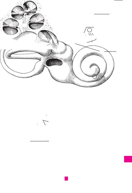
1
19
20
21
22
23
C Bony labyrinth, posterior wall
24
25

|
378 |
Sense organs |
|
|
|
|
|
||
|
|
1 |
Bony spiral lamina. Lamina spiralis ossea. |
15 |
MIDDLE EAR. Auris media. Part of the ear com- |
||||
1 |
|||||||||
|
|
Bilayered bony plate projecting from the modi- |
|
prising the tympanic (middle ear) cavity, audi- |
|||||
|
|
|
olus into the spiral canal of the cochlea in a spi- |
|
tory tube and mastoid cells. |
|
|||
2 |
|
|
ral fashion. Together with the cochlear duct to |
16 |
Tympanic cavity. Cavitas tympanica (cavum |
||||
|
|
|
which it is attached, it forms a complete parti- |
|
tympani). Obliquely oriented space medial to |
||||
|
|
|
tion between the scala vestibuli and scala tym- |
|
|||||
3 |
|
|
|
the tympanic membrane (eardrum). It contains |
|||||
|
|
pani. B |
|
|
|||||
|
|
|
|
the auditory ossicles and communicates post- |
|||||
|
2 |
Hook of spiral lamina. Hamulus laminae spi- |
|
||||||
|
|
erosuperiorly with the mastoid air cells and an- |
|||||||
4 |
|
|
ralis. Free hook-shaped upper end of the os- |
|
teroinferiorly with the nasopharyngeal cavity |
||||
|
|
|
seous spiral lamina at the apex of the cochlea. B |
|
via the auditory tube. |
|
|||
|
|
|
|
|
|||||
5 |
3 |
Helicotrema. Confluence between scala vesti- |
17 |
Tegmental wall (tegmen tympani). Paries |
|||||
|
|
|
buli and scala tympani at the apex of the |
|
tegmentalis. Thin roof of the tympanic cavity. It |
||||
|
|
|
cochlea. It exists because the bony spiral lamina |
|
lies lateral to the arcuate eminence of the |
||||
6 |
|
|
|
||||||
|
|
and the cochlear duct end before reaching the |
|
petrous part of the temporal bone. C |
|||||
|
|
|
apex of the cochlea. B |
|
18 |
Epitympanic recess (attic). Recessus epitym- |
|||
|
|
|
|
||||||
7 |
4 |
Secondary spiral lamina. Lamina |
spiralis |
||||||
|
|
secundaria. Bony ridge situated in the lower |
|
panicus. Dome |
of tympanic |
cavity located |
|||
|
|
|
|
above the upper margin of the tympanic mem- |
|||||
|
|
|
half of the basal turn. It projects from the outer |
|
|||||
8 |
|
|
|
brane and arching superiorly and laterally. C |
|||||
|
|
wall of the spiral canal across from the bony spi- |
|
||||||
|
|
19 |
Cupular part. Pars cupularis. Upper portion of |
||||||
|
|
|
ral lamina. The inferior part of the basilar mem- |
||||||
|
|
|
|||||||
|
|
|
brane extends between these two |
spiral |
|
epitympanic recess. C |
|
||
9 |
|
|
|
|
|||||
|
|
laminae. B |
|
20 |
Jugular wall. Paries jugularis. Floor of the tym- |
||||
|
|
|
|
||||||
|
|
5 Internal acoustic (auditory) meatus. Meatus |
|
panic cavity facing the jugular fossa. C |
|||||
10 |
|
|
|||||||
|
|
acusticus internus. Arises near the posterior |
21 |
Styloid prominence. Prominentia styloidea. |
|||||
|
|
|
wall of the petrous part of the temporal bone. It |
|
Elevation on the floor of the tympanic cavity |
||||
11 |
|
|
is about 1 cm long and transmits the vestibulo- |
|
produced by the styloid process. C |
||||
|
|
cochlear and facial nerves and the labyrinthine |
|
||||||
|
|
|
22 Labyrinthine wall. Paries labyrinthicus. Medial |
||||||
|
|
|
artery and vein. A |
|
|||||
12 |
|
|
|
|
wall of tympanic cavity. C |
|
|||
6 |
Porus acusticus. Outer opening of internal |
|
|
||||||
23 |
Oval window. Fenestra vestibuli [[ovalis]]. It is |
||||||||
|
|
|
acoustic meatus into the posterior wall of the |
||||||
|
|
|
|||||||
13 |
|
|
petrous part of the temporal bone above the |
|
closed by the base of the stapes. C |
||||
|
|
jugular foramen. A |
|
24 |
Fossula fenestrae vestibuli. Small depression |
||||
|
|
|
|
||||||
|
|
|
|
||||||
|
|
7 Fundus of internal auditory meatus. Fundus |
|
in medial wall of tympanic cavity between the |
|||||
14 |
|
|
|||||||
|
|
meatus acustici interni. Floor of meatus, which |
|
malleus and incus. C |
|
||||
|
|
|
is subdivided into several fields. A |
|
25 |
Promontory. |
Promontorium. |
Prominence |
|
15 |
|
|
|
||||||
8 |
Transverse crest. Crista transversa. Trans- |
||||||||
|
caused by the basal turn of the cochlea. C |
||||||||
|
|
|
versely oriented ridge dividing the fundus of |
26 |
Sulcus promontorii. Groove on the promontory |
||||
|
|
|
|||||||
16 |
|
|
the internal acoustic meatus into an upper and |
|
produced by the tympanic nerve from the tym- |
||||
|
|
lower part. A |
|
|
|||||
|
|
|
|
|
panic plexus. C |
|
|
||
|
|
9 Facial nerve area. Area nervi facialis. Region |
|
|
|
||||
|
|
27 |
Subiculum promontorii. Small bony ridge behind |
||||||
17 |
|
||||||||
|
|
containing the beginning of the facial nerve |
|||||||
|
|
|
canal. A |
|
|
the promontory and round window. C |
|||
|
|
|
|
|
|
|
|
||
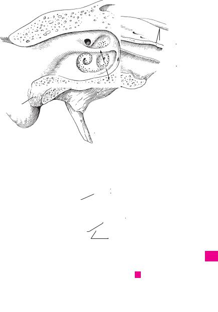
1810 Cochlear area. Area cochleae. Large region below the transverse crest. It contains the foraminous spiral tract. A
1911 Foraminous spiral tract. Tractus spiralis foraminosus. Area perforated by fibers of the spiral
20ganglion forming the cochlear part of the vestibulocochlear nerve. It corresponds to the spiral canal of the cochlea. A
2112 Superior vestibular area. Area vestibularis superior. Region lateral to the facial canal and per-
22forated by fibers of the utriculo-ampullar nerve. A
2313 Inferior vestibular area. Area vestibularis inferior. Region located lateral to foraminous spiral tract and perforated by fibers of the sacular
24nerve. A
14 Small opening for the posterior ampullar nerve.
25Foramen singulare. It lies behind the inferior vestibular area. A
|
|
|
|
|
|
|
|
|
|
|
|
|
|
|
Sense organs 379 |
|
|||||
|
|
|
|
|
|
|
|
|
|
|
|
|
|
|
|
|
|
|
|
|
|
|
|
|
|
|
|
|
|
|
|
|
|
|
|
|
|
|
|
|
|
|
1 |
|
|
|
|
|
|
|
|
|
|
|
|
|
|
|
|
|
|
|
|
|
|
|
|
|
|
|
|
|
|
|
|
|
|
|
|
|
|
|
|
|
|
|
|
|
|
|
|
|
|
|
|
|
|
|
|
|
|
|
|
|
|
|
|
|
2 |
|
|
|
|
|
|
|
|
|
|
|
|
|
9 |
12 |
|
|
|
|
|||
|
|
|
|
|
|
|
|
|
|
|
|
|
|
|
|||||||
|
|
|
|
|
|
|
|
|
|
|
|
|
|
3 |
|||||||
|
|
|
|
|
|
|
|
|
|
|
|
|
|
|
|
|
|
|
|
|
|
|
|
|
|
|
|
|
|
|
|
|
|
|
|
|
|
|
|
|
|
|
|
|
|
|
|
|
|
|
|
|
|
|
|
|
|
|
|
|
|
|
|
|
|
|
|
|
|
|
|
|
|
|
|
|
|
|
|
|
|
|
|
|
|
|
|
3 |
|
|
|
|
|
|
|
|
8 |
|
|
|
|
|
4 |
||||||
|
|
|
|
|
|
|
|
|
|
|
|
|
|||||||||
|
|
|
|
|
|
|
|
|
|
|
|
|
|
||||||||
|
|
|
|
|
|
||||||||||||||||
|
|
|
|
|
|
|
|
|
|
|
|
|
|
|
|
14 |
5 |
||||
|
|
|
|
|
|
|
|
|
|
|
|
|
|
|
|
||||||
|
|
|
|
|
|
|
|
|
|
|
|
|
|
|
|
|
|
|
|||
|
|
|
|
6 |
|
|
|
|
|
|
10 |
|
|
|
|||||||
|
|
|
|
|
|
|
|
|
|
|
|
|
|
|
6 |
||||||
|
|
|
|
|
|
|
|
|
|
|
|
|
|
|
|
|
|
|
|
||
|
|
|
|
|
|
2 |
|
|
|
|
|
|
|
|
|
|
|
|
|
|
|
|
|
|
|
|
|
|
|
|
|
||||||||||||
|
|
|
|
|
|
|
|
7 |
|||||||||||||
|
|
|
|
|
|
|
|
|
|
|
|
|
|
|
|
|
|
|
|
|
|
|
|
|
|
|
|
|
|
|
|
|
|
|
|
|
|
|
|
|
|
|
|
|
|
|
|
|
|
|
|
|
|
|
|
|
11 |
13 |
|
|
|
8 |
|||
1 |
|
|
|
|
|
|
|
|
|
|
|
|
|
|
|
|
|
||||
|
|
|
|
|
|
|
|
|
|
|
|
|
|
|
|
|
|
||||
|
|
|
|
|
|
|
|
|
|
|
|
Internal acoustic meatus |
|
|
|
|
|
|
|||
|
|
|
|
|
|
|
|
|
|
|
A |
|
|
|
|
|
9 |
||||
|
|
|
|
|
|
|
|
|
|
|
|
|
|
|
|
|
|
|
|
|
|
|
|
|
|
|
|
|
|
|
|
|
|
|
|
|
|
|
|
|
|
|
|
|
|
|
|
|
|
|
|
|
|
|
|
|
|
|
|
|
|
|
|
|
|
|
|
|
|
|
|
|
|
|
|
|
|
|
|
|
|
|
|
|
|
|
10 |
1 |
|
|
|
|
|
|
|
|
|
|
|
|
|
|
|
|
|||||
|
|
|
|
|
|
|
|
|
|||||||||||||
4 |
|
|
|
|
|
|
|
|
11 |
||||||||||||
|
|
|
|
|
|
|
|
|
|
|
|
|
|
|
|
||||||
|
|
|
|
|
|
|
|
|
|
||||||||||||
|
|
|
|
|
|
|
|
|
|
|
|
|
|
|
|
|
|
|
|
|
|
|
|
Section of cochlea |
|
|
|
|
|
|
|
|
12 |
||||||||||
|
B |
|
|
|
|
|
|
|
|
||||||||||||
|
|
|
|
|
|
|
|
|
|
|
|
|
|
|
|
|
|
|
|
|
|
|
|
|
|
|
|
|
|
|
|
|
|
|
|
|
|
|
|
|
|
|
|
|
|
|
|
|
|
|
|
|
|
|
|
|
|
|
|
|
|
|
|
|
13 |
|
|
|
|
|
|
|
|
|
|
|
|
|
|
|
|
|
|
|
|
|
|
|
|
|
|
|
|
|
|
|
|
|
|
|
|
|
|
|
|
|
|
|
|
19 |
|
|
|
|
|
|
|
|
|
|
14 |
||||||||||
|
|
|
|
|
|
|
|
|
|
|
|||||||||||
|
|
|
|
|
|
|
|
|
|
|
|
|
|
|
|
|
|
|
|
|
|
17 |
|
|
|
|
|
|
|
|
|
|
|
|
|
15 |
|||||||
|
|
|
|
|
|
|
|
|
|
|
|
|
|
|
|
|
|
|
|
|
|
|
|
|
|
|
|
|
|
|
|
|
|
|
|
|
|
|
|
|
|
|
|
18 |
|
|
|
|
|
|
|
|
|
|
|
|
|
16 |
|||||||
|
|
|
|
|
|
|
|
|
|
|
|
|
|||||||||
23 |
|
|
|
|
|
|
|
|
|
|
|||||||||||
|
|
|
|
|
|
|
|
|
|||||||||||||
|
|
|
|
|
|
|
|
17 |
|||||||||||||
24 |
|
|
|
|
|
|
|
|
|
|
|
||||||||||
|
|
|
|
|
|
|
|
|
|
|
|
||||||||||
|
|
|
|
|
|
|
|
|
|||||||||||||
25 |
|
|
|
|
|
|
|
|
|
||||||||||||
|
26 |
|
|
|
|
|
|
18 |
|||||||||||||
|
|
|
|
|
|
|
|||||||||||||||
|
|
|
|
|
|
|
|
|
|
|
|
|
|
|
|
|
|
|
|
|
|
|
|
|
|
|
|
|
|
|
|
|
|
|
|
|
|
|
|
|
|
|
|
27 |
|
|
|
|
|
|
|
|
|
|
|
|
|
|
19 |
||||||
21 |
|
|
|
|
|
|
|
|
|
|
|
|
|
||||||||
20 |
|
|
|
|
|
|
|
|
|
|
|
|
|
|
|
||||||
20
21
22
C Medial wall
of tympanic cavity 23
24
25

|
380 |
Sense organs |
|
|
|
|
|
|||
|
|
1 |
Tympanic sinus. Sinus tympani. Deep fossa be- |
|
17 |
Mastoid air cells. Cellulae mastoideae. Like |
||||
1 |
|
|||||||||
|
|
|
hind the promontory and round window. D |
|
|
the tympanic cavity, they are lined by squamous |
||||
|
2 |
Round window (fenestra of cochlea). |
|
|
and/or cuboidal epithelium. D |
|||||
2 |
|
18 |
Tympanic cells. Cellulae tympanicae. Small |
|||||||
|
|
|
Fenestra cochleae [[rotunda]]. Round opening at |
|
||||||
|
|
|
|
the end of the scala tympani. It is closed by the |
|
|
cell-like depressions in the floor of the tym- |
|||
|
|
|
|
|
|
|||||
3 |
|
|
|
secondary tympanic membrane. D |
|
|
|
panic cavity. D |
|
|
3 |
Fossula of round window. Fossula fenestrae |
|
19 |
Carotid wall. Paries caroticus. Anterior wall |
||||||
|
|
|||||||||
|
|
|
|
cochleae. Small fossa leading into the round |
|
|
formed partly by the carotid canal, partly by the |
|||
4 |
|
|
|
|
|
|||||
|
|
|
window. D |
|
|
|
|
opening of the auditory tube. D |
||
|
4 |
Crest of |
round window. Crista |
fenestrae |
|
20 |
Membranous |
wall. Paries membranaceus. |
||
|
|
|||||||||
5 |
|
|
|
cochleae. Bony ridge along the edge of the |
|
|
Lateral wall of |
tympanic cavity formed pri- |
||
|
|
|
|
|
marily by the tympanic membrane. B |
|||||
|
|
|
|
round window for attachment of the secondary |
|
|
||||
|
|
|
|
|
|
|||||
|
|
|
|
tympanic membrane. D |
|
|
21 |
Tympanic membrane (eardrum). Membrana |
||
6 |
|
|
|
|
|
|||||
5 |
Cochleariform process. Processus cochleari- |
|
|
tympanica. Obliquely oriented membrane at |
||||||
|
|
|
|
formis. Spoon-shaped bony process found |
|
|
the end of the external acoustic meatus. Diame- |
|||
7 |
|
|
|
|
|
ter: 9−11 mm. A B |
||||
|
|
|
above the promontory at the end of the semi- |
|
|
|||||
|
|
|
|
canal for the tensor tympani muscle. In combi- |
|
22 |
Pars flaccida [[Shrapnell’s membrane]]. Smaller, |
|||
|
|
|
|
|
||||||
|
|
|
|
nation with a connective tissue loop, it serves as |
|
|
more flaccid part of tympanic membrane lo- |
|||
8 |
|
|
|
|
|
|||||
|
|
|
a pulley for the muscle. D |
|
|
|
cated above the anterior and posterior mallear |
|||
|
|
|
6 Secondary tympanic membrane. Membrana |
|
|
folds. A B |
|
|||
9 |
|
|
|
|
|
|||||
|
|
|
23 |
Pars tensa. By far, the largest part of the tym- |
||||||
|
|
|
tympani secundaria. Sheath across the round |
|
||||||
|
|
|
|
window that forms a membranous partition be- |
|
|
panic membrane, situated within the tympanic |
|||
|
|
|
|
|
|
|||||
10 |
|
|
|
tween the scala tympani and the tympanic cav- |
|
|
ring. A B |
|
||
|
|
|
ity. |
|
|
|
24 |
Anterior malleolar fold (6). Plica mallearis |
||
|
|
|
|
|
|
|
||||
|
|
|
7 Mastoid wall. Paries mastoideus [adnexa mas- |
|
|
anterior. Fold on the inner surface of the tym- |
||||
11 |
|
|
|
|
||||||
|
|
|
toidea]. Components connected with the mas- |
|
|
panic membrane. It has a concave lower edge |
||||
|
|
|
|
toid process. |
|
|
|
and extends anteriorly from the base of the |
||
|
|
|
|
|
|
|
||||
12 |
|
|
|
|
|
|
manubrium of the malleus. B |
|||
8 |
Mastoid (posterior) wall. Paries mastoideus. |
|
|
|||||||
|
25 Posterior malleolar fold (6). Plica mallearis |
|||||||||
|
|
|
|
Posterior wall of middle ear cavity facing the |
|
|||||
|
|
|
|
|
|
posterior. Fold on the inner surface of the tym- |
||||
13 |
|
|
|
mastoid process. D |
|
|
|
|||
|
|
|
|
|
|
panic membrane. It has a concave lower edge |
||||
9 |
Mastoid antrum. Antrum mastoideum. Closed |
|
|
|||||||
|
|
|
and extends posteriorly from the root of the |
|||||||
|
|
|
|
space, the posterosuperior part of which is con- |
|
|
||||
14 |
|
|
|
|
|
manubrium of the malleus. B |
||||
|
|
|
tinuous with the tympanic cavity. The inferior |
|
|
|||||
|
|
|
|
26 |
Mallear prominence. Prominentia mallearis. |
|||||
|
|
|
|
edge communicates with the mastoid cells. D |
|
|||||
|
|
|
|
|
|
Small elevation on the outside of the tympanic |
||||
15 |
10 |
Aditus ad antrum. Entrance to the mastoid an- |
|
|
||||||
|
|
membrane caused by the lateral process of the |
||||||||
|
|
|
|
trum from the tympanic cavity. D |
|
|
|
maleus. A |
|
|
|
|
|
|
|
|
|
|
|||
16 |
11 |
Prominentia canalis semicircularis lateralis. Prom- |
|
27 |
Stria mallearis. A bright band that appears on |
|||||
|
|
|
|
inence on the wall of lateral semicircular canal, |
|
|
the outer surface of the tympanic membrane |
|||
|
|
|
|
situated above the prominence of the facial |
|
|
due to the underlying manubrium of the mal- |
|||
17 |
|
|
|
|
|
|||||
|
|
|
canal. D |
|
|
|
|
leus, which is fused with the tympanic mem- |
||
|
|
12 Prominence of facial nerve canal. Prominentia |
|
|
brane. A |
|
||||
|
|
|
|
|||||||
18 |
|
|
|
canalis facialis. Bulge lying between the oval |
|
28 |
Umbo of tympanic membrane. Umbo mem- |
|||
|
|
|
|
window and prominence of the lateral semi- |
|
|
branae tympani. Structure at the tip of the man- |
|||
|
|
|
|
circular canal. D |
|
|
|
ubrium of the malleus, where the tympanic |
||
19 |
|
|
|
|
|
|
||||
13 |
Pyramidal eminence (pyramid). Eminentia |
|
|
membrane is drawn inward. A |
||||||
|
|
|
|
pyramidalis. Small, bony pyramidal projection |
|
29 |
Cutaneous layer. Stratum cutaneum. The layer |
|||
20 |
|
|
|
at the level of the oval window. Its apex is per- |
|
|
of stratified squamous epithelium on the exter- |
|||
|
|
|
|
forated. The eminence contains the stapedius |
|
|
nal surface of the tympanic membrane. C |
|||
|
|
|
|
muscle, the tendon of which emerges from it. D |
|
30 |
Fibrocartilaginous ring. Anulus fibrocar- |
|||
21 |
|
|
|
|
||||||
14 |
Incudal fossa. Fossa incudis. Small depression |
|
|
tilagineus. Attaches the tympanic membrane in |
||||||
|
|
|
|
in the aditus ad antrum for the posterior liga- |
|
|
the tympanic sulcus. C |
|||
22 |
|
|
|
|
|
|||||
|
|
|
ment of the incus. D |
|
|
31 |
Radiate layer. Stratum radiatum. Outer group |
|||
|
|
15 |
Sinus posterior. Small groove between the in- |
|
|
of radially oriented fibers of the tympanic |
||||
23 |
|
|
|
cudal fossa and pyramid. D |
|
|
|
membrane. C |
|
|
|
|
|
|
|
32 |
Circular layer. Stratum circulare. Inner group of |
||||
|
|
16 |
Typmpanic aperture of canaliculus of |
|
||||||
24 |
|
|
|
chorda |
tympani. Apertura |
tympanica |
|
|
circularly oriented fibers of the tympanic mem- |
|
|
|
|
canaliculi chordae tympani. Opening of chorda |
|
|
brane. C |
|
|||
|
|
|
|
tympani canal into tympanic cavity. It lies at the |
|
33 |
Mucosal layer. Stratum mucosum. Layer of |
|||
25 |
|
|
|
posterior margin of the tympanic membrane at |
|
|
simple squamous epithelium covering the |
|||
|
|
|
|
the level of the pyramid. D |
|
|
|
inner surface of the tympanic membrane. C |
||
|
|
|
|
|
|
|
|
|
|
|
Sense organs 381
|
|
|
|
|
|
|
|
|
|
|
|
|
|
|
|
|
1 |
|
|
|
|
|
|
|
|
|
|
|
|
|
|
25 |
|
|
|
|
|
|
|
|
|
|
|
|
|
|
|
|
|
|
|
|
|
|
|
|
|
|
|
|
|
|
|
|
22 |
|
|
|
2 |
||
|
|
|
|
|
|
|
|
|
|
|
|
|
|
|
|||
|
|
|
|
|
|
|
|
|
|
|
|
|
|
|
|
|
|
|
|
|
|
|
|
|
|
|
|
|
|
|
|
|
|
|
|
23 |
|
|
|
22 |
|
|
|
|
|
|
|
|
|
|
|
|
3 |
26 |
|
|
|
|
|
|
|
|
24 |
|
|
|
|
|
|
||
|
|
|
|
|
|
|
|
|
|
|
|
|
|
4 |
|||
|
27 |
|
|
|
|
|
|
|
|
|
|
|
|
|
20 |
||
|
|
|
|
|
|
|
|
|
|
|
|
|
23 |
|
|||
|
|
|
|
|
|
|
|
|
|
|
|
|
|
||||
|
28 |
|
|
|
|
|
|
|
|
|
|
|
|
|
5 |
||
|
|
|
|
|
|
|
|
|
|
|
|
|
|
|
|||
|
|
|
|
|
|
|
|
|
|
|
|
|
|
|
|
||
|
|
|
|
|
|
|
|
|
|
|
|
|
|
|
|
||
|
|
|
|
|
|
|
|
|
|
|
|
|
|
|
|
|
6 |
|
|
|
|
|
|
|
|
|
|
|
|
|
|
|
|
|
|
|
|
|
|
|
|
|
|
|
|
|
|
|
|
|
|
|
|
|
|
|
|
|
|
|
|
|
|
31 |
|
|
|
|
|
|
7 |
|
|
Right eardrum, |
32 |
|
|
|
|
|
Lateral wall of |
|
|
|
|
||||
|
|
|
|
|
|
|
|
|
|
|
|
|
|
||||
|
|
|
|
|
|
|
|
|
|
|
|
|
|
||||
|
A |
|
|
|
|
29 |
|
B |
|
|
|
8 |
|||||
|
|
|
|
|
|
|
|||||||||||
|
|
external view |
33 |
|
|
|
|
|
|
|
|
tympanic cavity |
|
|
|
|
|
|
|
|
|
|
|
|
|
|
|
|
|||||||
|
|
|
|
|
|
|
|
|
|
|
|
|
|
|
|
||
9
10
11
30
12
C Attachment of eardrum
|
|
|
|
|
13 |
9 |
|
|
|
|
14 |
|
|
|
5 |
|
|
|
|
|
|
|
15 |
11 |
|
|
|
|
16 |
12 |
|
|
|
|
|
10 |
|
|
|
|
|
14 |
|
|
|
|
17 |
15 |
|
|
|
|
|
13 |
|
|
4 |
19 |
18 |
|
|
|
|||
|
1 |
3 |
2 |
18 |
|
8 |
|
19 |
|||
|
|
|
|
16 |
|
|
|
20 |
17 |
|
|
|
|
|
|
|
|
|
|
|
|
21 |
|
|
|
Medial wall of tympanic cavity |
|
|
|
|
|
|
|
|
|
|
|
|
|
D |
|
22 |
|
|
|
|
|
|
|
|
|
|
23 |
|
|
|
|
24 |
|
|
|
|
25 |
|
|
|
|
|