Материал: Атлас Ханц фениш

|
362 |
Sense organs |
|
|
|
|||
|
|
1 |
Posterior chamber. Camera posterior. It ex- |
14 |
Lens fibers. Fibrae lentis. Fibers corresponding |
|||
1 |
||||||||
|
|
tends from the iris and ciliary body to the ante- |
|
to the lens epithelium from which they develop. |
||||
|
|
|
rior surface of the vitreous. A |
|
They form the lens substance measuring 2.5− |
|||
2 |
2 |
Aqueous humor. Humor aquosus. Produced by |
|
12 µm thick and up to 10 mm long. C |
||||
|
|
|
the ciliary processes. It flows between the in- |
15 |
Epithelium of lens. Epithelium lentis. Part of |
|||
|
|
|
||||||
3 |
|
|
terstices of the suspensory ligaments of the lens |
|
the lens confined to the anterior surface and ex- |
|||
|
|
to the anterior surface of the lens and then be- |
|
tending as far as the equator. It is derived |
||||
|
|
|
tween the iris and lens to the pupil, through |
|
embryologically from the anterior epithelium |
|||
4 |
|
|
which it enters the anterior chamber. |
|
of the lens vesicle. C |
|||
|
3 |
Vitreous chamber. Camera vitrea. Space filled |
16 |
Lens capsule. Capsula lentis. Transparent mem- |
||||
|
||||||||
5 |
|
|
up by the vitreous body. B |
|
brane, up to 15 µm thick, covering the lens in- |
|||
4 |
Vitreous body. Corpus vitreum. It consists of |
|
cluding its epithelium. Its anterior pole is |
|||||
|
|
|||||||
|
|
|
about 98% water and primarily contains traces |
|
thicker than the posterior pole. It gives attach- |
|||
6 |
|
|
|
|||||
|
|
of protein and NaCl and a mixture of fine fibrils |
|
ment to the suspensory ligaments. C |
||||
|
|
|
which thicken near the surface to form a lim- |
17 |
Anterior pole. Polus anterior. D |
|||
|
|
|
||||||
7 |
|
|
iting membrane. It has a gelatinous consistency |
18 |
Posterior pole. Polus posterior. D |
|||
|
|
due to its high content of hyaluronic acid. A |
||||||
|
|
|
19 |
Anterior surface. Facies anterior. Less curved |
||||
|
5 |
Hyaloid |
artery. [A. hyaloidea]. Branch of the |
|||||
|
||||||||
8 |
|
|
ophthalmic artery supplying the vascular mem- |
|
lens surface with a radius of 8.3−10 mm. C |
|||
|
|
20 |
Posterior surface. Facies posterior. More |
|||||
|
|
|
brane of the lens. Present only during embry- |
|||||
9 |
|
|
onic development. The proximal portion per- |
|
curved lens surface with a radius of about |
|||
|
|
|
6.5 mm. C |
|||||
|
|
|
sists in the optic nerve as the central retinal |
|
||||
|
|
|
artery. B |
|
|
21 |
Axis. Line connecting anterior and posterior |
|
10 |
|
|
|
|
||||
6 |
Hyaloid canal. Canalis hyaloideus. Canal |
|
poles. D |
|||||
|
|
|
within the vitreous body formerly occupied by |
22 |
Equator. Margin of lens. D |
|||
11 |
|
|
the embryonic hyaloid artery which degener- |
23 |
Radii of lens. Suture line of the individual lens |
|||
|
|
|
ates in |
this region. |
The canal assumes a |
|
fibers. In the young it resembles a triradiate |
|
|
|
|
downward sagging corkscrew shape; it extends |
|
||||
12 |
|
|
|
seam. D |
||||
|
|
from the optic disc to the posterior surface of |
|
|||||
|
|
24 Ciliary zonule. Zonula ciliaris. Suspensory ap- |
||||||
|
|
|
the lens. Its wall is formed by condensed fibers. |
|||||
|
|
|
||||||
|
|
|
|
paratus together with its interstices. It encircles |
||||
13 |
|
|
A |
|
|
|
||
|
|
|
|
|
the lens equator and consists of a radially |
|||
7 |
Hyaloid (lenticular, patellar) fossa. Fossa hy- |
|
||||||
|
|
oriented system of fibers of variable length and |
||||||
|
|
|
aloidea. Fossa on the anterior surface of the vit- |
|
||||
14 |
|
|
|
the folds situated between them. C |
||||
|
|
reous body adjacent to the lens. A |
|
|||||
|
|
25 |
Zonular fibers (suspensory ligaments). Fi- |
|||||
|
8 |
Vitreous |
(hyaloid) |
membrane. Membrana |
||||
15 |
|
brae zonulares. Suspensory fibers attached to |
||||||
|
|
vitrea. Condensation of fibers on the surface of |
|
|||||
|
|
|
the equator and the adjacent anterior and post- |
|||||
|
|
|
the vitreous body. See (4), vitreous body. A |
|
||||
|
|
|
|
erior surfaces of the lens. They arise distally |
||||
16 |
9 |
Stroma of vitreous body. Stroma vitreum. |
|
from the basal lamina of the ciliary body and the |
||||
|
|
|
Fine network of fibers in the virtreous body. Its |
|
pars ciliaris retinae. C |
|||
|
|
|
surface thickens to form the vitreous mem- |
26 |
Zonular spaces. Spatia zonularia. Spaces be- |
|||
17 |
|
|
||||||
|
|
brane. |
|
|
||||
|
|
|
|
|
tween the zonule fibers filled with percolating |
|||
|
|
9 a Vitreous humor. Humor vitreus. Fluid part of |
|
|||||
|
|
|
aqueous humor. C |
|||||
18vitreous body. Primarily consists of mucupolysaccharides and is situated between the fibers of the stroma.
1910 LENS. Structure of the eye situated between the pupil and vitreous body. It is suspended by the
20ciliary zonule (suspensory ligaments), has a diameter of 9−10 mm and is about 4 mm thick. B C D
2111 Substantia lentis. Lens substance situated beneath the lens epithelium and comprising
22the lens nucleus and lens cortex with a refractive index of 1.44−1.55. C
2312 Lens cortex. Cortex lentis. External zone of the lens. It is softer owing to its high water content and blends into the lens nucleus without a
24sharp boundary. C
13 Nucleus of lens. Nucleus lentis. Harder core of
25the lens with a low water content, as is especially evident in the elderly. C

|
364 |
Sense organs |
|
|
|
|
|
||
|
|
1 |
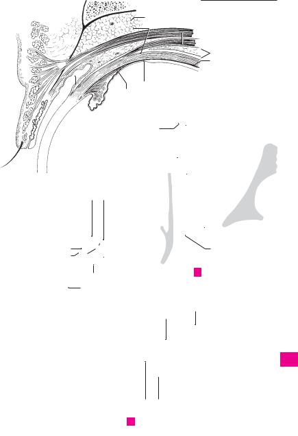
ACCESSORY ORGANS OF EYE. Organa oculi ac- |
15 |
Superficial lamina of levator tendon. |
||||
1 |
|||||||||
|
|
cessoria. |
|
|
|
|
Lamina superficialis. It passes between the tar- |
||
|
2 |
Muscles of eye. Musculi bulbi. Extrinsic ocular |
|
sus and orbicularis oculi to insert into the sub- |
|||||
2 |
|
cutaneous connective tissue of the upper eyelid. |
|||||||
|
|
muscles. |
|
|
|
|
|||
|
3 |
Orbital muscle. M. orbitalis. Thin layer of |
|
It is so broad that it extends mainly laterally to |
|||||
|
|
the wall of the orbit. A |
|||||||
3 |
|
||||||||
|
|
smooth muscle which bridges the inferior orbi- |
16 |
Deep lamina of levator tendon. Lamina pro- |
|||||
|
|
|
tal fissure. C |
|
|
|
|||
|
|
|
|
|
|
|
funda. It inserts into the upper margin and the |
||
|
4 |
Superior rectus. M. rectus superior. o: Common |
|
||||||
4 |
|
anterior surface of the tarsus. A |
|||||||
|
|
|
tendinous ring. i: Along an oblique line in front |
17 |
Orbital fasciae. Fasciae orbitales. |
||||
|
|
|
|||||||
5 |
|
|
of the equator, 7−8 mm posterior to the corneal |
||||||
|
|
18 |
Periosteum of orbit. Periorbita. It is delicate |
||||||
|
|
margin. A: Elevation and medial rotation of su- |
|||||||
|
|
|
perior pole of eyeball. I: Oculomotor nerve. B C |
|
and fused solidly to the bone at the inlet and |
||||
|
|
|
|
||||||
6 |
|
|
D |
|
|
|
|
outlet of the orbit. Anteriorly, it is continuous |
|
5 |
Inferior rectus. M. rectus inferior. o: Common |
|
with the adjacent periosteum, posteriorly with |
||||||
|
|
||||||||
|
|
the dura. A |
|||||||
7 |
|
|
tendinous ring. i: Along an oblique line about |
|
|||||
|
|
19 |
Orbital septum. Septum orbitale. Connective |
||||||
|
|
6 mm behind the corneal margin. A: Depression |
|||||||
|
|
|
and lateral rotation of superior pole of eyeball. I: |
|
tissue septum partly reinforced by tendon. It |
||||
8 |
|
|
Oculomotor nerve. B C D |
|
|
|
passes from the orbital margin below the orbic- |
||
|
6 |
Medial rectus. M. rectus medialis. o: Common |
|
ularis oculi to the external margins of the tarsi |
|||||
|
|
and forms the anterior end of the orbit. A |
|||||||
9 |
|
|
tendinous ring. i: About 5.5 mm from the cor- |
|
|||||
|
|
|
Muscular fasciae. Fasciae musculares. Sheaths |
||||||
|
|
|
neal margin. A: Adduction of corneal pole. I: |
20 |
|||||
|
|
|
Oculomotor nerve. B C |
|
|
|
of Tenon’s capsule enveloping the tendons and |
||
10 |
|
|
|
|
|
||||
7 |
Lateral rectus. M. rectus lateralis. o: Common |
|
muscular bellies of the 6 extrinsic ocular |
||||||
|
|
muscles. A |
|||||||
|
|
|
tendinous ring and lesser wing. i: 5.5 mm be- |
|
|||||
11 |
|
|
21 |
Tenon’s capsule (fascia bulbi). Vagina bulbi. |
|||||
|
|
hind corneal margin. A: Abduction of corneal |
|||||||
|
|
|
pole. I: Abducent nerve. B C D |
|
|
|
Connective tissue gliding membrane between |
||
|
|
|
|
|
|
||||
|
8 |
Tendon of lateral rectus at greater wing. |
|
the eyeball and orbital fat. It is fused to the |
|||||
12 |
|
||||||||
|
sclera posteriorly at the optic nerve. Anteriorly |
||||||||
|
|
|
Lacertus musculi recti lateralis. C |
|
|
|
it ends beneath the conjunctiva. It is separated |
||
|
|
|
|
|
|
||||
13 |
9 |
Common tendinous ring (common annular |
|
from the sclera primarily by the episcleral |
|||||
|
|
tendon). Anulus tendineus communis. Ten- |
|
space. A |
|||||
|
|
|
|
||||||
|
|
|
dinous ring for attachment of the recti ocular |
22 Episcleral space. Spatium episclerale [[inter- |
|||||
14 |
|
|
|||||||
|
|
muscles. It surrounds the optic canal and me- |
|||||||
|
|
|
vaginale]]. Gliding space between the eyeball |
||||||
|
|
|
dial part of the superior orbital fissure. C |
|
|
||||
|
|
|
|
|
and Tenon’s capsule. It is traversed by long, deli- |
||||
15 |
10 |
Superior oblique. M. obliquus superior. o: Body |
|
cate connective tissue fibers. A |
|||||
|
|
|
of sphenoid medial to common tendinous ring. |
23 Orbital fat body. Corpus adiposum orbitae. |
|||||
|
|
|
|||||||
16 |
|
|
i: Posterolateral |
aspect of sclera |
behind |
the |
|
Adipose tissue fills the spaces around the ocular |
|
|
|
equator after its |
tendon passes |
through |
the |
|
|||
|
|
|
muscles, the eyeball and the optic nerve and is |
||||||
|
|
|
trochlea and approaches sclera obliquely from |
|
bordered anteriorly by the orbital septum. A D |
||||
17the medial margin of orbit. A: Abduction, medial rotation and depression. I: Trochlear nerve. B
1811 Trochlea. Cartilaginous sling attached to the medial wall of the orbit [[trochlear spine]] and
19serving as a pulley for the tendon of the superior oblique muscle. B
2012 Tendon sheath of superior oblique muscle
(synovial bursa of trochlea). Vagina tendinis m. obliqui superioris [[bursa synovialis
21trochlearis]]. Synovial sheath (bursa) for the tendon of the superior oblique muscle separat-
22ing the tendon from the trochlea. B
13 Inferior oblique. M. obliquus inferior. o: Lateral to the nasolacrimal canal. i: Posterior to equa-
23tor. A: Elevation, abduction and lateral rotation. I: Oculomotor nerve. D
2414 M. levator palpebrae superioris. o: Bone above optic canal and dura of optic nerve. Its tendon
25broadens anteriorly and splits to form an upper and lower layer. I: Oculomotor nerve. A C D
 7
7
|
366 |
Sense organs |
|
|
|
|||
|
|
1 |
Eyebrow. Supercilium. The transverse eleva- |
19 |
Lateral palpebral raphe. [[Raphe palpebralis |
|||
1 |
||||||||
|
|
tion above the eyes, covered by thick, bristle- |
|
lateralis]]. Delicate band on the lateral palpebral |
||||
|
|
|
like hairs. A |
|
|
|
ligament. It is reinforced by the orbicularis oculi |
|
2 |
2 |
Eyelids. Palpebrae. |
|
|
muscle. D |
|||
|
20 |
Lateral palpebral ligament. Lig. palpebrale |
||||||
|
3 |
Upper eyelid. Palpebra superior. A |
||||||
3 |
|
laterale. Fibrous band that attaches the lateral |
||||||
|
|
|
|
|
|
|||
|
4 Lower eyelid. Palpebra inferior. A |
|
palpebral commissure to the lateral wall of the |
|||||
|
|
|
||||||
|
|
5 Anterior palpebral surface. Facies anterior pal- |
|
orbit in front of the orbital septum. B |
||||
4 |
|
|
||||||
|
21 |
Tarsal [[Meibomian]] glands. Glandulae tar- |
||||||
|
|
pebralis. The anterior external (skin-covered) |
||||||
|
|
|
||||||
|
|
|
surface of the eyelid. E |
|
|
sales. Elongated holocrine glands located in the |
||
5 |
|
|
|
|
||||
6 |
Epicanthus |
(mongolian fold). [Plica palpe- |
|
superior and inferior tarsal plates with openings |
||||
|
|
near the posterior edge of the free margin of the |
||||||
|
|
|
bronasalis] [[epicanthus]]. Vertical fold covering |
|
||||
|
|
|
|
eyelids. They produce a sebaceous secretion for |
||||
6 |
|
|
the medial angle of the eye. It is a continuation |
|
||||
|
|
|
lubrication of the lid margins. E |
|||||
|
|
|
of the upper eyelid at the lateral nasal wall. C |
22 Superior tarsal muscle. M. tarsalis superior. |
||||
|
|
|
||||||
7 |
7 |
Posterior palpebral surface. Facies posterior |
||||||
|
Smooth muscle fibers between the muscle-ten- |
|||||||
|
|
|
palpebralis. |
Surface lined |
by conjunctival |
|
don border of the levator palpebrae muscle and |
|
|
|
|
|
|||||
|
|
|
epithelium |
and containing |
dispersed goblet |
|
||
8 |
|
|
|
the superior tarsal plate. E |
||||
|
|
cells. E |
|
|
|
|||
|
|
|
|
23 |
Inferior tarsal muscle. M. tarsalis inferior. |
|||
|
8 |
Palpebral fissure. Rima palpebrarum. Space |
||||||
9 |
|
Smooth muscle fibers between the inferior for- |
||||||
|
|
between the margins of the upper and lower |
|
nix of the conjunctiva and the inferior tarsal |
||||
|
|
|
eyelids. A E |
|
|
|
plate. E |
|
10 |
|
|
|
|
|
|||
9 |
Lateral palpebral commissure. Commissura |
24 |
Tunica conjunctiva. The lining of the inner sur- |
|||||
|
|
|
palpebralis lateralis. Lateral junction of the |
|
face of the eyelids, which consists of two or more |
|||
|
|
|
|
|||||
11 |
|
|
upper and lower eyelids. A |
|
|
layers of columnar epithelium with goblet cells |
||
10 |
Medial palpebral commissure. Commissura |
|
and a loose, cell-rich lamina propria containing |
|||||
|
|
|||||||
|
|
multiple blood vessels. The tunica extends |
||||||
12 |
|
|
palpebralis medialis. Medial junction of the |
|
||||
|
|
|
around the fornix of the conjunctiva to the eye- |
|||||
|
|
upper and lower eyelids. A |
|
|
||||
|
11 Lateral angle (canthus) of eye. Angulus oculi |
|
ball, which it covers with a layer of stratified |
|||||
13 |
|
squamous epithelium that extends up to the cor- |
||||||
|
|
lateralis. Acute lateral angle of the eye; it is also |
|
neal margin. E |
||||
|
|
|
the lateral end of the palpebral fissure. A |
25 |
Semilunar fold of conjunctive. Plica semi- |
|||
14 |
|
|
||||||
12 |
Medial angle (canthus) of eye. Angulus oculi |
|||||||
|
lunaris conjunctivae. It lies in the medial angle of |
|||||||
|
|
|
medialis. More rounded medial end of the |
|
the eye between the fornix of the upper and |
|||
15 |
|
|
palpebral fissure which delimits a triangular |
|
lower eyelid. F |
|||
|
|
|
space, the lacrimal lake. A |
|
26 Lacrimal caruncle. Caruncula lacrimalis. Mu- |
|||
|
13 |
Limbi palpebrales anteriores. Anterior edges |
||||||
16 |
|
cosal mass in the medial angle of the eye covered |
||||||
|
|
|
of the free margins of the eyelids adjacent to the |
|
by stratified squamous or columnar epithelium. |
|||
|
|
|
external skin. E |
|
|
F |
||
1714 Limbi palpebrales posteriores. Posterior edges of the free margins of the eyelids adjacent to the
18conjunctiva. E
15 Eyelashes. Cilia. The 3−4 rows of hair growing
19near the anterior edge of the free margin of the eyelids. E F
2016 Superior tarsal plate. Tarsus superior. Curved plate about 10 mm high occupying the upper eyelid and consisting of compact, interwoven
21collagenous connective tissue with tarsal glands. B E
2217 Inferior tarsal plate. Tarsus inferior. Plate about
5 mm high within the lower eyelid. It likewise consists of firm, interwoven collagenous con-
23nective tissue with tarsal glands. B E
18 Medial palpebral ligament. [[Lig. palpebrale
24mediale]]. Band of connective tissue between the medial palpebral commissure and the medial
25wall of the orbit. It lies in front of the lacrimal sac. B D