Материал: Атлас Ханц фениш

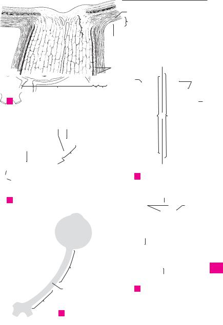
Autonomic nervous system |
353 |
|
|
|
|
|
|
21 |
|
1 |
|
|
|
|
|
|
|
|
2 |
|
1 |
|
|
22 |
|
A |
3 |
2 |
Cervical sympathetic trunk |
||
5 |
|
|
|
|
3 |
4 |
|
6 |
|
||
|
|
|
|
7 |
|
|
5 |
8 |
|
13 |
|
23 |
|
|
|
|
12 |
6 |
|
|
|
||
11 |
|
9 |
|
|
5 |
||
|
|
|
|
|
|
|
|
24 |
10 |
|
|
|
4 |
|
|
8 |
7 |
|
|
|
|
|
|
|
|
|||||||
|
|
|
|
|
|
|
|
|
|
|
||||
|
|
|
|
|
|
|
|
|
|
|
|
|
|
|
34 |
|
|
25 |
|
|
|
|
|
|
|
8 |
|||
|
|
|
|
|
|
|
|
|
||||||
|
|
|
|
|
|
|
|
|
|
|
|
|||
|
|
|
|
|
|
|
|
|
|
|||||
|
|
|
|
|
|
|
15 |
|
|
|
|
|
9 |
|
|
Lumbosacral sympathetic trunk |
|
|
|
|
|
|
|
||||||
|
|
|
|
|
|
|||||||||
|
|
|
|
|
|
|
|
|
|
|||||
B |
14 |
|
|
|
|
|
|
|
10 |
|||||
|
|
|
|
|
|
|
|
|
|
15 |
|
|
||
|
|
|
|
|
|
|
|
|
|
|
|
|
|
|
11
12
13
27 |
|
|
|
|
|
|
|
|
|
|
|
|
|
|
14 |
||
|
|
|
|
|
|
|
|
|
|
|
|
14 |
|
|
|||
|
|
|
|
|
|
|
|
|
|
|
|
|
|
||||
|
|
C |
Terminal nerves |
|
|
|
|
15 |
|||||||||
|
|
|
|
|
|
|
|
|
|
||||||||
|
|
|
|
|
|
|
|
|
|
|
|
|
|
|
|
|
|
|
|
|
|
|
|
|
|
|
|
|
|
|
|
|
|
|
|
|
|
|
|
|
|
|
|
|
|
|
|
|
|
|
|
|
16 |
|
|
|
|
|
|
|
|
|
|
|
|
|
|
|
|
|
|
|
|
|
|
|
|
|
|
|
|
|
|
|
|
|
|
|
|
29 |
|
|
|
|
|
|
|
|
|
|
|
|
17 |
||||
|
|
|
|
|
|
|
|
|
|
|
|
|
|
|
|
|
|
|
|
|
|
|
|
|
|
|
|
|
|
|
|
|
|
|
|
|
|
|
|
|
|
|
|
|
|
|
|
|
|
|
|
|
18 |
|
|
|
|
|
|
31 |
16 |
|
|
|
|
|
|
|
|
||
|
|
|
|
|
|
|
|
||||||||||
|
|
|
|
|
|
|
|
19 |
|||||||||
|
|
|
|
|
|
|
|
|
|
|
|
|
|
|
|
||
|
|
|
|
|
|
|
|
|
|
|
|
|
|
|
|
||
30 |
|
|
|
|
|
|
|
|
|
|
|
|
|
||||
17 |
|
|
|
|
|
|
|
20 |
|||||||||
|
|
|
|
|
|
|
|
|
|
|
|
|
|
|
|
||
|
|
|
|
|
|
|
|
|
18 |
|
|
|
|
|
|
|
|
|
|
|
|
|
|
|
|
|
|
|
|
|
|
21 |
|||
|
|
|
|
|
|
|
|
|
|
|
|
|
|
|
|
||
|
|
|
|
|
|
|
|
|
20 |
|
|
|
|
|
|
|
|
|
|
|
|
|
|
|
|
|
|
|
|
|
|
|
|
|
|
|
|
|
|
|
|
32 |
|
19 |
|
|
|
|
|
|
|
22 |
|
|
|
|
|
|
|
||||||||||||
|
|
|
|
|
|
|
|
|
|
|
|
|
|
|
|
|
|
|
|
|
|
|
|
|
|
|
|
|
|
|
|
|
|
|
23 |
|
|
|
|
|
|
|
|
|
|
|
|
|
|
|
|
|
24 |
|
Autonomic ganglia of the head |
|
|
|
Splanchnic nerves |
|
|
||||||||||
D |
|
E |
|
25 |
|||||||||||||
|
|
|
|
|
|
|
|
|
|
|
|
|
|
|
|
|
|
|
|
|
|
|
|
|
|
|
|
|
|
|
|
|
|
|
|

|
354 |
|
Sense organs |
|
|
|
|
|
|
|||||
|
|
1 |
|
|
|
Organa sensoria (sensuum). |
19 |
Meridians. Meridiani. Semicircles oriented at |
||||||
|
|
|
SENSE ORGANS. |
|||||||||||
1 |
|
|||||||||||||
|
|
|
|
In the narrow sense, the organs of vision, hear- |
|
right angles to the equator between the anterior |
||||||||
|
|
|
|
ing, smell and taste. |
|
|
|
and posterior poles. D. |
|
|||||
2 |
|
|
|
|
|
|
|
|||||||
2 |
|
ORGAN OF VISION. Organum visus (visuale). |
20 |
External axis of eyeball. Axis bulbi externus. |
||||||||||
|
3 |
|
EYE. Oculus. |
|
|
|
|
|
Line connecting anterior and posterior poles. C |
|||||
|
|
|
|
|
|
|
||||||||
3 |
4 |
|
Optic nerve. N. opticus. Fiber bundle beginning |
21 |
Internal axis of eyeball. Axis bulbi internus. |
|||||||||
|
|
|
|
in the retina and extending as far as the optic |
|
Distance from posterior surface of cornea to the |
||||||||
|
|
|
|
|
||||||||||
|
|
|
|
chiasm. |
Histologically |
and |
embryologically |
|
inner surface of retina measured along an im- |
|||||
4 |
|
|
|
|
||||||||||
|
|
|
speaking, it is the tract of the brain that is ac- |
|
aginary line (external axis of eyeball) through |
|||||||||
|
|
|
|
cordingly enclosed by meninges up to the pos- |
|
the anterior and posterior poles. C |
||||||||
5 |
|
|
|
terior aspect of the eyeball. Its axons have no |
22 |
Optic axis. Axis opticus. Line passing through |
||||||||
|
|
|
|
neurilemma (sheath of Schwann) but are myeli- |
|
the midline of the cornea and lens and bisecting |
||||||||
|
|
|
|
|
||||||||||
|
|
|
|
nated. The myelin sheath is formed by the oligo- |
|
the retina between the fovea centralis and optic |
||||||||
6 |
|
|
|
|
||||||||||
|
|
|
dendroglia. A C E |
|
|
|
disc. C |
|
|
|||||
|
5 |
Intracranial part. Pars intracranialis. Segment |
23 |
FIBROUS TUNIC OF EYEBALL. Tunica fibrosa |
||||||||||
7 |
|
|
|
of the optic nerve betweeen the optic canal and |
||||||||||
|
|
|
|
bulbi. External wall of eyeball comprising the |
||||||||||
|
6 |
|
the chiasm. E |
|
|
Pars |
intracanicularis. |
|
cornea and sclera. C |
|
||||
|
|
|
|
|
|
|||||||||
8 |
|
Intracanalicular part. |
24 |
Sclera. The bluish-white outer coat of the eye- |
||||||||||
|
|
|
Segment of the optic nerve located in the optic |
|||||||||||
|
|
|
|
|
ball, which |
consists of |
irregulatory arranged |
|||||||
|
|
|
|
canal. It is partially connected with the canal |
|
|||||||||
9 |
|
|
|
|
collagenous fibers visible through the conjunc- |
|||||||||
|
|
|
wall. E |
|
|
|
|
|
|
|||||
7 |
|
part. Pars orbitalis. Slightly tortuous |
|
tiva. A B C |
|
|
||||||||
|
Orbital |
25 Scleral sulcus. Sulcus sclerae. Shallow groove |
||||||||||||
10 |
|
|
|
segment of the optic nerve measuring about |
||||||||||
|
|
|
|
between the cornea and sclera caused by the |
||||||||||
|
|
|
3 cm in length and occupying the orbit. E |
|
||||||||||
|
|
|
|
|
greater curvature of the cornea. B C D |
|||||||||
|
|
8 Intraocular part. Pars intraocularis. Segment of |
|
|||||||||||
11 |
|
26 |
Corneoscleral junction. Limbus. The concave |
|||||||||||
|
|
|
optic nerve located in the wall of the eyeball. |
|||||||||||
|
9 |
|
Postlaminar |
part. Pars postlaminaris. In- |
|
border of the sclera adjacent to the cornea. B |
||||||||
|
|
|
||||||||||||
12 |
|
27 Trabecular meshwork (pectinate ligament). |
||||||||||||
|
|
|
traocular segment located behind the lamina |
|||||||||||
|
|
|
|
Reticulum |
trabeculare |
(lig. pectinatum) |
||||||||
|
|
|
|
cribrosa and thus at the site where the external |
|
|||||||||
|
|
|
|
|
[[spongium |
iridocorneale]]. Connective tissue |
||||||||
13 |
|
|
|
sheath of the optic nerve (dura) blends into the |
|
|||||||||
|
|
|
|
framework at the iridocorneal (filtration) angle. |
||||||||||
|
|
|
sclera. A |
|
|
|
|
|
|
|||||
|
10 |
|
Intralaminar part. Pars intralaminaris. In- |
28 |
Corneoscleral part. Pars corneoscleralis. Part |
|||||||||
14 |
|
|
of the meshwork attached to the sclera. B |
|||||||||||
|
|
|
traocular segment lying within the lamina cri- |
|
||||||||||
|
|
|
|
brosa. A |
|
|
|
|
|
29 |
Uveal part. Pars uvealis. Part of the trabecular |
|||
|
|
|
|
|
|
|
|
|
||||||
15 |
11 |
|
Prelaminar part. Pars preliminaris. Intraocular |
|
meshwork attached to the iris. B |
|||||||||
|
|
|
|
|
||||||||||
|
|
|
|
segment extending between the lamina cri- |
30 |
Canal of Schlemm. Sinus venosus sclerae. |
||||||||
|
|
|
|
brosa and the nerve fiber layer of the retina. A |
|
Circular vessel occupying the interior aspect of |
||||||||
16 |
|
|
|
|
||||||||||
12 |
|
External sheath. Vagina externa. Dural cover- |
|
the trabecular meshwork. It can be interrupted |
||||||||||
|
|
|
|
ing of the optic nerve extending up to the eye- |
|
or doubled and is involved in the discharge of |
||||||||
17 |
|
|
|
ball. A |
|
|
|
|
|
|
aqueous humor from the anterior chamber. B |
|||
|
13 |
|
Internal sheath. Vagina interna. Pia and |
31 |
Episclera. Lamina episcleralis. Delicate dis- |
|||||||||
|
|
|||||||||||||
18 |
|
|
|
arachnoid coverings acoompanying the optic |
|
placeable connective tissue between the outer |
||||||||
|
|
|
nerve to the eyeball. A |
|
|
|
surface of the sclera and [[Tenon’s capsule]] |
|||||||
|
|
|
|
|
|
|
||||||||
|
14 |
|
Intervaginal |
spaces. |
Spatia intervaginalia. |
|
(bulbar fascia). |
|
||||||
19 |
|
|
|
|||||||||||
|
|
|
Subarachnoid space accompanying the optic |
32 |
Substantia propria sclerae corneal stroma. |
|||||||||
|
|
|
|
nerve and the capillary space between the |
|
The proper substance, i. e., main part of the |
||||||||
20 |
|
|
|
arachnoid and dura. A |
|
|
|
sclera. It consists of irregularly arranged col- |
||||||
|
15 Eyeball. Bulbus oculis. Globe of the eye. It con- |
|
lagenous fibers with sparse elastic fibers. A B |
|||||||||||
|
|
|||||||||||||
21 |
|
|
|
sists of the cornea and sclera together with all of |
33 |
Lamina fusca sclerae. Layer of loose connective |
||||||||
|
|
|
the structures they enclose. D |
|
|
tissue connecting the sclera and the choroid |
||||||||
|
|
|
|
|
|
|||||||||
|
16 Anterior pole. Polus anterior (center of anterior |
|
lying below it. It appears yellowish owing to the |
|||||||||||
22 |
|
|
|
curvature) of the eyeball, which is determined |
|
pigment cells dispersed within it. A |
||||||||
|
|
|
|
by the corneal vertex. D |
|
|
34 |
Lamina cribrosa. Fine, perforated layer of the |
||||||
|
|
|
|
|
|
|||||||||
23 |
17 |
|
Posterior pole. Polus posterior (center of poste- |
|
slcera for the passage of optic nerve fibers from |
|||||||||
|
|
|
|
rior curvature) of the eyeball, which lies lateral |
|
the retina. A |
|
|
||||||
24 |
|
|
|
to the exit of the optic nerve and opposite to the |
|
|
|
|
||||||
|
|
|
anterior pole. D |
|
|
|
|
|
|
|||||
18 Equator. Aequator. Greatest circumference of
25the eyeball located equidistant from the anterior and posterior poles. D
|
|
|
|
Sense organs |
355 |
|
|
|
|
33 |
|
|
1 |
|
|
11 |
24 |
|
|
|
|
|
|
|
|
2 |
|
34 |
|
10 |
|
|
|
|
|
|
|
|
|
||
|
9 |
|
|
|
|
|
|
|
32 |
|
|
3 |
|
|
|
|
|
|
||
|
|
|
|
|
|
4 |
|
|
|
14 |
|
|
5 |
|
|
|
|
|
|
|
|
|
|
25 |
|
23 |
6 |
|
|
|
|
|
||
|
|
|
|
|
|
|
4 |
|
13 |
12 |
|
|
7 |
A Optic nerve with coverings |
|
|
|
|
24 8 |
|
at point of exit |
|
|
|
|
|
|
|
|
|
21 |
22 |
20 |
9 |
30 |
25 |
|
|
|
|
10 |
|
26 |
|
|
|
|
11 |
32 |
|
|
|
|
|
12 |
|
28 |
|
|
|
|
|
|
29 |
|
4 |
|
|
13 |
|
|
|
|
|
|
|
24 |
|
|
C Eye, schematic |
14 |
||
|
|
|
|
|
||
|
|
|
|
16 |
|
15 |
|
|
|
|
|
|
|
B Iridocorneal angle |
|
|
|
|
|
16 |
|
|
|
19 |
|
25 |
|
|
|
|
|
|
|
17 |
|
|
|
18 |
|
|
18 |
|
|
|
|
|
|
|
|
|
|
|
|
|
19 |
|
|
|
|
|
|
20 |
|
7 |
|
|
|
|
21 |
|
|
|
|
17 |
|
22 |
|
|
|
|
|
|
|
|
6 |
|
D Eye, lines of orientation |
23 |
||
|
|
|
|
|
|
|
5 |
24 |
|
E Segments of optic nerve
25

|
356 |
Sense organs |
|
|
|
|
|
|
|
|
|
|
|
|
|||
|
|
1 |
Cornea. The transparent anterior part (1/6) of |
|
16 |
Vascular lamina. Lamina vasculosa. It contains |
|||||||||||
1 |
|
||||||||||||||||
|
|
|
the eyeball with an anterior convex curvature |
|
|
the branchings of the short posterior ciliary ar- |
|||||||||||
|
|
|
|
and a posterior concave curvature. It is 0.9 mm |
|
|
teries. A |
|
|
|
|||||||
2 |
|
|
|
thick in the middle, 1.2 mm thick at its margins. |
|
17 |
Choriocapillaris. |
Lamina |
choroidocapillaris. |
||||||||
|
|
|
|
B D |
|
|
|
|
|
|
|
|
Pigment-free layer of connective tissue with a |
||||
|
|
|
2 Conjunctival ring. Anulus conjunctivae. Junc- |
|
|
||||||||||||
3 |
|
|
|
|
dense network of capillaries extending as far as |
||||||||||||
|
|
|
tion between bulbar conjunctival epithelium |
|
|
the ora serrata. It is often delimited from the |
|||||||||||
|
|
|
|
|
|
||||||||||||
|
|
|
|
and the anterior epithelium of the cornea. D |
|
|
vascular lamina by a special connective tissue |
||||||||||
4 |
|
|
|
|
|
||||||||||||
3 |
Corneoscleral junction. Limbus corneae. D |
|
|
layer. A |
|
|
|
|
|||||||||
|
4 |
Vertex corneae. The most prominent point on |
|
18 |
Basal lamina [[Bruch’s membrane]]. Com- |
||||||||||||
5 |
|
||||||||||||||||
|
|
|
the anterior surface of the cornea. |
|
|
|
|
plexus (lamina) basalis. Homogeneous zone |
|||||||||
|
|
|
5 Anterior surface. Facies anterior. Corneal sur- |
|
|
about 2−4 mm thick between the choriocapil- |
|||||||||||
|
|
|
|
|
laris and the pigment epithelium of the retina. A |
||||||||||||
6 |
|
|
|
|
|||||||||||||
|
|
|
face facing the outside air. D |
|
|
|
|
19 Ciliary body. Corpus ciliare. Enlarged uveal seg- |
|||||||||
|
|
|
6 Posterior surface. Facies posterior. Corneal sur- |
|
|||||||||||||
|
|
|
|
|
ment situated between the ora serrata and root |
||||||||||||
7 |
|
|
|
face facing the anterior chamber. D |
|
|
|
|
|||||||||
|
|
|
|
|
|
|
of the iris. It contains ciliary muscles and |
||||||||||
|
|
|
7 Anterior |
epithelium. Epithelium |
anterius. |
|
|
processes. C |
|
|
|
||||||
|
|
|
|
|
|
|
|
||||||||||
8 |
|
|
|
Stratified |
(about |
5 layers) |
squamous |
|
20 |
Pars plicata (Corona ciliaris). Circular zone oc- |
|||||||
|
|
|
epithelium covering the anterior surface of the |
|
|||||||||||||
|
|
|
|
|
|
cupied by ciliary processes. C |
|
||||||||||
|
|
|
|
cornea with a very smooth surface. B D |
|
|
|
||||||||||
9 |
8 |
|
21 |
Ciliary |
processes. |
Processus ciliares. 70−80 |
|||||||||||
Anterior |
limiting |
(Bowman’s) |
membrane. |
|
|||||||||||||
|
|
|
radially |
oriented, |
capillary-rich folds, |
0.1− |
|||||||||||
|
|
|
|
Lamina limitans |
anterior [[Bowman]]. Basal |
|
|
0.2 mm wide, 1 mm high and 2−3 mm long. |
|||||||||
10 |
|
|
|
membrane of the anterior epithelium, about |
|
|
|||||||||||
|
|
|
|
|
Their epithelium produces aqueous humor. C |
||||||||||||
|
|
|
|
10−20 mm thick. It is continuous posteriorly |
|
22 |
Ciliary folds. Plicate ciliares. Low folds in the re- |
||||||||||
|
|
|
|
|
|||||||||||||
|
|
|
|
with the substantia propria. B |
|
|
|
|
|||||||||
11 |
|
|
|
|
|
|
|
||||||||||
9 |
Substantia propria. Predominant part of the |
|
|
gion of the corona ciliaris and between the cili- |
|||||||||||||
|
|
|
ary processes. C |
|
|
|
|||||||||||
|
|
|
|
avascular cornea consisting of highly organized |
|
|
|
|
|
||||||||
12 |
|
|
|
|
23 |
Pars plana. Orbiculus ciliaris. Circular zone |
|||||||||||
|
|
|
lamellar connective tissue embedded within a |
|
|||||||||||||
|
|
|
|
mucopolysaccharide substance. The state of |
|
|
lying between the corona and ora serrata. It is |
||||||||||
|
|
|
|
|
|
||||||||||||
|
|
|
|
turgescence of its fibers and the distribution of |
|
|
occupied by ciliary folds. C |
|
|
||||||||
13 |
|
|
|
|
|
|
|
||||||||||
|
|
|
its colloidal matrix affect the transparency of |
|
24 Ciliary muscle. M. ciliaris. Smooth muscle oc- |
||||||||||||
|
|
|
|
|
|||||||||||||
|
|
|
|
the cornea. B |
|
|
|
|
|
|
|
cupying the ciliary body. It pulls the choroid for- |
|||||
14 |
|
|
|
|
|
|
|
|
|
|
|||||||
10 |
Posterior |
limiting |
(Descemet’s) |
membrane. |
|
|
ward and, in so doing, relaxes the zonule fibers |
||||||||||
|
|
|
|
Lamina limitans posterior [[Descemet]]. Basal |
|
|
so that the lens can become more strongly |
||||||||||
|
|
|
|
|
|
||||||||||||
15 |
|
|
|
membrane of the |
corneal |
(posterior) en- |
|
|
curved for accomodation of near objects. D |
||||||||
|
|
|
dothelium. At its lateral margin it divides into |
|
25 |
Meridional (longitudinal) fibers. Fibrae mer- |
|||||||||||
|
|
|
|
|
|||||||||||||
|
|
|
|
fibers which radiate into the trabecular mesh- |
|
||||||||||||
16 |
|
|
|
|
|
idionales [fibrae longitudinales]. Larger muscle |
|||||||||||
|
|
|
work of the sclera and iris. Aqueous humor |
|
|
||||||||||||
|
|
|
|
|
fibers oriented meridionally (longitudinally). |
||||||||||||
|
|
|
|
passes through its interstices to drain into the |
|
|
Anteriorly they are attached to the posterior |
||||||||||
17 |
|
|
|
sinus venosus sclerae. B D |
|
|
|
|
|
limiting lamina above the trabecular mesh- |
|||||||
|
|
|
|
|
|
|
|
|
|
|
|
||||||
|
11 |
Posterior epithelium (endothelium). Epithe- |
|
|
work; posteriorly, they insert into the choroid. |
||||||||||||
|
|
|
|||||||||||||||
|
|
|
|
lium posterius. Simple squamous epithelium |
|
|
D |
|
|
|
|
||||||
18 |
|
|
|
|
|
|
|
|
|
||||||||
|
|
|
lining the posterior surface of the cornea. B D |
|
26 |
Circular fibers. |
Fibrae |
circulares. Circular |
|||||||||
|
|
12 VASCULAR TUNIC OF EYEBALL (UVEAL TRACT). |
|
|
muscle lying internal to the meridional fibers. D |
||||||||||||
19 |
|
|
|||||||||||||||
|
|
|
Tunica vasculosa bulbi (tractus uvealis). It rep- |
|
27 |
Radial |
fibers. Fibrae radiales. Muscle |
fibers |
|||||||||
|
|
|
|
resents the middle layer of the wall of the eye- |
|
||||||||||||
|
|
|
|
|
|
crossing perpendicular to the two other muscle |
|||||||||||
20 |
|
|
|
ball and consists of the choroid, ciliary body and |
|
|
|||||||||||
|
|
|
|
|
systems and coursing outwardly. |
|
|||||||||||
|
|
|
iris. |
|
|
|
|
|
|
|
|
|
|||||
|
|
|
|
|
|
|
|
|
|
|
28 Basal lamina. Lamina basalis. Continuation of |
||||||
|
13 |
Choroid. Choroidea. The vascular coat lying be- |
|
||||||||||||||
21 |
|
|
the basal membrane of the choroid. It supports |
||||||||||||||
|
|
|
tween the retina and sclera. A |
|
|
|
|
|
|||||||||
|
|
|
|
|
|
|
|
the epithelium. D |
|
|
|
||||||
|
|
14 Suprachoroid lamina (lamina fusca). Lamina |
|
|
|
|
|
|
|
||||||||
22 |
|
|
|
|
|
|
|
|
|||||||||
|
|
|
suprachoroidea. |
Displaceable |
layer |
directly |
|
|
|
|
|
|
|
||||
|
|
|
|
beneath the sclera. It contains only a few vessels |
|
|
|
|
|
|
|
||||||
23 |
|
|
|
and pigment; its fibers are partly covered by en- |
|
|
|
|
|
|
|
||||||
|
|
|
dothelium. A |
|
|
|
|
|
|
|
|
|
|
|
|
||
|
|
|
|
|
|
|
|
|
|
|
|
|
|
|
|
||
24 |
|
15 Perichoroidal space. Spatium perichoroideale. |
|
|
|
|
|
|
|
||||||||
|
|
|
Spatial system in the suprachoroid lamina, part |
|
|
|
|
|
|
|
|||||||
|
|
|
|
of which forms lymph pathways. It houses the |
|
|
|
|
|
|
|
||||||
25 |
|
|
|
ciliary nerves, long and short posterior ciliary |
|
|
|
|
|
|
|
||||||
|
|
|
|
arteries and the vorticose veins. A |
|
|
|
|
|
|
|
|
|
||||
|
|
|
|
|
|
|
|
|
|
|
|
|
|
|
|
|
|