Материал: Атлас Ханц фениш

|
358 |
Sense organs |
|
|
||
|
|
1 |
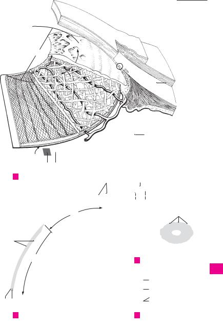
Iris. Frontally-located, round, variably colored |
16 |
Lesser arterial circle of iris. Circulus arteriosus |
|
1 |
||||||
|
|
disk about 10−12 mm in diameter, with a cen- |
|
iridis minor. Ringlike vascular system in the vi- |
||
|
|
|
tral aperture (pupil). The iris forms the poste- |
|
cinity of the pupillary margin formed by anas- |
|
2 |
|
|
rior border of the anterior chamber of the eye. |
|
tomoses between the radial branches of the |
|
|
|
|
Its lateral margins become continuous with the |
|
greater arterial circle. A |
|
|
|
|
ciliary body. A |
17 |
Pupillary membrane. [Membrana pupillaris]. |
|
3 |
|
|
||||
2 |
Pupillary margin. Margo pupillaris. Medial (in- |
|
Anterior part of embryonical vascular mem- |
|||
|
|
|
ternal) margin of the iris bordering the pupil. A |
|
brane around the lens that is situated behind |
|
4 |
|
|
B |
|
the pupil. It is fused to the pupillary margin and |
|
|
3 |
Ciliary margin. Margo ciliaris. Lateral (external) |
18 |
receives blood vessels from there. |
||
|
||||||
|
|
|
margin of iris attached to ciliary body at the ir- |
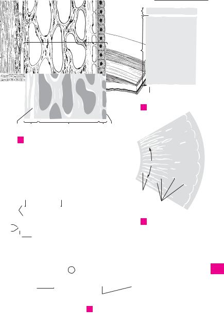
INTERNAL (SENSORY) TUNIC OF EYEBALL. |
||
5 |
|
|
||||
|
|
idocorneal angle. B |
|
Tunica interna bulbi. It comprises the retina |
||
|
4 |
Anterior surface. Facies anterior. It faces the |
|
with its pigment epithelium. |
||
6 |
|
|
anterior chamber. B |
19 |
Retina. Inner lining of eyeball developed from |
|
|
5 |
Posterior surface. Facies posterior. Surface |
|
the two layers of the optic cup. Most of it is |
||
|
|
|||||
7 |
|
|
facing the posterior chamber. A B |
|
light-sensitive (pars optica). B |
|
|
|
20 Pars optica retinae. Retinal segment capable of |
||||
6 |
Greater ring (circle) of iris. Anulus iridis major. |
|||||
|
||||||
|
|
|
Ciliary segment of the iris, and outer cirucular |
|
transforming light stimuli into nerve impulses. |
|
|
|
|
|
|||
8 |
|
|
|
It lines the posterior aspect of the eyeball and |
||
|
|
zone on the anterior surface of the iris. It is |
|
|||
|
|
|
coarser and broader than the lesser ring. A |
|
extends as far anteriorly as the ora serrata. B |
|
|
|
|
|
|||
9 |
7 |
Lesser ring (circle) of iris. Anulus iridis minor. |
21 |
Pigmented part. Pars pigmentosa. Pigment |
||
|
epithelium arising from the external layer of the |
|||||
|
|
|
Pupillary segment of iris. Narrow, circular inner |
|
||
|
|
|
zone on the anterior surface of iris. Its structure |
|
optic cup. B |
|
10 |
|
|
|
|||
|
|
22 |
Nervous part. Pars nervosa. Retina proper con- |
|||
|
|
is finer than that of the greater ring. A |
||||
|
8 |
Iridial folds. Plicae iridis. Folds passing around |
|
sisting essentially of three nuclear layers lying |
||
|
|
|||||
11 |
|
|
the pupillary margin on the anterior side of the |
|
internal to the pigment epithelium. B |
|
|
|
23 |
Neuroepithelial (photosensitive) layer. Stratum |
|||
|
|
|
iris. They make the pupillary margin appear |
|||
|
|
|
slightly serrated. A |
|
neuroepitheliale (photosensorium). Outer layer |
|
12 |
|
|
|
|||
9 |
Pupil. Pupilla. Aperture in the iris surrounded |
|
of the cerebral stratum. It consists of rods and |
|||
|
|
|
by the pupillary margin of the iris. Its diameter |
|
cones, the outer segments of which affect the |
|
|
|
|
|
|||
13 |
|
|
varies depending upon the intensity of light and |
|
transformation of light stimuli into nerve im- |
|
|
|
|
pulses. Cell bodies of rods and cones form the |
|||
|
|
the focal distance of the observed object. A |
|
|||
|
10 |
M. sphincter pupillae. Network of spirally |
|
outermost layer of the retinal nuclei (external |
||
14 |
|
nuclear layer). D |
||||
|
|
coursing muscle fibers the longitudinal axes of |
|
|||
|
|
24 |
Internal nuclear layer. [[Stratum ganglionare reti- |
|||
|
|
|
which run approximately parallel to the pupil- |
|||
15 |
|
|
lary margin when the pupil is dilated. It is in- |
|
nae]]. Middle layer of cell nuclei mainly con- |
|
|
|
|
sisting of the cell bodies of bipolar and amacrine |
|||
|
|
nervated by parasympathetic fibers from the |
|
|||
|
|
|
oculomotor nerve. B |
|
cells. D |
|
16 |
|
|
|
|||
|
|
25 |
Ganglion cell layer. [[Stratum ganglionare n. op- |
|||
11 |
M. dilator pupillae. Thin layer of smooth |
|||||
|
|
|
muscle mainly comprised of radially oriented |
|
tici]]. Internal layer of nuclei consisting of multi- |
|
|
|
|
|
|||
17 |
|
|
fibers. It is innervated by sympathetic fibers |
|
polar cell bodies of initially non-myelinated |
|
|
|
|
ganglion cells the axons of which form the optic |
|||
|
|
from the carotid plexus. |
|
|||
|
12 |
Stroma iridis. Vascular framework of the iris in- |
|
nerve. D |
||
18 |
|
|||||
26 |
Ora serrata. Serrated margin between the |
|||||
|
|
filtrated by pigmented connective tissue cells. |
||||
|
|
|
Its anterior and posterior portions are thicker |
|
light-sensitive and light-insensitive parts of the |
|
|
|
|
|
|||
19 |
|
|
than the rest and are divided by a fine fibrous |
|
neural retina. B C |
|
|
|
27 |
Pars ciliaris retinae. Light-insensitive retinal |
|||
|
|
|
network. A B |
|||
|
13 |
Pigmented (posterior) epithelium. Epithelium |
|
segment consisting of a bilayered cuboidal |
||
20 |
|
|||||
|
epithelium (ciliary epithelium) forming the |
|||||
|
|
pigmentosum. Bilayered epithelium on the |
|
|||
|
|
|
posterior surface of the iris. It is so heavily pig- |
|
posterior surface of the ciliary body. Its outer |
|
|
|
|
|
|||
21 |
|
|
mented that no nuclei are visible on the surface |
|
layer of epithelium is continuous with the pig- |
|
|
|
|
ment epithelium of the retina and is pigmented, |
|||
|
|
facing the posterior chamber. A |
|
|||
|
14 |
Spaces of iridocorneal angle [spaces of Fon- |
|
whereas the innermost epithelium is continu- |
||
22 |
|
ous with the pars nervosa of the retina and is |
||||
|
|
tana]. Spatia anguli iridocornealis. Interstices |
|
|||
|
|
|
devoid of pigment. B |
|||
|
|
|
between the fibers of the trabecular meshwork. |
28 Pars iridica retinae. Light-insensitive retinal |
||
23 |
|
|
They form passageways that convey aqueous |
|||
|
|
|
segment on the posterior surface of the iris. It is |
|||
|
|
fluid to the sinus venosus sclerae. A |
|
|||
|
15 |
Greater arterial circle of iris. Circulus arterio- |
|
continuous with the pars ciliaris retinae and |
||
24 |
|
forms the bilayered posterior epithelium of the |
||||
|
|
sus iridis major. Ringlike vascular system with |
|
|||
|
|
|
iris. Both layers are heavily pigmented. B |
|||
|
|
|
radiating branches. It is formed by anastomoses |
|
|
|
25between the long and short posterior ciliary arteries. A


 2
2 28
28 

360 |
Sense organs |
|
|
|||
|
|
1 |
Optic disc (papilla). Discus nervi optici [papilla |
16 |
Iridocorneal angle. Angulus iridocornealis. |
|
1 |
||||||
|
|
nervi optici]. Beginning of the optic nerve as |
|
Angle between the iris and cornea. It houses the |
||
|
|
|
visualized in the fundus about 3−4 mm medial |
|
trabecular meshwork, the interstices of which |
|
2 |
|
|
to the macula. It is about 1.6 mm in diameter. C |
|
serve as passageways that drain aqueous humor |
|
|
|
2 Physiological cup. Excavatio disci. Depression |
|
into the sinus venosus sclerae. A |
||
|
|
17 |
Aqueous humor. Humor aquosus. Fluid pro- |
|||
3 |
|
|
in the middle of the optic disc with the stems of |
|||
|
|
the central retinal artery and vein. C |
|
duced by the epithelium of the ciliary processes |
||
|
|
|
|
|||
|
|
3 Macula [[lutea]]. Transversely oval, yellowish |
|
(total quantity: 0.2−0.3 cm3). The clear fluid |
||
4 |
|
|
consists of 98% water, 1.4% NaCl and traces of |
|||
|
|
area, 2−4 mm in diameter, at the posterior pole |
|
|||
|
|
|
of the retina. C |
|
protein and sugar. It has a refractive index of |
|
|
|
|
|
|||
|
|
|
|
1.336. |
||
|
|
|
|
|
||
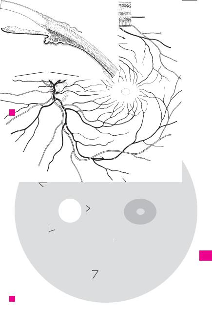
54 Fovea centralis. Central fovea, a small depres-
sion in the macula caused by thinning of the upper retinal layers. Its diameter, measured
6from the beginning of the decrease in retinal thickness from one side to the other, is approxi-
7mately 1−2 mm. B C
5Foveola. Thinnest area of fovea centralis with a
8diameter of about 0.2−0.4 mm. Here, the retina is comprised entirely of approx. 2500 closely packed cones. B
96 Retinal blood vessels. Vasa sanguinae retinae. Branches of the central retinal artery and vein
10 |
located on the internal aspect of the retina. |
7Circle of arteries around the optic nerve. Cir-
11culus vasculosus nervi optici. Small vascular ring penetrating the sclera around the optic nerve.
128 Superior temporal arteriole/venule or retina.
Arteriola/venula temporalis retinae superior.
13Lateral upper branch of the central retinal artery and vein. C
149 Inferior temporal arteriole/venule of retina.
Arteriola/venula temporalis retinae inferior. Lateral lower branch of the central retinal artery
15and vein. C
10 Superior nasal arteriole/venule of retina.
16Arteriola/venula nasalis retinae superior. Upper medial branch of the central retinal artery and
vein. C
17
11 Inferior nasal arteriole/venule of retina. Arteriola/venula nasalis retinae inferior. Lower me-
18dial branch of the central retinal artery and vein. C
1912 Superior macular arteriole/venule. Arteriola/ venula macularis superior. They supply and drain the upper part of the macula. C
2013 Inferior macular arteriole/venule. Arteriola/ venula macularis inferior. They supply and
21drain the lower part of the macula. C
14 Medial arteriole/venule of retina. Arteriola/
22venula medialis retinae. Small branches that supply and drain the medial part of retina proximal to the optic disc. C
2314 a CHAMBERS OF THE EYE. Camerae bulbi.
15 Anterior chamber. Camera anterior. Space that
24extends from the anterior surface of the iris to the posterior surface of the cornea and com-
25municates with the posterior chamber via the pupil. A