Материал: Атлас Ханц фениш

|
312 |
Brain |
|
|
|
|
|
|||
|
|
|
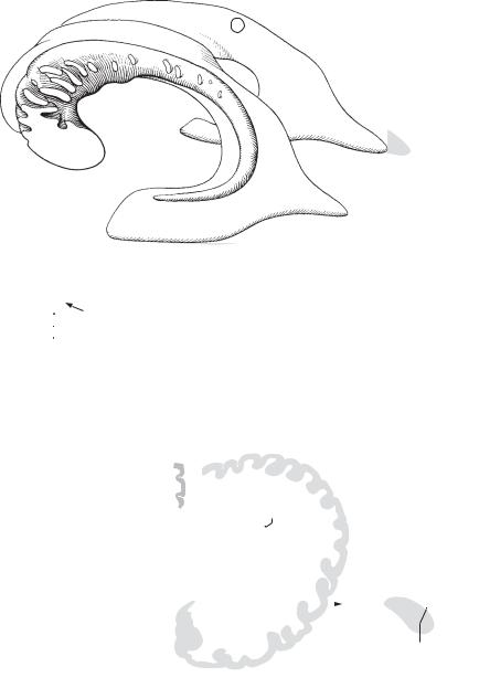
Olfactory brain. Its comprised of the substantia |
18 |
Gyrus fasciolaris. Convolution that passes |
|||||
1 |
|
1 |
||||||||
|
|
|
perforata anterior, stria diagonalis, area subcal- |
|
around the splenium of the corpus callosum and |
|||||
|
|
|
|
losa and gyrus paraterminalis. |
|
forms a connection between the longitudinal |
||||
2 |
|
2 |
Anterior |
perforated |
substance. Substantia |
|
striae, including the indusium griseum and den- |
|||
|
|
|
|
perforata anterior. Area posterior to the ol- |
|
tate gyrus. B |
||||
|
|
|
|
19 |
Lamina terminalis. Thin walled, anterior border |
|||||
3 |
|
|
|
factory trigone with perforations for the passage |
||||||
|
|
|
of cerebral vessels. A |
|
|
of the 3rd ventricle. A B |
||||
4 |
|
|
3 Diagonal stria (band) of Broca. Stria diagonalis |
20 |
Anterior commissure. Commissura anterior. |
|||||
|
|
|
[Broca]. Bundle of myelinated fibers often |
|
Anterior, transverse connection between the |
|||||
|
|
|
|
coursing obliquely over the anterior perforated |
|
right and left halves of the cerebrum. It lies be- |
||||
5 |
|
|
|
substance. It connects the precommissural sep- |
|
hind the lamina terminalis and is visible in the |
||||
|
|
|
|
tum with the uncus. A |
|
|
most anterior segment of the 3rd ventricle. A |
|||
6 |
|
4 |
Subcallosal area. Area subcallosa. Area on the |
21 Fornix. Curved bundle of fibers that pass in both |
||||||
|
|
|
medial surface of the frontal lobe situated below |
|
directions between the mamillary body and |
|||||
|
|
|
|
|
||||||
7 |
|
|
|
the genu and rostrum of the corpus callosum. A |
|
hippocampus. B |
||||
|
5 |
Paraterminal |
gyrus. |
Gyrus paraterminalis. |
22 |
Crus of fornix. Crus. The posterior limb of the |
||||
|
|
|
|
Convolution on the medial surface below the |
|
fornix that arises from the hippocampus as the |
||||
8 |
|
|
|
rostrum and in front of the laminal terminalis. A |
|
hippocampal fimbria, circles around the pulvi- |
||||
|
|
|
|
nar and unites with the contralateral limb to |
||||||
|
|
6 |
Corpus |
callosum. Massive transverse fibers |
|
|||||
|
|
|
form the body of the fornix. B |
|||||||
9 |
|
|
|
connecting the right and left hemispheres at the |
|
|||||
|
|
|
23 |
Body of fornix. Corpus. Unparied middle part of |
||||||
|
|
|
base of the longitudinal fissure of the cerebrum. |
|||||||
|
|
|
|
|
fornix situated below the corpus callosum and |
|||||
|
|
|
|
A B C |
|
|
|
|
||
10 |
|
|
|
|
|
|
|
formed by the union of both crura. B |
||
|
|
7 Splenium. Thick, free posterior end of the cor- |
|
|||||||
|
|
|
24 Tenia. Taenia. Thin, lateral margin of the fornix |
|||||||
|
|
|
|
pus callosum. B |
|
|||||
11 |
|
|
|
|
|
that gives attachment to the choroid plexus of |
||||
|
|
8 Trunk. Truncus. Portion of corpus callosum be- |
|
|||||||
|
|
|
the lateral ventricle. B |
|||||||
|
|
|
|
|||||||
12 |
|
|
|
tween the splenium and genu. B |
25 Column. Columna. Anterior part of the fornix lo- |
|||||
|
9 |
Genu. Bend in the corpus callosum located ante- |
||||||||
|
|
cated partly in the lateral wall of the 3rd ven- |
||||||||
13 |
|
|
|
riorly above the rostrum. B |
|
tricle. It extends as far as the mamillary body. B |
||||
|
10 |
Rostrum. Anterior end of corpus callosum that |
26 Commissure. Commissura. Triangular connect- |
|||||||
|
|
|
|
tapers inferiorly to a point where it joins the |
|
ing plate situated between the crura of the for- |
||||
14 |
|
|
|
lamina terminalis. B |
|
|
nix below the posterior part of the corpus callo- |
|||
|
11 |
Radiation of |
corpus |
callosum. Radiatio cor- |
|
sum. It contains fibers crossing from the hippo- |
||||
|
|
|
||||||||
15 |
|
|
|
poris callosi. Fibers radiating from the corpus |
|
campal fimbriae of both sides. B |
||||
|
|
|
|
|
||||||
|
|
|
callosum to the cerebral cortex. A D |
27 |
Septum pellucidum (lucidum). Bilayered, thin |
|||||
|
|
12 |
Forceps |
minor. Forceps frontalis (minor). U- |
|
plate extending between the corpus callosum |
||||
16 |
|
|
and fornix. It separates the anterior horns of the |
|||||||
|
|
|
shaped fibers passing through the genu of the |
|
||||||
|
|
|
|
lateral ventricles from one another. B |
||||||
|
|
|
|
corpus callosum and connecting the frontal |
|
|||||
|
|
|
|
28 Cavity of septum pellucidum. Cavum septi pel- |
||||||
|
|
|
|
|||||||
17 |
|
|
|
lobes. D |
|
|
|
|||
13 |
Forceps major. Forceps occipitalis (major). U- |
|
lucidi. Enclosed cavity of variable size between |
|||||||
|
|
|||||||||
|
|
the two laminae of the septum pellucidum. B |
||||||||
|
|
|
|
shaped fibers passing through the splenium of |
|
|||||
18 |
|
|
|
|
||||||
|
|
|
29 |
Lamina of septum pellucidum. Lamina septi |
||||||
|
|
|
the corpus callosum and connecting the poste- |
|||||||
|
|
|
|
rior parts of the occipital lobes. D |
|
pellucidi. Paired sheet forming the septum pel- |
||||
19 |
14 |
Tapetum. Continuous layer of fibers arching |
|
lucidum and the lateral wall of its cavity. B |
||||||
30 |
Precommissural septum. Septum precommis- |
|||||||||
|
|
|
|
laterally and inferiorly from the corpus callosum |
||||||
|
|
|
|
|||||||
20 |
|
|
|
and forming the lateral wall of the inferior and |
|
surale. Area on the free medial surface of the |
||||
|
|
|
posterior horns of the lateral ventricle as well as |
|
frontal lobe in front of the lamina terminalis. |
|||||
|
|
|
|
|
||||||
|
|
|
|
the roof of the posterior horn. C |
|
|
||||
2115 Indusium griseum. Thin layer of gray matter on the superior surface of the corpus callosum. B C
2216 Medial longitudinal stria. Stria longitudinalis medialis. A medial longitudinal band of white
23fibers in the indusium griseum. It is part of the olfactory brain. B C
17 Lateral longitudinal stria. Stria longitudinalis
24lateralis. Paired longitudinal stripe embedded in the indusium griseum and covered laterally
25by the cingulate gyrus. It is part of the olfactory brain. B C

|
314 |
Brain |
|
|
|
|
|
|
|
|
||
|
|
1 |
Lateral ventricle. Ventriculus lateralis. Paired |
15 |
Collateral trigone. Trigonum collaterale. |
|||||||
1 |
||||||||||||
|
|
ventricle which communicates with the third |
|
Broadened triangular area near the beginning of |
||||||||
|
|
|
ventricle via the interventricular foramen. It |
|
the collateral eminence at the border between |
|||||||
2 |
|
|
consists of the pars centralis and three horns |
|
the inferior and posterior horns. C |
|||||||
|
|
|
(anterior, posterior, inferior). A |
|
|
16 |
Hippocampus. Enlongated elevation in the infe- |
|||||
|
|
|
|
|
||||||||
|
2 |
Central part. Pars centralis. Middle portion of |
|
rior horn formed by the hippocampal sulcus. It is |
||||||||
3 |
|
|||||||||||
|
|
lateral ventricle located above the thalamus and |
|
a specifically structured part of the rhinen- |
||||||||
|
|
|
below the corpus callosum. It contains a part of |
|
cephalon. C |
|||||||
4 |
|
|
the choroid plexus. A |
|
|
|
|
17 |
Pes. Foot like elevation at the anterior end of the |
|||
|
|
3 Interventricular |
foramen. |
Foramen inter- |
|
hippocampus. C |
||||||
|
|
|
||||||||||
5 |
|
|
ventriculare. Passage behind |
and |
below |
the |
18 |
Alveus. Thin layer of white matter on the hippo- |
||||
|
|
genu of the fornix through which the lateral and |
|
campus. C |
||||||||
|
|
|
|
|||||||||
|
|
|
third ventricles communicate. D |
|
|
19 |
Fimbria. Bundle of white fibers emanating from |
|||||
6 |
|
|
|
|
||||||||
4 |
Anterior horn. Cornu frontale (anterius). Part of |
|||||||||||
|
the alveus and passing medially and upward on |
|||||||||||
|
|
|
the lateral ventricle that extends forward from |
|
the hippocampus to continue into the fornix as |
|||||||
|
|
|
|
|||||||||
7 |
|
|
the interventricular foramen. It is bounded me- |
|
its crus. C |
|||||||
|
|
dially by the septum pellucidum, laterally by the |
19 a |
Sections of the telencephalon. Sectiones telen- |
||||||||
|
|
|
||||||||||
|
|
|
head of the caudate nucleus, superiorly by the |
|
cephalici. |
|||||||
8 |
|
|
trunk of the corpus callosum, anteriorly and in- |
|
||||||||
|
|
20 |
Archicortex (archipallium). Archaecortex. Phy- |
|||||||||
|
|
feriorly by the genu and rostrum of the corpus |
||||||||||
|
|
|
callosum. A |
|
|
|
|
|
|
logenetically speaking, this is an older part of |
||
9 |
|
|
|
|
|
|
|
|
the cerebral cortex. Unlike the neocortex, it has |
|||
5 |
Posterior |
horn. Cornu |
occipitale |
(posterius). |
|
|||||||
|
three instead of six layers and is formed by the |
|||||||||||
|
|
|
Part of the lateral ventricle that extends into the |
|
hippocampus and dentate gyrus. |
|||||||
10 |
|
|
occipital lobe. A |
|
|
|
|
|
|
|||
|
|
|
|
|
|
|
21 |
Paleocortex. Palaeocortex. Oldest part of cere- |
||||
6 |
Inferior horn. Cornu temporale (inferius). Part |
|||||||||||
|
|
bral cortex originally derived from the piriform |
||||||||||
11 |
|
|
of the lateral ventricle that accompanies the |
|
lobe. |
|||||||
|
|
hippocampus laterally and contains a part of the |
|
|||||||||
|
|
|
22 Neocortex. The largest part of the cerebral cor- |
|||||||||
|
|
|
choroid plexus. A |
|
|
|
|
|||||
12 |
|
|
|
|
|
|
|
tex comprised of six layers. |
||||
7 |
Stria terminalis. Longitudinal band of myeli- |
|
||||||||||
22 a Mesocortex. Incompletely differentiated zone |
||||||||||||
|
|
|
nated fibers located in the angle between the |
|||||||||
|
|
|
|
in the region of the insular cortex with visceral |
||||||||
13 |
|
|
thalamus |
and |
caudate |
nucleus |
above |
the |
|
|||
|
|
|
functions. |
|||||||||
|
|
thalamostriate vein. It arises from the amyg- |
|
|||||||||
|
|
|
|
|
||||||||
|
|
|
daloid body. B |
|
|
|
|
|
|
|
||
148 Lamina affixa. Floor of lateral ventricle between the stria terminalis and tenia choroidea. B
159 Choroid fissure. Fissura choroidea. Cleft between the thalamus and fornix for passage of the choroid plexus into the lateral ventricle. In
16the inferior horn it lies between the fimbria of the hippocampus and the stria terminalis. B
1710 Tenia choroidea. Taenia choroidea. Line of attachment of the lateral choroid plexus to the thalamus. It becomes visible after removal of the
18choroid plexus. B
|
11 Choroid plexus of lateral ventricle. Plexus |
|
19 |
choroideus ventriculi lateralis. Highly vascu- |
|
|
larized, fringelike villous folds that protrude |
|
|
into the lateral ventricle through the choroid fis- |
|
20 |
||
sure. They extend anteriorly to the interventric- |
||
|
ular foramen and posteriorly into the inferior |
|
|
||
21 |
horn. B |
|
12 Bulb of posterior horn. Bulbus cornus occipi- |
talis (posterioris). Enlargement on the medial
22side of the posterior horn caused by fibers of the splenium of the corpus callosum. C
2313 Calcar avis. Enlargement on the medial side of the posterior horn produced by the calcarine fissure. C
2414 Collateral eminence. Eminentia collateralis. Elevation in the lateral floor of the inferior horn
25near the hippocampus. It is caused by the collateral sulcus. C
|
|
|
|
|
|
|
|
|
|
|
|
|
|
|
|
|
|
Brain 315 |
|
||
|
|
|
|
|
|
|
|
|
|
|
|
|
|
|
|
|
|
|
|
|
|
|
|
|
|
1 |
|
|
|
|
|
|
|
|
|
|
|
|
|
|
1 |
||
|
|
|
|
|
|
|
|
|
|
|
|
|
|
|
|
|
|
|
|||
|
|
|
|
2 |
|
|
|
|
|
|
|
|
|
|
|
|
|
|
2 |
||
|
|
|
|
|
|
|
|
|
|
|
|
|
|
|
|
|
|
|
|
|
3 |
4 |
|
|
|
|
|
|
|
|
|
|
|
|
|
|
|
|
|
|
|
4 |
|
|
|
|
|
|
|
|
|
|
|
|
|
|
|
|
|
|
|
|
5 |
||
|
|
|
|
|
|
|
|
|
|
|
|
|
|
|
|
|
|
|
|
|
|
|
|
|
|
|
|
|
|
|
|
|
|
|
|
|
|
|
|
|
|
|
6 |
|
|
|
|
|
|
|
|
|
|
|
|
|
|
|
|
|
|
|
|
|
7 |
|
|
|
|
|
|
|
|
|
|
5 |
|
|
|
8 |
|||||||
|
|
|
|
6 |
|
|
|
|
|
|
|
|
|
|
|
|
|
|
9 |
||
|
|
|
|
|
|
|
|
|
|
|
|
|
|
|
|
|
|
|
|
|
|
|
|
|
|
|
|
|
|
|
|
Right and left lateral ventricles |
|
|
|||||||||
|
|
|
|
|
|
|
|
A |
|
10 |
|||||||||||
|
|
|
|
|
|
|
|
|
|
with left caudate nucleus |
|
||||||||||
|
|
|
|
|
|
|
|
|
|
|
11 |
||||||||||
|
|
|
|
|
|
|
|
|
|
|
|
|
|
|
|
|
|
|
|
|
|
7 |
|
|
|
|
9 |
|
|
|
|
|
|
|
|
|
|
|
|
|
|
12 |
|
|
|
|
|
|
|
|
|
|
|
|
|
|
|
|
|||||||
8 |
|
|
|
11 |
|
|
|
|
|
|
|
|
|
|
|
|
|
|
|
||
|
|
|
|
|
|
10 |
|
|
|
3 |
|
|
|
|
|
|
13 |
||||
|
|
|
|
|
|
|
|
|
|
|
|
||||||||||
|
|
|
|
|
|
|
|
|
|
|
|
|
|
|
|
||||||
|
|
|
|
|
|
|
|
|
|
|
|
|
|
|
|
||||||
|
|
|
|
|
|
|
|
|
|
|
|
|
|
|
|
|
|
|
|
|
14 |
|
|
|
|
|
|
|
|
|
|
|
|
|
|
|
|
|
|
|
|
|
15 |
|
|
|
|
|
|
|
|
|
|
|
|
Interventricular foramen |
|
|
|||||||
|
|
|
|
|
Thalamus with fornix |
|
D |
|
16 |
||||||||||||
|
|
|
|
|
|
|
|
|
|
|
|
|
|
|
|
||||||
|
|
|
|
|
|
|
|
|
|
|
|
|
|
|
|
|
|||||
|
|
B |
|
|
|
|
|
|
|
|
|
|
|
|
|
||||||
|
|
|
|
|
|
|
|
|
|
|
|
||||||||||
|
|
|
|
|
|
|
|
|
|
|
17 |
||||||||||
|
|
|
|
|
|
|
|
|
|
|
|
|
|
|
|
|
|
|
|
|
|
|
|
|
|
|
|
|
|
|
|
|
|
|
|
|
|
|
|
|
|
|
|
|
|
|
|
|
|
|
|
|
|
|
|
|
|
|
|
|
|
|
|
|
|
|
|
|
|
|
|
|
|
|
|
|
|
|
|
|
|
|
|
|
|
|
18 |
|
|
|
|
|
|
|
|
|
|
|
|
|
|
|
|
|
|
|
|
|
|
|
|
|
|
|
|
|
|
|
|
|
|
|
|
|
|
|
|
|
|
|
|
|
|
|
|
12 |
|
|
|
|
|
|
|
|
|
|
|
|
|
19 |
|||
|
|
|
|
|
|
|
|
|
|
|
|
|
|
|
|
|
|
|
|
|
|
|
|
|
|
13 |
|
|
|
|
|
|
|
|
|
|
|
|
20 |
||||
|
|
|
|
15 |
|
|
|
|
|
|
|
|
|
|
|
|
|
||||
|
|
|
|
|
|
|
|
|
|
|
|
|
|
|
21 |
||||||
|
|
|
|
|
|
|
|
|
|
|
|
|
|
|
|
|
|
|
|
|
|
|
|
|
|
14 |
|
|
|
|
|
|
|
|
|
|
|
|
|
||||
|
|
|
|
|
|
|
|
|
|
|
|
|
|
|
|
||||||
|
|
|
|
19 |
16 |
|
|
22 |
|||||||||||||
|
|
|
|
|
|
|
|
|
|
|
|
||||||||||
|
|
|
|
16 |
|
|
|
|
|
|
|
|
|
|
|
|
|
|
|
||
|
|
|
|
|
|
|
|
|
|
|
|
|
|
|
|
|
|
23 |
|||
|
|
|
|
17 |
|
|
|
|
|
|
|
|
|
|
|
|
|
|
|||
|
|
|
|
|
|
|
|
|
|
|
|
|
|
|
|
|
|
|
|
|
|
|
|
|
|
|
Left hippocampus |
|
|
|
|
|
|
|
|
18 |
|
|
24 |
||||
|
|
C |
|
|
|
|
|
|
|
|
|
|
|
||||||||
25

|
316 |
Brain |
|
|
|
|
|
|
|
|
|
|
|
|
|||
|
|
|
Sections through telencephalon. Sectiones tel- |
16 |
Superior |
longitudinal |
fasciculus. Fasciculus |
||||||||||
1 |
|
1 |
|||||||||||||||
|
|
|
encephali. |
|
|
|
|
|
|
|
|
longitudinalis superior. Largest bundle of asso- |
|||||
|
|
|
2 Cerebral cortex. Cortex cerebralis (pallium). |
|
ciation fibers in the cerebrum, extending from |
||||||||||||
2 |
|
|
|
the frontal lobe to the temporal lobe via the |
|||||||||||||
|
|
|
Gray |
matter, |
1.5−4.5 mm |
thick, |
consisting |
|
|||||||||
|
|
|
|
mainly of the following six layers. A |
|
|
|
occipital lobe. E |
|
|
|||||||
3 |
|
|
|
|
|
17 |
Inferior |
longitudinal |
fasciculus. |
Fasciculus |
|||||||
|
3 |
Molecular (plexiform) layer. 1st layer. Lamina |
|||||||||||||||
|
|
longitudinalis inferior. Bundle of |
association |
||||||||||||||
|
|
|
|
molecularis (plexiformis). It contains a few tan- |
|
fibers connecting the temporal and occipital |
|||||||||||
|
|
|
|
gential cells and a thick network of tangential |
|
||||||||||||
4 |
|
|
|
|
lobes. E |
|
|
|
|||||||||
|
|
|
fibers from dendrites of pyramidal cells and |
|
|
|
|
||||||||||
|
|
|
|
18 |
Uncinate fasciculus. Fasciculus uncinatus. As- |
||||||||||||
|
|
|
|
axons of other cells. None of its projections ex- |
|||||||||||||
|
|
|
|
|
sociation fibers connecting the inferior surface |
||||||||||||
5 |
|
|
|
tend beyond the cerebral cortex. A |
|
|
|
||||||||||
|
|
|
|
|
|
of the frontal lobe and the anterior part of the |
|||||||||||
|
4 |
External granular layer. 2nd layer. Lamina |
|
||||||||||||||
|
|
|
temporal lobe. E |
|
|
||||||||||||
6 |
|
|
|
granularis externa. A layer of small cells in a fine |
19 |
Radiation of corpus callosum. Radiatio cor- |
|||||||||||
|
|
|
|
fiber network. A |
|
|
|
|
|
|
poris callosi. Fibers connecting the right and left |
||||||
|
|
|
|
|
|
|
|
|
|
|
|
|
|
||||
7 |
|
5 |
External |
pyramidal |
layer |
(pyramidal |
cell |
|
cerebral cortices. See also p. 312.11−14. C |
||||||||
|
|
|
layer). 3rd layer. Lamina pyramidalis externa. It |
20 |
Basal nuclei. Nuclei basales. Basal ganglion. |
||||||||||||
|
|
|
|
||||||||||||||
8 |
|
|
|
contains medium-sized pyramidal cells which |
21 Corpus striatum. The striate body, which is |
||||||||||||
|
|
|
do not form long tracts. A |
|
|
|
|
||||||||||
|
|
|
|
|
|
|
|
comprised of basal ganglia (caudate nucleus and |
|||||||||
|
|
6 |
Internal granular layer. 4th layer. Lamina granu- |
|
putamen) united by bundles of gray matter. |
||||||||||||
9 |
|
|
|
laris interna. It consists predominantly of |
|
Central synaptic station of the extrapyramidal |
|||||||||||
|
|
|
|
closely packed stellate cells and receives im- |
|
system. D |
|
|
|
||||||||
10 |
|
|
|
pulses primarily from thalamocortical fibers. Its |
22 |
Caudate nucleus. Nucleus caudatus. Elongated, |
|||||||||||
|
|
|
stria is comprised of densely stratified tangen- |
|
arched nucleus that arises from the ganglionic |
||||||||||||
|
|
|
|
tial fibers. A |
|
|
|
|
|
|
|
mass of the telencephalon and curves around |
|||||
11 |
|
7 |
Internal pyramidal (ganglionic) layer. 5th layer. |
|
the thalamus. |
|
|
||||||||||
|
|
|
|
Lamina pyramidalis interna (ganglionaris). It |
23 |
Head of caudate nucleus. Caput [[nuclei cau- |
|||||||||||
12 |
|
|
|
contains larger pyramidal cells and is the exit of |
|
dati]]. Anteriorly situated structure that forms |
|||||||||||
|
|
|
the corticonuclear and corticospinal tracts in |
|
the lateral wall of the anterior horn of the lateral |
||||||||||||
|
|
|
|
|
|||||||||||||
13 |
|
|
|
the corresponding regions of areas 4 and 6. A |
|
ventricle. B D |
|
|
|||||||||
|
8 |
Multiform (fusiform) layer. 6th |
layer. Lamina |
24 |
Body of caudate nucleus. Corpus [[nuclei cau- |
||||||||||||
|
|
|
|
multiformis. Poorly defined layer made up of |
|
dati]]. Middle part of caudate nucleus lying on |
|||||||||||
14 |
|
|
|
|
the thalamus. B D |
|
|
||||||||||
|
|
|
many, mostly small, fusiform cells extending |
|
|
|
|||||||||||
|
|
|
25 |
Tail of caudate nucleus. Cauda [[nuclei cau- |
|||||||||||||
|
|
|
|
into the white matter. A |
|
|
|
|
|||||||||
15 |
|
9 |
Tangential fibers. Neurofibrae tangentiales. Su- |
|
dati]]. It accompanies the inferior horn and |
||||||||||||
|
|
forms the tapering posterior and inferior seg- |
|||||||||||||||
|
|
|
|
perficial fiber complex comprised of the follow- |
|
ments of the caudate nucleus. D |
|
||||||||||
|
|
|
|
ing four layers. |
|
|
|
|
|
|
|
|
|||||
16 |
|
|
|
|
|
|
|
|
|
26 Lentiform nucleus. Nucleus lentiformis (lentic- |
|||||||
|
10 |
Stria of molecular layer. Stria laminae |
|||||||||||||||
|
|
ularis). Part of the corpus striatum that arises |
|||||||||||||||
|
|
||||||||||||||||
|
|
|
|
molecularis (plexiformis). Band of tangential |
|
from the telencephalon and diencephalon. D |
|||||||||||
17 |
|
|
|
|
|||||||||||||
|
|
|
fibers in the 1st layer of the cerebral cortex. A |
27 |
Putamen. Lateral, telencephalic portion of the |
||||||||||||
|
11 |
Stria of external granular layer. Stria laminae |
|
lentiform nucleus. B |
|
|
|||||||||||
18 |
|
|
|
||||||||||||||
|
|
|
granularis externa. Thinner band of tangential |
28 |
Lateral medullary lamina. Lamina medullaris |
||||||||||||
|
|
|
|
fibers in the 2nd layer of the cerebral cortex. A |
|
lateralis. Medullary layer of the corpus striatum |
|||||||||||
|
|
|
|
|
|||||||||||||
19 |
12 |
Stria of internal granular layer. Stria laminae |
|
situated between the globus pallidus and puta- |
|||||||||||||
|
men. B |
|
|
|
|||||||||||||
|
|
|
|
granularis interna. Band of tangential fibers in |
|
|
|
|
|||||||||
|
|
|
|
29 |
Lateral globus pallidus. Globus pallidus later- |
||||||||||||
|
|
|
|
the 4th layer of the cerebral cortex [[outer stripe |
|||||||||||||
20 |
|
|
|
||||||||||||||
|
|
|
of Baillarger]]. A |
|
|
|
|
|
|
alis. Part of the diencephalic globus pallidus lo- |
|||||||
|
13 |
Stria |
of |
internal |
pyramidal |
layer. |
Stria |
|
cated between the lateral and medial medullary |
||||||||
21 |
|
laminae. B |
|
|
|||||||||||||
|
|
|
laminae pyramidalis interna (ganglionaris). |
|
|
|
|||||||||||
|
|
|
30 |
Medial medullary lamina. Lamina medullaris |
|||||||||||||
|
|
|
|
Band of tangential fibers in the 5th layer of the |
|||||||||||||
|
|
|
|
||||||||||||||
22 |
|
|
|
cerebral cortex [[inner stripe of Baillarger]]. A |
|
medialis. Medullary layer between the medial |
|||||||||||
|
|
|
|
and lateral parts of the globus pallidus. B |
|||||||||||||
|
14 Arcuate fibers of cerebrum. Fibrae arcuatae |
|
|||||||||||||||
|
|
31 |
Medial globus pallidus. Globus pallidus medi- |
||||||||||||||
|
|
|
|
cerebri. Arcuate fibers connecting the adjacent |
|||||||||||||
23 |
|
|
|
|
alis. Part of the globus pallidus located medial to |
||||||||||||
|
|
|
cerebral gyri. F |
|
|
|
|
|
|
|
|||||||
|
|
|
|
|
|
|
|
|
|
|
the medial medullary lamina. B |
|
|||||
|
15 |
Cingulum. Fiber bundle lying in the medulla of |
|
|
|||||||||||||
24 |
32 |
Claustrum. Layer of gray matter between the |
|||||||||||||||
|
|
|
the cingulate gyrus. It arises from the area sub- |
|
lentiform nucleus and the insular cortex. B |
||||||||||||
|
|
|
|
|
|||||||||||||
|
|
|
|
callosa, arches |
around the |
corpus |
callosum, |
|
|
|
|
|
|||||
25passes the splenium and extends anteriorly up to the uncus. C