Материал: Атлас Ханц фениш

|
302 |
Brain |
|
|
|
|
|
|
|
|
|
|
|
|
|
|
|
|
|
|
||
|
|
|
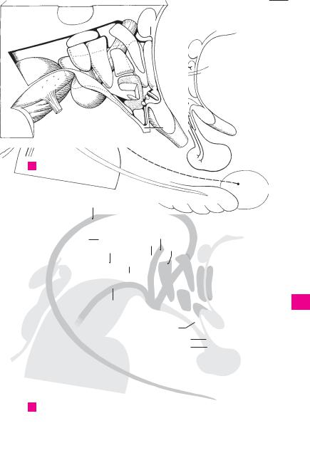
Anterior thalamic radiations. Raditiones 13 |
Sections of the hypothalamus. Sectiones hy- |
||||||||||||||||||
1 |
|
1 |
||||||||||||||||||||
|
|
|
thalamicae anteriores. Fibers of the anterior nu- |
|
pothalami. |
|
|
|
|
|
|
|
|
|||||||||
|
|
|
|
cleus passing to and from the cingulate gyrus |
14 |
Dorsal (posterior) hypothalamic region. Regio |
||||||||||||||||
2 |
|
|
|
and likewise reciprocal connections between |
|
(area) hypothalamica dorsalis. Area of the hy- |
||||||||||||||||
|
|
|
|
the lateral nucleus and frontal lobe. The fibers |
|
pothalamus next to the apex. |
|
|
|
|
||||||||||||
3 |
|
|
|
run in the anterior limb of the internal capsule. A |
15 |
Nucleus of ansa lenticularis. Nucleus ansae |
||||||||||||||||
|
2 |
Central |
thalamic |
radiations. |
Radiationes |
|||||||||||||||||
|
|
lenticularis. Groups of cells dispersed in the |
||||||||||||||||||||
|
|
|
|
thalamicae centrales. Reciprocal fibers passing |
|
ansa lenticularis. |
|
|
|
|
|
|
|
|||||||||
4 |
|
|
|
fan-like through the posterior limb of the inter- |
16 |
Anterior |
(ventral) region of |
hypothalamus. |
||||||||||||||
|
|
|
|
nal capsule from the posterior lateral, anterior |
|
Regio hypothalamica anterior. |
|
|
|
|
||||||||||||
|
|
|
|
ventral, lateral ventral and posterior ventral nu- |
|
|
|
|
|
|||||||||||||
5 |
|
|
|
17 |
Medial/lateral |
preoptic |
nucleus. |
Nucleus |
||||||||||||||
|
|
|
clei to the preand postcentral gyri in addition |
|||||||||||||||||||
|
|
|
|
to the connecting fields of the cortex. A |
|
|
preopticus medialis/lateralis. Group of nuclei |
|||||||||||||||
|
|
|
|
|
|
located beneath the anterior commissure and |
||||||||||||||||
6 |
|
3 |
Posterior |
thalamic |
raditaions. |
Radiationes |
|
|||||||||||||||
|
|
along the lamina terminalis with projections to |
||||||||||||||||||||
|
|
|
thalamicae posteriores. They lie in the occipital |
|
||||||||||||||||||
|
|
|
|
|
the |
stria terminalis, |
medial |
telencephalic |
||||||||||||||
|
|
|
|
region of the posterior limb of the internal cap- |
|
|||||||||||||||||
7 |
|
|
|
|
fasciculus and medial thalamic nuclei. D |
|
||||||||||||||||
|
|
|
sule and contain fibers from the lateral genicu- |
|
|
|||||||||||||||||
|
|
|
18 |
Supraoptic |
nucleus. |
Nucleus supraopticus. |
||||||||||||||||
|
|
|
|
late body (optic radiation) and the pulvinar for |
|
Nucleus lying above the optic chiasm with neu- |
||||||||||||||||
8 |
|
|
|
the occipital lobes and adjacent regions. A |
|
|
||||||||||||||||
|
|
|
|
|
rosecretory fibers (oxytocin and vasopressin) |
|||||||||||||||||
|
4 |
Dentatothalamic |
tract. |
Tractus |
denta- |
|
||||||||||||||||
|
|
|
projecting to the posterior pituitary. D |
|
|
|||||||||||||||||
|
|
|
|
tothalamicus. It arises from the cerebellum and |
|
|
|
|||||||||||||||
9 |
|
|
|
19 |
Paraventricular nuclei. Nuclei paraventricu- |
|||||||||||||||||
|
|
|
radiates into the thalamic fasciculus and to the |
|||||||||||||||||||
|
|
|
|
lateral ventral nucleus. C |
|
|
|
|
|
lares. Group |
of |
autonomic nuclei |
with neu- |
|||||||||
|
|
|
|
|
|
|
|
|
rosecretory fibers (oxytocin and vasopressin) |
|||||||||||||
10 |
|
5 |
Thalamic fasciculus. Fasciculus thalamicus. It |
|
||||||||||||||||||
|
|
projecting to the posterior lobe of the hypophy- |
||||||||||||||||||||
|
|
|
lies below the thalamus, next to and above the |
|
||||||||||||||||||
|
|
|
|
|
sis. They lie superiorly near the base of the hy- |
|||||||||||||||||
|
|
|
|
zona incerta in field H1 and is composed of the |
|
|||||||||||||||||
11 |
|
|
|
|
pothalamic sulcus and behind the anterior hy- |
|||||||||||||||||
|
|
|
ventricular fasciculus, ansa |
lenticularis |
and |
|
||||||||||||||||
|
|
|
|
pothalamic nucleus. D |
|
|
|
|
|
|
||||||||||||
|
|
|
|
fibers from the cerebellum. It is a conveyor of |
|
|
|
|
|
|
|
|||||||||||
|
|
|
|
20 |
Anterior hypothalamic nucleus. Nucleus hy- |
|||||||||||||||||
12 |
|
|
|
impulses for the anterior ventral and lateral |
||||||||||||||||||
|
|
|
|
pothalamicus anterior. Located behind the pre- |
||||||||||||||||||
|
|
|
ventral nuclei. C |
|
|
|
|
|
|
|||||||||||||
|
|
6 |
Subthalamic fasciculus. Fasciculus subthalami- |
|
optic nucleus with projections to the hemi- |
|||||||||||||||||
13 |
|
|
spheres, stria terminalis and thalamus, its effer- |
|||||||||||||||||||
|
|
|
cus. Fiber bundle extending from the globus pal- |
|
||||||||||||||||||
|
|
|
|
ent fibers communicate with motor and auton- |
||||||||||||||||||
|
|
|
|
lidus to the subthalamic nucleus. C |
|
|
|
|
||||||||||||||
|
|
|
|
|
|
|
|
omic nuclei. It influences heat regulation, glan- |
||||||||||||||
|
|
|
7 Mamillothalamic fasciculus. Fasciculus mamil- |
|
||||||||||||||||||
14 |
|
|
|
dular activity and circulation. D |
|
|
|
|
||||||||||||||
|
|
|
lothalamicus. Fiber |
tract extending |
from the |
|
|
|
|
|
||||||||||||
|
|
|
|
21 Intermediate hypothalamic region. Regio hy- |
||||||||||||||||||
|
|
|
|
mamillary body to the anterior nucleus of the |
||||||||||||||||||
15 |
|
|
|
|
pothalamica intermedia. Area situated between |
|||||||||||||||||
|
|
|
thalamus. D |
|
|
|
|
|
|
|||||||||||||
|
8 |
|
|
Pedunculus |
|
the |
anterior |
and posterior hypothalamic |
re- |
|||||||||||||
|
|
Inferior |
thalamic |
peduncle. |
|
gions. |
|
|
|
|
|
|
|
|
|
|||||||
16 |
|
|
|
thalamicus inferior. Fibers between |
the hy- |
|
|
|
|
|
|
|
|
|
|
|||||||
|
|
|
22 |
Arcuate |
nucleus. [[Nucleus arcuatus]]. Mural |
|||||||||||||||||
|
|
|
pothalamus and thalamus. According to some |
|||||||||||||||||||
|
|
|
|
|
nucleus situated in the entrance to the infun- |
|||||||||||||||||
|
|
|
|
anatomists, it consists of fibers of the pulvinar |
|
|||||||||||||||||
|
|
|
|
|
dibulum. It belongs to the tuberal nuclei, i. e., it |
|||||||||||||||||
|
|
|
|
|
||||||||||||||||||
17 |
|
|
|
from and to the occipital lobes and its vicinity, as |
|
|||||||||||||||||
|
|
|
|
regulates |
the release |
of |
hormones from |
the |
||||||||||||||
|
|
|
|
well as fibers of the auditory tract. |
|
|
|
|
anterior lobe by delivering an active substance |
|||||||||||||
|
|
|
|
|
|
|
|
|||||||||||||||
18 |
|
|
9 Ansa lenticularis and fasciculus lenticularis. |
|
(neurohormone) to blood vessels of the hy- |
|||||||||||||||||
|
|
|
Ansa et |
fasciculus |
lenticulares. |
Two |
fiber |
|
||||||||||||||
|
|
|
|
|
pophysial stalk where its processes (axons) are |
|||||||||||||||||
|
|
|
|
bundles from the lentiform nucleus to the ven- |
|
found. D |
|
|
|
|
|
|
|
|
|
|||||||
|
|
|
|
|
|
|
|
|
|
|
|
|
|
|||||||||
19 |
|
|
|
tral nuclei of the thalamus. One part passes |
|
|
|
|
|
|
|
|
|
|
||||||||
|
|
|
23 |
Tuberal nuclei. Nuclei tuberales. Groups of nu- |
||||||||||||||||||
|
|
|
|
around the anterior margin of the internal cap- |
|
clei in the posterior wall of the infundibulum. |
||||||||||||||||
|
|
|
|
|
||||||||||||||||||
|
|
|
|
sule (ansa lenticularis); the other part passes |
|
|||||||||||||||||
20 |
|
|
|
|
They function similar to the arcuate nucleus. D |
|||||||||||||||||
|
|
|
through the internal capsule. Both tracts are |
|
||||||||||||||||||
|
|
|
|
united in the thalamic fasciculus. C |
|
|
|
24 |
Lateral |
hypothalamic |
region. |
Regio |
hy- |
|||||||||
|
|
|
|
|
|
|
||||||||||||||||
21 |
10 |
Ansa peduncularis and fasciculus peduncu- |
|
pothalamica lateralis. Area separated from the |
||||||||||||||||||
|
medial hypothalamus |
by |
the |
fornix, |
mamil- |
|||||||||||||||||
|
|
|
|
laris. Ansa et fasciculus pedunculares. Fiber |
|
lothalamic fasciculus and medial telencephalic |
||||||||||||||||
|
|
|
|
|
||||||||||||||||||
|
|
|
|
tract connecting the thalamus and claustrum, |
|
|||||||||||||||||
22 |
|
|
|
|
fasciculus. It is occupied by the lateral preoptic |
|||||||||||||||||
|
|
|
thereby extending between the lentiform nu- |
|
||||||||||||||||||
|
|
|
|
cleus and the amygdaloid body. B C |
|
|
|
|
nucleus and the supraoptic nucleus including its |
|||||||||||||
|
|
|
|
|
|
|
|
lateral portion. D |
|
|
|
|
|
|
|
|||||||
23 |
11 |
Intrathalamic fibers. Fibrae |
intrathalamicae. |
|
|
|
|
|
|
|
|
|||||||||||
|
|
|
|
|
|
|
|
|
|
|
|
|||||||||||
|
|
|
|
Connections of individual thalamic nuclei. |
|
|
|
|
|
|
|
|
|
|
|
|
|
|||||
|
|
|
|
|
|
|
|
|
|
|
|
|
|
|
|
|
||||||
12 Periventricular fibers. Fibrae periventriculares.
24Fibers coursing beneath the ependyma of the third ventricle between the medial nucleus and
25the hypothalamic nucleus to enter the posterior longitudinal fasciculus.

 17
17 18
18
|
304 |
Brain |
|
|
|
|
|
|
|
|
|
|
|
||
|
|
|
Ventromedial hypothalamic nucleus. Nucleus |
14 |
Posterior |
(dorsal) |
longitudinal |
fasciculus. |
|||||||
1 |
|
1 |
|||||||||||||
|
|
|
hypothalamicus ventromedialis. Lies in and |
|
Fasciculus |
longitudinalis dorsalis |
[[Schütz]]. |
||||||||
|
|
|
|
above the entrance into the infundibulum. This |
|
Cranial continuation of a large portion of the |
|||||||||
2 |
|
|
|
nucleus belongs to the group of tuberal nuclei |
|
ventricular fibers. In the midbrain they lie close |
|||||||||
|
|
|
|
and, like them, controls the release of regulating |
|
to the cerebral aqueduct and connect the hy- |
|||||||||
3 |
|
|
|
hormones for the anterior lobe via the hypophy- |
|
pothalamus with the rest of the brainstem. B |
|||||||||
|
|
|
sial stalk. A |
|
|
|
|
|
15 |
Mamillotegmental |
fasciculus. |
Fasciculus |
|||
|
|
2 |
Dorsomedial hypothalamic nucleus. Nucleus |
|
mamillotegmentalis. Dissectible fiber bundle |
||||||||||
4 |
|
|
|
hypothalamicus dorsomedialis. It lies near the |
|
between the mamillary body and the tegmental |
|||||||||
|
|
|
|
apex of the ventromedial hypothalamic nucleus |
|
nuclei of the midbrain. It arises as a common |
|||||||||
5 |
|
|
|
and has similar functions. A |
|
|
|
|
trunk together with the mamillothalamic |
||||||
|
3 |
Dorsal hypothalamic nucleus. Nucleus hy- |
|
fasciculus and branches off into the mesen- |
|||||||||||
|
|
|
|
pothalamicus dorsalis. Group of cells located |
|
cephalic tegmentum. B |
|
||||||||
6 |
|
|
|
below |
the |
dorsal |
hypothalamic |
area |
(see |
16 |
Mamillothalamic fasciculus. Fasciculus mamil- |
||||
|
|
|
|
p. 302.14). A |
|
|
|
|
|
|
lothalamicus. It arises together with the mamil- |
||||
7 |
|
4 |
Posterior periventricular |
nucleus. Nucleus |
|
lotegmental fasciculus and passes to the ante- |
|||||||||
|
|
|
periventricularis posterior. Cell group located |
|
rior thalamic nuclei. B |
|
|||||||||
|
|
|
|
below the ependyma in the posterior segment |
17 Fornix. It conveys fibers from the hippocampal |
||||||||||
8 |
|
|
|
of the 3rd ventricle. A |
|
|
|
|
formation to the medial thalamic nuclei and hy- |
||||||
|
|
5 |
Infundibular (arcuate) nucleus. Nucleus infun- |
|
pothalamus, and projects fibers to the lateral |
||||||||||
9 |
|
|
|
dibularis (arcuatus). It lies near the apex of the |
|
nuclei of the mamillary body. B |
|
||||||||
|
|
|
funnel of the infundibulum and has functions |
18 Fibers of stria terminalis. Fibrae striae termi- |
|||||||||||
|
|
|
|
||||||||||||
|
|
|
|
similar to those of the tuberal nuclei. A |
|
|
nalis. Fibers from the amygdaloid body which |
||||||||
10 |
|
6 |
Posterior hypothalamic area. Regio hy- |
|
communicate with the stria terminalis in the |
||||||||||
|
|
|
|
pothalamica posterior. It contains the lateral |
|
hypothalamus. B |
|
|
|||||||
11 |
|
|
|
and medial nuclei of the mamillary body and |
19 |
Medial prosencephalic fasciculus. Fasciculus |
|||||||||
|
|
|
other structures. |
|
|
|
|
|
prosencephalicus medialis. Fibers lying be- |
||||||
|
|
|
|
|
|
|
|
|
|||||||
12 |
|
|
7 Medial and lateral nuclei of mamillary body. |
|
tween the medial and lateral hypothalamus. |
||||||||||
|
|
|
Nuclei corporis mamillaris mediales/laterales. |
|
They connect individual hypothalamic nuclei |
||||||||||
|
|
|
|
The medial nucleus forms the mamillary body |
|
with one another and continue toward the oc- |
|||||||||
13 |
|
|
|
and is |
the |
origin |
of the |
mamillothalamic |
|
ciput in the posterior longitudinal fasciculus. B |
|||||
|
|
|
fasciculus. The lateral nucleus lies ventrolateral |
20 |
Hypothalamohypophysial tract. Tractus hy- |
||||||||||
|
|
|
|
||||||||||||
14 |
|
|
|
and receives the fornix. A B |
|
|
|
|
pothalamohypophysialis. Bundle of neu- |
||||||
|
8 |
Posterior hypothalamic nucleus. Nucleus hy- |
|
rosecretory fibers that arises after the union of |
|||||||||||
|
|
|
|
pothalamicus posterior. It lies occipital to the |
|
the fiber groups from the supraoptic and para- |
|||||||||
15 |
|
|
|
dorsomedial |
and |
ventromedial |
nuclei |
and |
|
ventricular nuclei. B |
|
|
|||
|
|
|
above the mamillary body up to the hy- |
21 |
Supraoptic fibers. Fibrae supraopticae. Fibers |
||||||||||
|
|
|
|
||||||||||||
16 |
|
|
|
pothalamic sulcus and influences circulation, |
|
that arise in the supraoptic nucleus. B |
|||||||||
|
|
|
peristalsis and the blood sugar level. A B |
|
22 |
Paraventricular fibers. Fibrae paraventricu- |
|||||||||
|
|
|
9 Neurohypophysis. In contrast to the two other |
|
lares. Fibers that arise in the paraventricular nu- |
||||||||||
|
|
|
|
||||||||||||
17 |
|
|
|
posterior lobes of the hypophysis, it is of neuro- |
|
cleus. B |
|
|
|
||||||
|
|
|
genic origin; so is the continuation of the infun- |
23 |
Supraopticohypophysial tract. Tractus su- |
||||||||||
|
|
|
|
||||||||||||
|
|
|
|
dibulum. B |
|
|
|
|
|
|
praopticohypophysialis. Fibers that arise in the |
||||
18 |
|
|
|
|
|
|
|
|
|
||||||
10 |
Hypothalamic tract and fasciculi. Tractus et |
|
supraoptic nucleus and form part of the hy- |
||||||||||||
|
|
|
|
fasciculi hypothalamici. Tracts and fiber |
|
pothalamohypophysial tract. |
|
||||||||
19 |
|
|
|
bundles of the hypothalamus. |
|
|
24 |
Paraventriculohypophysial tract. Tractus para- |
|||||||
|
11 |
Periventricular fibers. Fibrae periventriculares. |
|
ventriculohypophysialis. Fibers that arise in the |
|||||||||||
|
|
||||||||||||||
|
|
|
|
Fiber tract directly under the ependyma of the |
|
paraventricular nucleus and form part of the hy- |
|||||||||
20 |
|
|
|
|
|||||||||||
|
|
|
3rd ventricle. It is permeated by cells, connects |
|
pothalamohypophysial tract. |
|
|||||||||
|
|
|
|
the thalamus with the hypothalamus and con- |
|
|
|
|
|
||||||
21tinues posteriorly into the posterior longitudinal fasciculus. B
22 |
12 Dorsal supraoptic commissure. Commissura |
|
supraoptica dorsalis [[Meynert]]. Decussation |
||
|
lying directly above the chiasm. Passes to the |
|
23 |
other side and may connect the subthalamic nu- |
|
|
cleus with the contralateral globus pallidus. |
|
|
13 Ventral supraoptic commissure. Commissura |
|
24 |
||
supraoptica ventralis [[Gudden]]. Crossing fibers |
||
|
lying partially in the chiasm. Among other |
|
25 |
things, it may connect the medial geniculate bo- |
|
|
dies with one another. |
 2
2

|
306 |
Brain |
|
|
|
|
|
|
||
|
|
|
ENDBRAIN. Telencephalon. The endbrain, which |
19 |
Ascending ramus. Ramus ascendens. Short |
|||||
1 |
|
1 |
||||||||
|
|
|
is formed by invagination of the prosencephalon |
|
branch of the lateral sulcus ascending into the |
|||||
|
|
|
|
(forebrain). It consists of the cerebral cortex to- |
|
frontal lobe. A |
||||
2 |
|
|
|
gether with the corpus callosum, corpus stri- |
20 |
Posterior ramus. Ramus posterior. Long poste- |
||||
|
|
|
|
atum and olfactory brain. |
|
|
|
rior branch of the lateral sulcus terminating |
||
3 |
|
|
2 CEREBRUM. In the present context, it comprises |
|
near the supramarginal gyrus. A |
|||||
|
|
|
the two cerebral hemispheres and their con- |
21 |
Interlobar sulci. Sulci interlobares. Furrows |
|||||
|
|
|
|
|||||||
|
|
|
|
tents. |
|
|
|
|
||
4 |
|
|
|
|
|
|
|
|
which separate the cerebral lobes from one |
|
|
3 |
Cerebral cortex. Cortex cerebralis (pallium). |
|
|||||||
|
|
another. They include the central and parieto- |
||||||||
|
|
|
|
Paired portion of the hemispheres covering |
|
occipital sulci and the lateral sulcus together |
||||
5 |
|
|
|
most of the brainstem. |
|
|
|
|
with its posterior ramus. |
|
|
|
4 |
Cerebral gyri. Gyri cerebrales. Convolutions of |
22 |
Frontal lobe. Lobus frontalis. Lobe extending |
|||||
6 |
|
|
|
the cerebral hemispheres, about 1 cm wide. |
|
from the frontal pole to the central sulcus. A |
||||
|
5 |
Cerebral sulci. Sulci cerebrales. Fissures be- |
23 |
Frontal pole. Polus frontalis. Anterior end of the |
||||||
|
|
|||||||||
7 |
|
|
|
tween gyri. |
|
|
|
|
|
frontal lobe. A |
|
6 |
Cerebral lobes. Lobi cerebrales. The four lobes |
24 |
Precentral sulcus. Sulcus precentralis. Furrow |
||||||
|
|
|||||||||
8 |
|
|
|
of the cerebrum: frontal, parietal, temporal and |
|
in front of the precentral gyrus. A |
||||
|
|
|
occipital. |
|
|
|
|
25 Precentral gyrus. Gyrus precentralis. Convolu- |
||
|
|
7 |
Longitudinal |
fissure |
of |
cerebrum. Fissura |
||||
9 |
|
|
tion of the frontal lobe lying in front of the cen- |
|||||||
|
|
|
longitudinalis |
cerebralis. |
Deep |
longitudinal |
|
|||
|
|
|
|
tral sulcus. Motor area of the cerebral cortex. A |
||||||
|
|
|
|
groove between the |
right and |
left cerebral |
26 Superior frontal gyrus. Gyrus frontalis superior |
|||
10 |
|
|
|
hemispheres. It lodges the falx cerebri. B |
||||||
|
|
|
|
(primary motor area ???). A |
||||||
|
|
8 Transverse fissure of cerebrum. Fissura trans- |
|
|||||||
|
|
|
27 |
Superior frontal sulcus. Sulcus frontalis super- |
||||||
11 |
|
|
|
versa cerebralis [[fissura |
telodiencephalica]]. |
|||||
|
|
|
Fissure beneath the corpus callosum and fornix |
|
ior. Furrow below the superior frontal gyrus. A |
|||||
|
|
|
|
as well as above the thalamus and roof of the 3rd |
28 |
Middle frontal gyrus. Gyrus frontalis medius. A |
||||
12 |
|
|
|
ventricle. B |
|
|
|
|
29 |
Inferior frontal sulcus. Sulcus frontalis inferior. |
|
|
9 Lateral fossa of cerebrum. Fossa lateralis cere- |
||||||||
|
|
|
|
Furrow lying between the middle and inferior |
||||||
13 |
|
|
|
bralis. Space deep within the lateral sulcus. B |
|
frontal gyri. A |
||||
|
10 Superior (superomedial) margin. Margo su- |
30 Inferior frontal gyrus. Gyrus frontalis inferior. |
||||||||
|
|
|||||||||
|
|
|
|
perior (superomedialis). Superior border of a |
||||||
14 |
|
|
|
31 |
Opercular part (frontal operculum). Pars |
|||||
|
|
|
hemisphere between the superolateral and me- |
|||||||
|
|
|
|
dial surface. B |
|
|
|
|
|
opercularis [operculum frontale]. Part of infe- |
15 |
|
11 |
Inferior (inferolateral) margin. Margo inferior |
|
rior frontal gyrus lying behind the ascending |
|||||
|
|
ramus and covering the insula. A |
||||||||
|
|
|
|
(inferolateralis). Inferolateral border of a hemi- |
32 |
Orbital part. Pars orbitalis. Part of the inferior |
||||
16 |
|
|
|
sphere between the superolateral and inferior |
||||||
|
|
|
surfaces. B |
|
|
|
|
|
frontal gyrus located below the anterior ramus |
|
|
|
12 Medial (inferomedial) margin. Margo medialis |
|
of the lateral sulcus. A |
||||||
|
|
|
||||||||
17 |
|
33 |
Triangular part. Pars triangularis. Portion of |
|||||||
|
|
|
(inferomedialis). Inferomedial border of either |
|||||||
|
|
|
|
hemisphere between the inferior and medial |
|
the inferior frontal gyrus located between the |
||||
|
|
|
|
|
||||||
18 |
|
|
|
surfaces. B |
|
|
|
|
|
anterior and descending rami of the lateral sul- |
13 |
[[Fissura limitans]]. Fissure between the insula |
|
cus. Region of the motor speech center of Broca. |
|||||||
|
|
A |
||||||||
19 |
|
|
|
and opercula. The floor of this cleft, the sulcus |
|
|||||
|
|
|
|
|
||||||
|
|
|
limitans, receives the insula. |
|
|
|
||||
|
14 |
Cerebral hemisphere. Hemispharium (cere- |
|
|
||||||
20 |
|
|
||||||||
|
|
|
bralis). Half of the telencephalon. B |
|
|
|||||
|
|
|
|
|
|
|
|
|
|
|
15 Superolateral surface of hemisphere. Facies
21superolateralis hemispherii. Upper and lateral surface of the hemisphere. B
2216 Central sulcus. Sulcus centralis. Furrow located between the preand postcentral gyri and between the frontal and parietal lobes. A
2317 Lateral sulcus. Sulcus lateralis. Deep cleft passing superiorly between the temporal and frontal
24lobes and inferiorly between the temporal and parietal lobes.
2518 Anterior ramus. Ramus anterior. Short anteriorly directed branch of the lateral sulcus. A