Материал: Атлас Ханц фениш
|
|
|
|
|
|
|
|
|
|
|
|
|
|
|
|
Brain |
297 |
|
|
22 |
|
|
|
|
|
|
|
|
|
|
|
|
|
|
|
|
|||
|
|
|
|
|
|
|
|
|
|
|
|
|
|
|
1 |
||||
|
|
|
|
|
|
|
|
|
|
|
|
|
|
|
|
|
|
|
|
|
|
|
|
|
|
|
|
|
|
|
|
|
|
|
|
|
|
||
|
|
|
|
|
|
|
|
|
|
|
|
24 |
|
2 |
|||||
|
|
|
|
|
|
|
|
|
|
|
|
|
|
|
|||||
|
|
|
|
|
|
|
|
|
|
|
|
|
|
|
|
4 |
|
|
|
|
|
|
|
|
|
|
|
|
|
|
|
|
|
|
|
||||
|
|
|
|
|
|
24 |
|
|
|
|
|
|
|
3 |
|||||
|
|
|
|
|
|
|
|
|
|
|
|
|
|
|
|
|
|||
4 |
|
|
|
5 |
|
18 |
|
|
|
21 |
|
|
|
|
7;13 |
|
|||
|
|
|
|
|
|
|
|
|
|
|
|
|
|
||||||
|
|
|
|
|
|
|
|
|
|
|
|
|
|||||||
|
6 |
|
|
|
|
|
|
|
|
|
|
4 |
|||||||
14 |
|
|
|
|
|
|
|
|
|
|
|
|
|
|
|||||
9 |
25 |
|
31 |
|
|
|
|
|
9 |
|
|
|
|||||||
|
|
|
|
|
|
|
|
|
|||||||||||
13 |
|
|
|
|
|
30 |
|
|
|
|
5 |
||||||||
|
|
|
|
|
|
|
|
||||||||||||
|
|
|
|
|
|
|
|
|
|
|
|
|
|
|
|||||
27 |
28 |
32 |
|
8;16 |
|
|
|
|
|
|
|
||||||||
|
|
|
|
|
|
|
|
||||||||||||
|
|
|
|
|
|
|
|
|
|
6 |
|||||||||
|
|
|
|
|
|
|
19 |
|
|
||||||||||
|
|
|
|
|
|
|
|
|
|
|
|
|
|||||||
|
|
|
26 |
38 37 36 |
|
|
|
|
|
|
|
|
|||||||
|
|
|
|
|
|
|
|
|
|
7 |
|||||||||
|
|
|
|
|
|
|
|
|
|
|
|
|
|
|
|||||
|
|
|
|
|
|
|
|
|
|
|
|
|
|
|
|
|
|
||
|
|
|
|
|
|
|
|
|
|
|
|
|
|
|
|
|
|
|
|
|
|
|
|
|
|
|
|
|
|
|
|
|
|
|
|
|
|
||
|
|
|
|
|
|
|
|
|
|
|
|
|
|
|
|
|
|
8 |
|
|
|
|
|
|
|
|
|
39 |
|
|
|
|
|
|
|
|
|||
|
|
|
|
|
|
|
|
|
|
|
|
|
|
|
|
|
|||
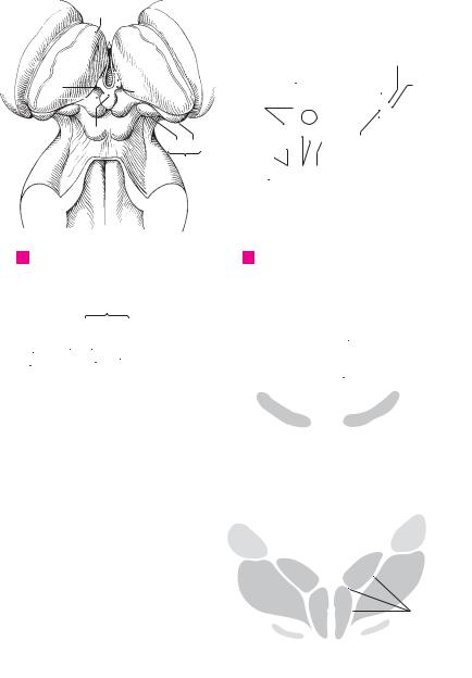
9
A Brain stem, dorsal view B Brain stem, sagittal section 10
11
|
|
|
|
|
|
|
|
|
26 |
|
|
|
|
|
|
|
|
12 |
33 |
34 |
28 |
27 |
|
|
|
|
|
|
|
||||||||
|
|
|
|
|
|
|
|
|||||||||||
|
|
|
|
|
|
|
||||||||||||
|
|
|
|
|
|
|
|
|
|
|
|
|
|
|
|
11 |
13 |
|
|
|
|
|
|
|
|
|
|
|
9 |
|
|
|
|
|
|||
|
|
|
|
|
|
|
|
|
|
|
||||||||
|
|
|
|
|
|
|
|
|
|
14 |
|
|
|
|
|
|
|
|
|
|
|
|
|
|
|
|
|
|
|
|
|
|
12 |
14 |
|||
|
|
|
|
|
|
|
|
|
|
|
|
|
|
|
|
|
||
|
|
|
|
|
|
|
|
|
|
|
|
|
|
|
|
|
||
|
|
|
|
|
|
35 |
|
|
|
|
|
|
|
|
||||
|
|
|
|
|
|
|
|
|
|
|
||||||||
|
|
|
|
|
|
|
|
|
15 |
|||||||||
32 |
|
|
|
|
|
|
|
|
|
|
|
|
|
|
||||
|
|
|
|
|
|
|
|
|
|
|
|
|
|
|
|
|
|
|
|
|
|
|
|
|
|
|
|
|
|
|
|
|
|
|
|
|
|
|
|
|
|
|
|
|
|
|
|
|
|
|
|
|
|
|
|
16 |
|
|
|
|
|
|
|
|
|
|
|
|
|
|
|
|
|
|
|
|
|
|
|
|
|
|
|
|
|
|
|
|
|
|
|
|
|
|
|
|
|
|
|
|
|
|
|
|
|
|
|
|
|
|
|
|
17 |
|
|
|
|
|
|
|
|
|
|
|
|
|
|
|
|
|
|
|
|
|
|
|
|
|
|
|
|
|
|
|
|
|
|
|
|
|
|
|
Termination of optic tract |
|
|
|
|
|
|
18 |
||||||||||
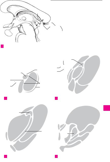
C |
|
D |
Oblique section through midbrain |
|||||||||||||||
|
|
|||||||||||||||||
|
|
|
|
|
|
|
|
|
|
|
|
|
|
|
|
|
|
|
|
|
|
|
|
|
|
|
|
|
|
|
|
|
|
|
|
|
19 |
|
|
|
|
|
|
|
|
|
|
|
|
|
|
|
|
|
|
|
|
|
|
|
|
|
|
|
|
|
|
|
|
|
|
|
|
|
|
|
|
|
|
|
|
|
|
|
|
|
|
|
|
|
|
|
|
20 |
|
|
|
|
|
|
|
|
|
|
|
|
|
|
|
|
|
|
|
|
|
|
|
|
|
|
|
|
|
|
|
|
|
|
|
|
|
|
|
|
|
|
|
|
|
|
|
|
|
|
|
|
|
|
|
|
21 |
|
|
|
|
|
|
|
|
|
|
|
|
|
|
|
|
|
|
|
|
|
|
|
|
|
|
|
|
|
|
|
|
|
|
|
|
|
|
|
|
|
|
|
|
|
|
|
|
|
|
|
|
|
|
|
|
22 |
|
|
|
|
|
|
|
|
|
|
|
|
|
|
|
|
|
|
|
|
|
|
|
|
|
|
|
|
|
|
|
|
|
|
|
|
|
|
|
|
|
|
|
|
|
|
|
|
|
|
23 |
23 |
|||||
|
|
|
|
|
|
|
|
|
|
|
|
|
|
|
|
|
|
|
|
|
|
|
|
|
|
|
|
|
|
|
|
|
|
|
|
|
|
|
|
|
|
|
|
|
|
|
|
|
|
Section through |
24 |
|||||
|
|
|
|
|
|
|
|
|
|
|
E |
|||||||
|
|
|
|
|
|
|
|
|
|
|
|
|||||||
|
|
|
|
|
|
|
|
|
|
|
|
|
|
|
|
|
|
|
diencephalon
25

|
298 |
Brain |
||
|
|
1 |
Third ventricle. Ventriculus tertius. Dien- 16 |
|
1 |
||||
|
|
cephalic portion of the cerebral ventricular sys- |
||
|
|
|
||
|
|
|
tem. It extends from the lamina terminalis to |
|
2the beginning of the cerebral aqueduct. A C
2Hypothalamic sulcus. Sulcus hypothalamicus. 17
3Furrow extending from the interventricular foramen to the entrance into the cerebral aque-
|
|
duct. It separates the dorsal and ventral |
|
4 |
|
||
|
thalami. A |
||
|
3 |
Interventricular foramen. Foramen inter- |
|
|
|||
5 |
|||
|
ventriculare. Opening between the lateral ven- |
||
|
|
tricle and third ventricle behind the genu of the |
|
|
|
||
6 |
|
fornix. A |
|
4 |
Optic recess. Recessus opticus. Recess of third 19 |
||
|
|||
|
|||
|
|
ventricle above the optic chiasm. A |
75 Recess of infundibulum. Recessus infundibuli 20
(infundibularis). Recess of third ventricle
8within the infundibulum. A
6Pineal recess. Recessus pinealis. Recess of third
9 |
ventricle extending partially into the epiphysis. |
|
|
A |
20 a |
|
|
107 Supraspinal recess. Recessus supraspinalis. Recess between the roof of the third ventricle
and the epiphysis. A
11
8Tela choroidea. Forms the thin, narrow roof of
12 |
|
third ventricle and its choroid plexus. B C |
21 |
9 |
Tenia of thalamus. Taenia thalami. Lateral at- |
||
|
|
tachment line of the upper wall of the third |
|
13ventricle along the stria medullaris of the thalamus. B C
22
1410 Choroid plexus. Plexus choroideus. Paired, highly vascularized villous infolding which
hangs down from the thin roof of the third ven23
15tricle and is continuous anteriorly with the choroid plexus of the 4th ventricle via the inter-
16ventricular foramen. B C
|
11 Sections of thalamus and metathalamus. Sec- |
|
17 |
tiones thalamici et metathalamici. See p. 409. |
|
|
12 Reticular nucleus of thalamus. Nucleus reticu- |
|
|
||
18 |
latus [thalami]. Thin layer lying mainly laterally |
|
along the thalamus between the posterior limb |
||
|
||
|
of the internal capsule and external medullary |
|
19 |
lamina of the thalamus. It receives tributaries 25 |
|
|
from the entire cerebral cortex, globus pallidus |
|
|
||
|
and reticular formation of the brainstem and |
20gives off efferent fibers to the reticular formation of the midbrain and thalamus. B
2113 Anterior nuclei of thalamus. Nuclei anteriores
|
|
[thalami]. Cell group in the apex of the |
|
22 |
|
thalamus. They receive fibers from the mamil- |
27 |
|
|
lothalamic tract and have projections to the |
|
|
|
cingulate gyrus. |
|
23 |
|
|
|
14 |
Anterodorsal nucleus. Nucleus anterodorsalis |
28 |
|
24 |
|
(anterosuperior). Narrow cell plate anterosupe- |
|
|
riorly. B |
|
15 Anteroventral nucleus. Nucleus anter29
25oventralis (anteroinferior). Main nucleus of the anterior nuclei. B


|
300 |
Brain |
|
|
|
|
|
|
|
|
|
|
|
|
|
|
|
|
|||
|
|
|
Nuclei ventrolaterales [thalami]. Ventrolateral |
|
15 |
Lateral geniculate nucleus [ventral part]. Nu- |
|||||||||||||||
1 |
|
1 |
|
||||||||||||||||||
|
|||||||||||||||||||||
|
|
|
nuclei, the group of nuclei lateral to the internal |
|
|
cleus corporis geniculati lateralis [pars |
|||||||||||||||
|
|
|
|
medullary lamina. B |
|
|
|
|
|
|
ventralis]. Small group of cells with fibers from |
||||||||||
2 |
|
2 |
Posterior lateral nucleus. Nucleus lateralis |
|
|
the retina: part of a light reflex tract. C |
|
||||||||||||||
|
|
|
|
posterior. Portion of the lateral nucleus situated |
|
16 |
Medial geniculate nucleus [ventral part]. Nu- |
||||||||||||||
3 |
|
|
|
between the pulvinar and dorsal lateral nu- |
|
|
cleus corporis geniculati medialis [pars |
||||||||||||||
|
|
|
cleus with connections to the parietal lobe. A |
|
|
ventralis]. Possibly the true acoustic part of the |
|||||||||||||||
|
|
3 |
Dorsal lateral nucleus. Nucleus lateralis |
|
|
geniculate nucleus. C |
|
|
|
|
|||||||||||
4 |
|
|
17 |
Subthalamic nucleus. Nucleus subthalamicus |
|||||||||||||||||
|
|
|
dorsalis. Anterosuperior portion of the lateral |
|
|||||||||||||||||
|
|
|
|
nucleus with projections to the region of the |
|
|
[corpus Luysii]. It lies between the lower end of |
||||||||||||||
|
|
|
|
|
|
the internal capsule and the zona incerta. Of |
|||||||||||||||
5 |
|
|
|
posterior cingulum segment and the lower part |
|
|
|||||||||||||||
|
|
|
|
|
clinical importance is its reciprocal connection |
||||||||||||||||
|
|
|
of the parietal lobe. A |
|
|
|
|
|
|
||||||||||||
|
|
|
|
|
|
|
|
|
|
with the globus pallidus. B |
|
|
|||||||||
|
|
4 |
Anterior ventral nucleus. Nucleus ventralis |
|
|
|
|
||||||||||||||
6 |
|
|
18 |
Reticular nuclei of thalamus. Nuclei reticulares |
|||||||||||||||||
|
|
|
anterior. Anterior portion of the ventral nucleus |
|
|||||||||||||||||
|
|
|
|
with |
projections |
to the |
interlaminar |
nuclei, |
|
|
[thalami]. Disaggregated |
cell |
layer on |
the |
|||||||
|
|
|
|
|
|
lateral surface of the thalamus between the ex- |
|||||||||||||||
7 |
|
|
|
globus pallidus and dentate nucleus and recip- |
|
|
|||||||||||||||
|
|
|
|
|
ternal medullary lamina and internal capsule. B |
||||||||||||||||
|
|
|
rocal connections to the precentral gyrus and |
|
|
||||||||||||||||
|
|
|
|
|
19 |
Zona incerta. Basal continuation of the reticu- |
|||||||||||||||
|
|
|
|
the area anterior to it. It plays a role in Parkin- |
|
||||||||||||||||
8 |
|
|
|
son’s disease. A |
|
|
|
|
|
|
|
lar nucleus of the thalamus and other struc- |
|||||||||
|
|
|
|
|
|
|
|
|
|
tures. It lies in the path of the globus pallidus to |
|||||||||||
|
|
5 |
Intermediate |
ventral |
nucleus. |
Nucleus |
|
|
|||||||||||||
|
|
|
|
the tegmentum of the diencephalon. B |
|
||||||||||||||||
9 |
|
|
|
ventralis intermedius. Portion of the ventral |
|
|
|
||||||||||||||
|
|
|
|
20 |
Nuclear |
regions |
H, |
H1 |
and |
H2. Nuclei re- |
|||||||||||
|
|
|
|
nucleus situated behind the anterior |
|
ventral |
|
|
gionum H, H1 and H2. Dispersed neurons in the |
||||||||||||
|
|
|
|
nucleus; it is a synaptic station connecting the |
|
|
|||||||||||||||
10 |
|
|
|
|
|
corresponding Forel’s fields. Field H lies medial |
|||||||||||||||
|
|
|
cerebellum, red nucleus and motor cortex. A |
|
|
||||||||||||||||
|
|
|
|
|
to the zona incerta and in front of the red nu- |
||||||||||||||||
|
|
6 |
Medial ventral nucleus. Nucleus ventralis me- |
|
|
||||||||||||||||
|
|
|
|
cleus, H1 between the thalamus and zona in- |
|||||||||||||||||
11 |
|
|
|
dialis. Poorly demarcated nuclear region sit- |
|
|
certa, H2 between the zona incerta and sub- |
||||||||||||||
|
|
|
|
uated anterior to the posterior ventral nuclei; |
|
|
thalamic nucleus. B |
|
|
|
|
||||||||||
12 |
|
|
|
its function is unclear. A |
|
|
|
|
|
21 |
Thalamic |
tract |
and |
fasciculi. Tractus |
et |
||||||
|
7 |
Posterior ventral nuclei. Nuclei ventrales |
|
||||||||||||||||||
|
|
|
fasciculi thalamici. |
|
|
|
|
||||||||||||||
|
|
|
|
|
|
|
|
||||||||||||||
|
|
|
|
posteriores. Collective term for the following |
|
22 |
Lateral lemniscus. Lemniscus lateralis. Audi- |
||||||||||||||
13 |
|
|
|
two nuclei. |
|
|
|
|
|
|
|
|
tory pathway passing into the medial genicu- |
||||||||
|
|
|
8 Posterolateral ventral nucleus. Nucleus ventralis |
|
|
late body. A |
|
|
|
|
|
||||||||||
14 |
|
|
|
posterolateralis. The lateral part of the poste- |
|
23 |
Medial lemniscus. Lemniscus |
medialis. Con- |
|||||||||||||
|
|
|
rior ventral nucleus that receives the medial |
|
|
tinuation of the tract from the posterior |
|||||||||||||||
|
|
|
|
|
|
||||||||||||||||
15 |
|
|
|
lemniscus and spinothalamic tract and relays |
|
|
funiculus radiating into the posterolateral ven- |
||||||||||||||
|
|
|
their impulses to the postcentral gyrus via the |
|
|
tral nucleus. A |
|
|
|
|
|
||||||||||
|
|
|
|
thalamocortical tract. A |
|
|
|
|
|
24 |
Spinal lemniscus. Lemniscus spinalis. Pain |
||||||||||
16 |
|
9 |
Posteromedial ventral nucleus. Nucleus ventralis |
|
|
pathway extending into the posterolateral ven- |
|||||||||||||||
|
|
|
tral nucleus. A |
|
|
|
|
|
|||||||||||||
|
|
|
|
posteromedialis. Part located between the cen- |
|
|
|
|
|
|
|
||||||||||
|
|
|
|
tromedian and posterolateral nuclei. It receives |
|
25 |
Trigeminal lemniscus. Lemniscus trigeminalis. |
||||||||||||||
17 |
|
|
|
|
|||||||||||||||||
|
|
|
the trigeminal lemniscus. A |
|
|
|
|
|
Fibers of the sensory trigeminal nucleus. They |
||||||||||||
|
|
10 Posterior nuclei of thalamus. Nuclei posteri- |
|
|
pass into the posteromedial ventral nucleus. A |
||||||||||||||||
|
|
|
|
||||||||||||||||||
|
|
|
26 Brachium of inferior colliculus. Brachium col- |
||||||||||||||||||
18 |
|
|
|
ores [thalami]. Collective term for the following |
|
||||||||||||||||
|
|
|
|
|
liculi inferioris. Outwardly visible connection |
||||||||||||||||
|
|
|
|
three parts of the thalamus. |
|
|
|
|
|
||||||||||||
19 |
|
11 |
Pulvinar nuclei. Nuclei pulvinares. Nuclei that |
|
|
between the inferior colliculus and the medial |
|||||||||||||||
|
|
|
geniculate body. C |
|
|
|
|
|
|||||||||||||
|
|
|
|
occupy the posterior portion of the thalamus; |
|
27 |
Acoustic radiation. Radiatio acustica. Portion of |
||||||||||||||
|
|
|
|
they begin at the habenulae, receive tributar- |
|
||||||||||||||||
|
|
|
|
|
|
auditory pathway extending from the medial |
|||||||||||||||
20 |
|
|
|
ies from the auditory and visual pathways as |
|
|
|||||||||||||||
|
|
|
|
|
geniculate body to the transverse temporal gyn. |
||||||||||||||||
|
|
|
|
well |
as |
from |
other thalamic |
nuclei |
and are |
|
|
||||||||||
|
|
|
|
|
|
It passes through the occipital part of the poste- |
|||||||||||||||
21 |
|
|
|
connected with the visual cortex, optic and |
|
|
|||||||||||||||
|
|
|
|
|
rior limb of the internal capsule. A |
|
|||||||||||||||
|
|
|
acoustic control centers, and other structures. |
|
|
|
|||||||||||||||
|
|
|
|
A |
|
|
|
|
|
|
|
|
|
28 Brachium of superior colliculus. Brachium col- |
|||||||
22 |
|
|
|
|
|
|
|
|
|
|
|
|
|
liculi superioris. Externally visible connection |
|||||||
|
12 |
Lateral |
geniculate nucleus |
[dorsal |
part]. |
|
|
||||||||||||||
|
|
|
between the superior colliculus and the lateral |
||||||||||||||||||
|
|
|
|
Nucleus |
[corporis geniculati] |
lateralis [pars |
|
|
|||||||||||||
|
|
|
|
|
|
geniculate body. Connection of the visual path- |
|||||||||||||||
|
|
|
|
dorsalis]. Part of the visual pathway. A |
|
|
|
|
|||||||||||||
23 |
|
|
|
|
|
|
|
way with the extrapyramidal system. C |
|
||||||||||||
|
13 |
Medial geniculate nucleus [dorsal part]. Nu- |
|
|
|
||||||||||||||||
|
|
29 Optic radiation. Radiatio optica [[Gratiolet]]. |
|||||||||||||||||||
|
|
|
|
cleus |
[corporis |
geniculati] |
medialis |
[pars |
|
||||||||||||
24 |
|
|
|
|
|
Portion of the visual pathway emanating from |
|||||||||||||||
|
|
|
dorsalis]. Part of medial geniculate body con- |
|
|
||||||||||||||||
|
|
|
|
|
the lateral geniculate body. It passes through the |
||||||||||||||||
|
|
|
|
taining small cells. A |
|
|
|
|
|
|
occipital part of the posterior limb of the inter- |
||||||||||
|
|
|
|
|
|
|
|
|
|
|
|
|
|
|
|||||||
25 |
|
14 |
Sections of ventral thalamus. Sectiones |
|
|
nal capsule and around the posterior horn of the |
|||||||||||||||
|
|
|
|
thalami ventralis. |
|
|
|
|
|
|
|
lateral ventricle to the area striata. A |
|
||||||||
|
|
|
|
|
|
|
|
|
|
|
|
|
|
|
|
|
|
|
|
|
|
|
|
|
|
|
|
|
|
|
|
|
|
Brain |
301 |
|
|
||
|
|
|
|
|
|
|
|
|
|
|
|
|
|
|
|
|
|
|
|
|
|
|
|
|
|
|
|
298.13 |
|
|
|
1 |
|||
|
|
|
|
|
|
|
|
|
|
|
|
|
|
|
|
2 |
|
|
|
|
|
|
|
298.21 |
|
3 |
|
|
|
|
3 |
||||
|
|
|
|
|
|
|
|
|
|
4 |
|
|
|||||
|
|
|
|
|
|
|
|
|
|
|
|
|
|
||||
|
|
|
|
|
|
|
|
|
|
2 |
|
|
|
|
|
4 |
|
|
|
|
|
|
|
|
|
|
|
5 |
|
|
|
|
|
||
|
|
|
|
|
|
9 |
|
6 |
|
|
|
|
|
|
5 |
||
|
|
|
|
|
|
|
|
|
|
|
|
|
|||||
|
|
|
11 |
|
|
|
|
|
|
8 |
|
|
|
|
|
|
6 |
|
|
|
|
|
|
|
|
|
|
|
|
|
|
|
|||
|
|
|
|
|
|
27 |
|
|
|
|
|
|
|
7 |
|||
|
|
|
|
|
|
|
|
|
|
|
|
|
|
|
|||
|
|
|
|
|
|
24 |
|
|
|
|
|
|
|
8 |
|||
13 |
|
|
|
|
|
|
|
|
|
|
|
|
9 |
||||
|
12 |
23 |
|
|
|
|
|
|
|
|
|||||||
|
|
|
|
|
|
|
|
|
|
||||||||
22 |
|
25 |
|
|
|
|
|
|
|
|
10 |
||||||
|
|
|
|
|
|
|
|
|
|
|
|
|
|
|
|||
|
|
|
|
|
|
|
|
|
|
|
|
|
|
|
|
||
|
|
|
29 |
|
|
|
|
|
|
|
|
|
|
|
|
11 |
|
|
|
|
|
|
|
|
|
|
|
|
|
|
|
|
|
||
|
|
|
|
|
|
|
|
|
|
|
|
|
|
|
|
12 |
|
|
|
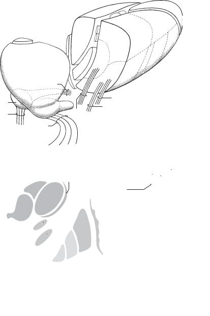
Thalamic nuclei and pathways |
|
|
|
|
|||||||||||
|
A |
28 16 15 |
|
|
|
13 |
|||||||||||
|
|
|
|
|
|
18 |
|
|
|
|
|
|
|
|
|||
|
|
|
|
|
|
|
|
|
|
|
|
|
14 |
||||
|
|
|
|
|
|
|
|
|
|
|
|
|
|
||||
|
|
|
|
|
|
|
|
|
|
|
|
|
|
15 |
|||
|
|
|
|
|
|
|
|
|
|
|
|
|
|
|
|
||
|
|
|
|
|
|
26 |
|
|
|
|
|
|
|
|
16 |
||
|
|
|
1 |
|
|
|
|
|
|
|
|
|
|||||
|
|
|
|
|
|
|
|
|
|
|
|
|
|
||||
|
|
|
|
|
|
|
|
|
|
|
|
|
|
|
|
||
|
|
|
|
|
|
|
|
|
|
|
|
|
|
|
|
|
|
|
|
|
|
|
|
|
|
|
|
|
|
|
|
|
|
17 |
|
|
|
|
H1 |
|
|
|
|
|
|
|
|
|
|
|
|
|
|
|
|
|
|
|
|
|
|
|
|
|
|
|
|
|
|||
20 |
|
|
|
19 |
|
|
|
|
|
|
|
18 |
|||||
|
|
|
|
|
|
|
|
|
|
||||||||
|
|
|
|
|
|
|
|
|
|
|
|
|
|
|
|||
H2 |
|
|
|
17 |
|
|
|
|
|
|
|
|
19 |
||||
|
|
H |
|
|
|
|
|
|
|
|
|
||||||
|
|
|
|
|
|
|
|
|
|||||||||
20 |
|
|
|
|
|
|
|
|
|
|
|
|
|
|
|
||
|
|
|
|
|
|
|
|
|
|
|
|
|
|
20 |
|||
|
|
|
|
|
|
|
|
|
|
|
|
|
|
|
|
||
|
|
|
|
|
|
|
|
|
|
|
|
|
|
|
|
|
|
|
|
|
|
|
|
|
|
|
|
|
|
|
|
|
|
||
|
|
|
|
|
|
|
|
|
|
|
|
|
|
|
|
21 |
|
|
|
|
|
|
|
|
|
|
|
|
|
|
|
|
|
|
|
|
|
|
|
|
|
|
|
|
|
|
|
|
|
|
|
||
|
|
|
|
|
|
|
|
|
|
|
|
|
|
|
|
22 |
|
|
|
|
|
|
|
|
|
|
|
|
|
|
|
|
|
|
|
|
|
|
|
|
|
|
|
|
|
|
|
|
|
|
|
||
|
|
|
|
|
|
|
|
|
|
|
|
|
|
|
|
23 |
|
|
B |
Subthalamic region |
|
|
|
C |
Geniculate body |
|
|
|
|||||||
|
|
|
|
|
|
|
|
||||||||||
|
|
|
|
|
|
|
|
|
|
|
|
|
|
|
|
|
|
24
25