Материал: Атлас Ханц фениш


 8 10
8 10
|
290 |
Brain |
|
|
|
|
|
|
|
|||
|
|
|
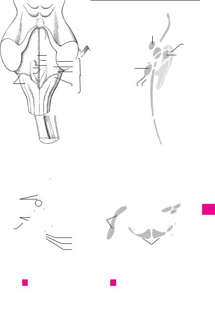
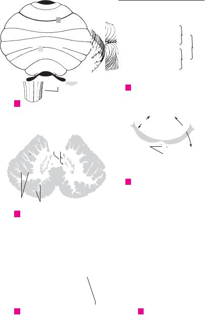
1 Flocculonodular lobe. Lobus flocculonodularis. |
19 Hilum of dentate nucleus. Hilum nuclei dentati. |
||||||||
1 |
|
|
||||||||||
|
|
|
Small archicerebellar portion of the cerebellum |
|
Opening of dentate nucleus from which |
|||||||
|
|
|
|
located caudal to the dorsolateral fissure. |
|
emerges most of the superior cerebellar |
||||||
2 |
|
2 |
Nodulus. Medial protuberance of the vermis |
|
peduncle. C |
|
||||||
|
20 |
Emboliform nucleus. Nucleus emboliformis. It |
||||||||||
|
|
|
|
united with the flocculus by the peduncles. E |
||||||||
3 |
|
3 |
Flocculus. Claw-like portion of the cerebellum |
|
is found just in front of the hilum of the dentate |
|||||||
|
|
nucleus. C |
|
|
||||||||
|
|
|
|
between the inferior cerebellar peduncle and |
|
|
|
|||||
|
|
|
|
21 |
Globose nucleus. Nucleus globosus. It lies me- |
|||||||
4 |
|
|
|
biventral lobule. E |
|
|
||||||
|
|
4 Peduncle of flocculus. Pedunculus floccularis. |
|
dial to the dentate nucleus. C |
|
|||||||
|
|
|
|
|
||||||||
|
|
|
22 |
Fastigial nucleus. Nucleus fastigii. The most |
||||||||
5 |
|
|
|
Band of nerve fibers connecting the flocculus to |
||||||||
|
|
|
the nodulus. Part of it extends into the inferior |
|
medial of the deep cerebellar nuclei. C |
|||||||
6 |
|
|
|
medullary velum. E |
|
|
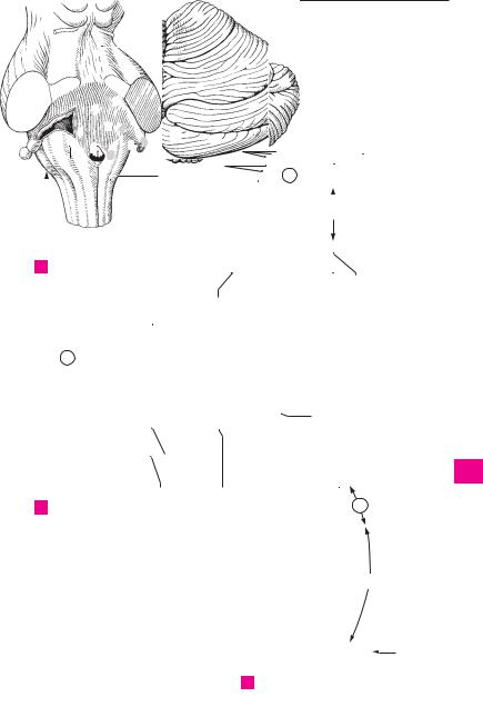
23 Cerebellar peduncles. Pedunculi cerebellares. |
|||||
|
|
|
|
|
|
|
||||||
|
5 |
Paraflocculus. In humans, a small insignificant |
|
Fibers that provide connections to and from the |
||||||||
|
|
|
|
part of the caudal lobe of the cerebellum that |
|
cerebellum. |
|
|||||
7 |
|
|
|
communicates with the flocculus. |
|
24 |
Inferior |
cerebellar peduncle |
(restiform |
|||
|
|
6 |
Archicerebellum. Archaeocerebellum. Phylo- |
|
body). Pedunculus cerebellaris inferior. Infe- |
|||||||
8 |
|
|
|
genetically the oldest part of the cerebellum; it |
|
rior connection to the cerebellum formed by |
||||||
|
|
|
|
fibers of the posterior spinocerebellar tract and |
||||||||
|
|
|
|
consists of the lingula and the flocculonodular |
|
|||||||
9 |
|
|
|
lobe. A |
|
|
|
|
olive. E F |
|
|
|
|
|
7 Paleocerebellum. An old part of the cerebel- |
25 Middle cerebellar peduncle (brachium pon- |
|||||||||
|
|
|
||||||||||
|
|
|
|
lum consisting of the central lobule, culmen, |
|
tis). Pedunculus cerebellaris medius (pon- |
||||||
10 |
|
|
|
pyramid, uvula, ala of the central lobule and |
|
tinus). Large peduncle containing fibers origi- |
||||||
|
|
|
|
quadrangular lobule. A |
|
|
|
nating from the pontocerebellar tract. E F |
||||
11 |
|
8 |
Neocerebellum. Phylogenetically young por- |
26 Superior |
cerebellar peduncle |
(brachium |
||||||
|
|
|
|
tion of the cerebellum; it comprises the declive, |
|
conjunctivum). Pedunculus cerebellaris su- |
||||||
|
|
|
|
|
perior. Superior, paired (right and left) connect- |
|||||||
12 |
|
|
|
folium, tuber, lobulus simplex, cranial and |
|
|||||||
|
|
|
|
ing fibers extending from the cerebellum to the |
||||||||
|
|
|
caudal semilunar lobules, paramedian lobule |
|
||||||||
|
|
|
|
and tonsil. A |
|
|
|
brain stem. The superior medullary velum ex- |
||||
13 |
|
|
|
|
|
|
tends between them. E F |
|
||||
|
9 |
CEREBELLAR SECTIONS. Sectiones cerebellares. |
|
|
||||||||
|
27 MIDBRAIN. Mesencephalon. It consists of cere- |
|||||||||||
|
|
|
|
Anatomical subdivisions of the cerebellum. |
||||||||
14 |
|
|
|
|
bral crura, tegmentum and quadrigeminal plate |
|||||||
|
10 |
Arbor vitae [cerebelli]. Treelike pattern of the |
|
|||||||||
|
|
(tectal lamina). |
|
|||||||||
|
|
|
|
white matter seen in prepared sections of the |
28 Cerebral peduncle. Pedunculus cerebri (cere- |
|||||||
15 |
|
|
|
cerebellum. C |
|
|
|
bralis). It comprises the cerebral crura and the |
||||
|
11 |
Medullary body. Corpus medullare. White |
|
|||||||||
|
|
|
tegmentum and extends up to cerebral aque- |
|||||||||
16 |
|
|
|
matter consisting of myelinated fibers. C |
|
duct. D |
|
|
||||
|
12 |
White laminae. Laminae albae. White matter |
29 Anterior part (cerebral crus, basis pedunculi). |
|||||||||
|
|
|||||||||||
|
|
|||||||||||
17 |
|
|
|
extending |
into the folia |
from the |
medullary |
|
Part anterior (crus cerebri). It consists of the |
|||
|
|
|
body. C |
|
|
|
|
previously mentioned cerebral crura. D |
||||
|
|
|
|
|
|
|
|
|||||
|
|
13 Cerebellar cortex. Cortex cerebellaris. Superfi- |
30 |
Part posterior. Posterior part of cerebral |
||||||||
18 |
||||||||||||
|
|
|
cial gray matter of the cerebellum, about 1 mm |
|
peduncle or tegmentum. See p. 292.10 D |
|||||||
|
|
|
|
thick, that consists pirmarily of nerve cells. B C |
31 Oculomotor sulcus. Sulcus oculomotorius. Fur- |
|||||||
19 |
|
|
|
|||||||||
|
|
|
|
|
|
|
||||||
14 |
Molecular |
(plexiform) |
layer. |
Stratum |
|
row on the medial surface of the cerebral crus, |
||||||
|
|
|
|
moleculare (plexiforme). External cortical layer |
|
exit site of the oculomotor nerve. D |
||||||
20 |
|
|
|
rich in dendrites and axons, poor in cell bodies. |
32 |
Interpeduncular fossa. Fossa interpeduncu- |
||||||
|
|
|
|
The nuclei of the Purkinje cells are found at its |
|
laris. Fossa situated between the cerebral crura. |
||||||
|
|
|
|
border with the granular layer. |
|
|
||||||
21 |
|
|
|
|
|
D |
|
|
||||
|
|
|
|
|
|
|
|
|
|
|||
15 |
Purkinje cell layer. Stratum neurium piri- |
33 |
Interpeduncular (posterior) perforated sub- |
|||||||||
|
|
|
|
formium. The layer in which the perikarya of |
|
stance. Substantia perforata interpeduncularis |
||||||
22 |
|
|
|
|
||||||||
|
|
|
the Purkinje cells are located. B |
|
|
[posterior]. Perforated floor of interpeduncular |
||||||
|
16 |
Stratum granulosum. Internal nuclear layer |
|
fossa produced by openings for numerous ves- |
||||||||
23 |
|
|
|
containing numerous closely packed small |
|
sels. D |
|
|
||||
|
|
|
|
neurons. B |
|
|
|
|
|
|
|
|
|
|
|
|
|
|
|
|
|
|
|
||
2417 Nuclei of cerebellum. Nuclei cerebellaris.
18 Dentate nucleus. Nucleus dentatus. Large
25cerebellar nucleus located in the medullary body and resembling a folded pouch. C
 6
6


 19
19


 18
18
 15 13
15 13