Материал: Атлас Ханц фениш

Внимание! Если размещение файла нарушает Ваши авторские права, то обязательно сообщите нам

Spinal cord 277

7


4


3


2


5


12


16


14


6


26

11


10


13

8


19


1

A Cross section of spinal cord

9;11

B

Columns of spinal cord

28


7


3


26

16

14


29


16a

12

13


27


18


19

33


22

20


24


25


23


32


19


C Cross section of spinal cord with tracts

1

2

3

4

5

6

7

8

9

10

11

12

13

14

15

16

17

18

19

20

21

22

23

24

25

278 Spinal cord, Brain

1

Spinotectal tract. Tractus spinotectalis. Its

1

fibers arise from the posterior horn of the op-

posite side, run in the lateral spinothalamic

2tract and end in the tectal lamina. A

2Spinothalamic tract. Tractus spinothalamicus. Lies in the lateral funiculus and arises primarily

3from the opposite side. Its fibers transmit pain and temperature sensations. A

4 3 Anterior spinocerebellar tract. Tractus spino-


cerebellaris anterior [[Gowers]]. It extends

5

partly from the opposite side and transmits to

the cerebellum information necessary for the


coordination of movements concerning muscle

6extension and limb position. A


4 Posterior spinocerebellar tract. Tractus spino-

7

cerebellaris posterior [[Flechsig]]. Its fibers are

uncrossed and function similar to those of the


anterior spinocerebellar tract. A

85 Dorsolateral tract. Tractus dorsolateralis [[Lissauer]]. Bundle of fine myelinated and nonmy-

9elinated fibers between the apex of the posterior horn and the surface. The fibers are short and some form lateral branches of posterior

10 root fibers. A

6 Spino-olivary tract. Tractus spino-olivaris

11[[Hellweg]]. Predominantly crossed fibers to the olive. They lie by the anterior root fibers together with the olivospinal tract. A

127 Spinoreticular tract. Tractus spinoreticularis. Its fibers lie beside the spino-olivary tract and

13end in the reticular substance of the medulla oblongata. A

8Posterior funiculus. Funiculus posterior. White

14

matter between the posterior horns. A


9Posterior fasciculi proprii. Fasciculi proprii

15posteriores. They lie directly on the gray matter and unite individual spinal cord segments for

16coordinated activity.

Septomarginal fasciculus. Fasciculus septo-10

marginalis. Bundle of mostly descending fibers

17situated along the posterior median septum in the lower half of the thoracic spinal cord. A

1811 Interfascicular fasciculus. Fasciculus interfascicularis. Bundle of mostly descending fibers [comma tract of Schultze] located chiefly in the

19upper half of the thoracic spinal cord between the fasciculi gracilis and cuneatus. A

2012 Fasciculus gracilis [[Goll]]. Medial fasciculus that contains fibers from the lower half of the spinal cord which function in the transmission

21of tactile and proprioceptive sensory information. A

2213 Fasciculus cuneatus [[Burdach]]. It begins with the upper half of the thoracic cord an likewise contains fibers for transmission of tactile and

23proprioceptive impulses. A

14 BRAIN. Encephalon.

2415 BRAINSTEM. Truncus encephali. It extends from the medulla oblongata to the midbrain.

25According to some, the diencephalon and insula likewise belong to the brainstem.

16HINDBRAIN. Rhombencephalon. It includes the brain substance around the rhomboid fossa, thus the cerebellum, pons with dorsally attached substance and medulla oblongata. B

17MEDULLA OBLONGATA (BULBUS). Beginning at the lower margin of the pons, it extends up to the root fibers of C1. In the rhomboid fossa it arises from the medullary striae of the 4th ventricle. B

18Anterior median fissure. Fissura mediana anterior. Anterior groove continuous with that of the spinal cord. The pyramidal decussation crosses over it. C

19Pyramid. Pyramis [medullae oblongatae]. Longitudinal elevation of pyramidal tract fibers to the right and left of the anterior median fissure. It ends with the pyramidal decussation. C

20Pyramidal decussation. Decussatio pyramidum (dec. pyramidalis anterior motoria). 3−5 bundles of lateral pyramidal tract fibers crossing to the opposite side at the end of the medulla. C

21Anterolateral sulcus. Sulcus anterolateralis. Furrow lateral to the pyramid for transmission of the hypoglossal nerce. C

22Lateral funiculus. Funiculus lateralis. Continuation of the lateral funiculus of the spinal cord up to the olive. C

23Olive. Oliva. Bean-shaped prominence about 1.5 cm long between the roots of cranial nerves X and XII. it is produced by the nuclei lying beneath it. C

24Anterior external arcuate fibers. Fibrae arcuatae externae anteriores. Fibers from the arcuate nucleus coursing over the caudal end of the olive to the caudal cerebellar peduncle. Part of the pontocerebellar tract. C

25Retro-olivary sulcus. Sulcus retro-olivaris. Furrow behind the olive. C

26Retro-olivary area. Area retro-olivaris. Area behind the olive. C

Spinal Cord, Brain 279

8

12


13


10


5


4

11

9


2

3

1

67

A Cross section of spinal cord

18


26


23


19


25


21


21


24


22

20


C Medulla oblongata from above

16

17

B Rhombencephalon

1

2

3

4

5

6

7

8

9

10

11

12

13

14

15

16

17

18

19

20

21

22

23

24

25


280

Brain


1

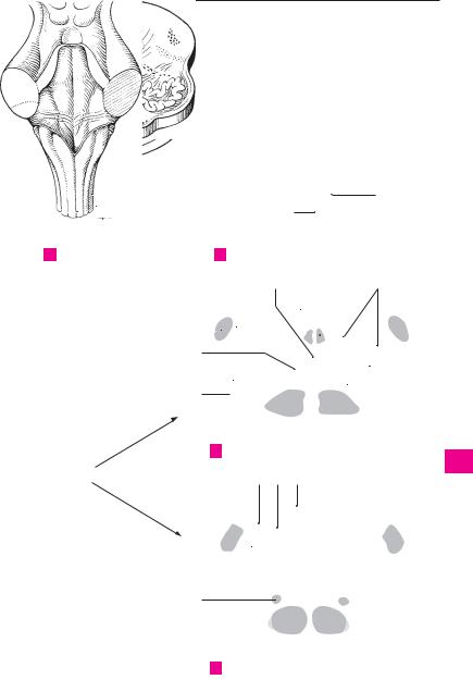
Posterolateral sulcus. Sulcus posterolateralis.

19

Internal arcuate fibers. Fibrae arcuatae inter-

1


Furrow reaching up to the lateral recess of the


nae. Fiber component of the medial lemniscus


4th ventricle. Site of exit of cranial nerves IX, X


arising from the nucleus of the posterior

2


and XI. A


funiculus. C


2

Inferior cerebellar peduncle. Pedunculus cere-

20

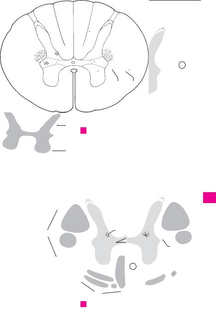
Decussation of medial lemniscus. Decussatio


3


bellaris inferior. Inferior connection to the cere-


lemniscorum medialium (d. sensoria). Formed


bellum with fibers of the posterior spinocere-


by fibers of nuclei gracilis ans cuneatus, second


bellar tract and olive. A


order neurons of posterior funiculus. C D

4


3

Trigeminal tubercle (tuber cinereum). Tuber-

21

Medial lemniscus. Lemniscus medialis. Con-


culum trigeminale. Low lateral elevation above


tinuation of second order neuron of posterior

5


the spinal tract of the trigeminal nerve. B


funiculus after decussation of lemniscus [bul-


4

Cuneate fasciculus. Fasciculus cuneatus.


bothalamic tract]. D


22

Tectospinal tract. Tractus tectospinalis. Decus-

6


Lateral part of posterior funiculus coming from


the upper half of the body. A


sated connection between the quadrigeminal


5 Cuneate tubercle. Tuberculum cuneatum. Ob-


plate and the spinal cord. It lies between the fa-

7


cial and trigeminal nuclei. D


long prominence at the end of the cuneate


23

Medial longitudinal fasciculus. Fasciculus


fasciculus produced by the nucleus cuneatus. A

8

6

Fasciculus gracilis. Medial part of posterior


longitudinalis medialis. Nerve fiber bundle for


reciprocal connection to the motor nuclei of


funiculus coming from the lower half of the


ocular muscles, as well as vestibular, accessory

9


body. A


and cervical muscle nuclei. C

7Gracile tubercle (clava). Tuberculum gracile. 24 Posterior longitudinal fasciculus. Fasciculus

10


Oblong bulge over the nucleus gracilis. A


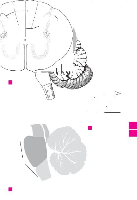
longitudinalis posterior [[Schütz]]. Connection


8

Posterior

median sulcus. Sulcus

medianus


between the hypothalamus, III, VII, XII cranial


nerve nuclei, nucleus ambiguus, tractus solitar-

11


posterior. Posterior furrow continued from the


ius and tractus salvatorius in the floor of the


spinal cord and closed above by a medullary


rhomboid fossa. C


lamella (obex). A


12


25

Spinal tract of trigeminal nerve. Tractus spi-

9

Sections through the medulla oblongata. Sec-


tiones medullae oblongatae.


nalis nervi trigeminalis. Descending fibers of


13


the trigeminal nerve for transmission of pain

10

Pyramidal fasciculus (tract). Fasciculus py-


and temperature stimuli. C D


ramidalis. Nerve tract for the transmission of

26 Spinal nucleus of trigeminal nerve. Nucleus

14


impulses concerned with conscious move-


spinalis nervi trigeminalis. Continuous with the


ments. C D


substantia gelatinosa in the spinal cord; this nu-


11

Corticospinal fibers. Fibrae corticospinales.


15


cleus receives the fibers of the spinal tract of the


Fibers from the precentral gyrus of the cortex to


trigeminal nerve. C D


the anterior horn cells of the spinal cord.

27

Reticular formation (substance). Formatio

16


12

Corticonuclear fibers. Fibrae corticonu-


(substantia) reticularis. Cells lying scattered in


cleares. Fibers from the precentral gyrus of the


the tegmentum in the vicinity of the vagus, ves-

17


cortex to the motor nuclei of cranial nerves.


tibular and facial nuclei with a regulatory effect


13

Decussation of the pyramids. Decussatio py-


on the muscles of the pharyngeal arch and other


muscles of the body. It extends cranially and


18


ramidum (dec. motoria). Fibers of the lateral py-


caudally. C


ramidal tract with 3−5 bundles crossing at the

28

Inferior olivary nucleus. Nucleus olivaris infe-

19


end of the medulla oblongata. B


14

Fasciculus

gracilis. Medial part of

posterior


rior. Main olviary nucleus lying below the olive.


It is shaped like a thick-walled pouch that opens


funiculus coming from the lower half of the


20


medially and is connected with the spinal cord


body.


and cerebullum. C


15

Nucleus gracilis. Nucleus of fasciculus gracilis

29

Amiculum of the olive. Amiculum olivare.

21


medial to cuneate nucleus. D


Fibrous sheath that surrounds and contains af-


16

Cuneate

fasciculus. Fasciculus

cuneatus.


ferent and efferent fibers of the olivary nucleus. C

22


Lateral part of posterior funiculus coming from

30

Hilum of inferior olivary nucleus. Hilum nu-


the upper half of the body.


clei olivaris inferioris. Opening of the medially

23


17

Cuneate nucleus. Nucleus cuneatus. Nucleus of


oriented pouch-like olivary nucleus. C


Medial accessory olivary nucleus. Nucleus


fasciculus cuneatus lateral to the nucleus 31

24


gracilis. D


olivaris accessorius medialis. It is located in front


18

Accessory cuneate nucleus. Nucleus cuneatus


of the hilum of the olivary nucleus. C


accessorius. Gray matter lateral to the upper

32

Posterior accessory olivary nucleus. Nucleus

25


part of cuneate nucleus. Origin of external ar-


olivaris accessorius posterior. It is situated be-


cuate fibers which pass to the cerebellum. D


tween the olive and reticular formation. C


Brain 281

2

7 5


8


1


6


4


A Rhomboid fossa

3

13

B Pons and medulla oblongata

26

20

24

23


27


25


19


31


28


21


32


30


29

10

C Section through medulla oblongata

18 17 15

25

26

21 20

22

10

D Section through medulla oblongata

1

2

3

4

5

6

7

8

9

10

11

12

13

14

15

16

17

18

19

20

21

22

23

24

25