Материал: Атлас Ханц фениш

Внимание! Если размещение файла нарушает Ваши авторские права, то обязательно сообщите нам

Lymphatic system 267


2


4


3


9

10


8


7


11

12


9


A Lymph nodes in the female pelvis

1

2

3

4

5

6

7

8

9

10

11

12

13

16

15

20


14


19


15


18


17


16


17


18


19


20


B Lymph nodes in the inguinal region

C Lymph nodes in the popliteal fossa

21


22

23

24

25

268 Spleen, meninges

1SPLEEN. Splen (lien). Lymphoreticular organ 1 within the circulatory system. Actions: phagocytosis and destruction of red blood cells,

2lymphopoiesis, blood filtration and synthesis of antibodies. A B

3

2 Accessory spleen. [Splen accessorius]. Small is-

lands of splenic tissue mostly in the greater

omentum or gastrosplenic ligament.

43 Diaphragmatic surface. Facies diaphragmatica. Convex surface facing the diaphragm.

54 Visceral surface. Facies visceralis. Concave surface facing the viscera. A

6

5

Renal surface. Facies renalis. Lower surface in


contact with the kidney. A


6 Gastric surface. Facies gastrica. Upper surface

7in contact with the stomach. A

7Colic surface. Facies colica. Surface in contact

8with the colon. A

8Posterior end. Extremitas posterior. A

99 Anterior end. Extremitas anterior. A


10

Upper margin. Margo superior. Border be-

10


tween gastric and diaphragmatic surfaces. A

11

Lower margin. Margo inferior. Border between


diaphragmatic and renal surfaces. A

11


12

Hilum of spleen. Hilum spenicum. Entry and


exit site of vessels between the gastric and

12


renal surfaces. A


13

Peritoneal covering. Tunica serosa. B


1314 Connective tissue capsule of spleen. Tunica fibrosa. B

15 Trabeculae of spleen. Trabeculae splenicae

14[lienales]. Connective tissue partitions penetrating into the spleen from the hilum and cap-

15sule and containing blood vessels. B

16

Splenic pulp. Pulpa splenica [lienalis]. It com-

16


prises white pulp (lymphoreticular tissue in


form of arterial sheaths) and red pulp (venous


sinuses with erythrocytes, reticular tissue). B

17


17

Splenic sinus. Sinus splenica [lienalis]. Thin-


walled venous spaces with multiple anasto-

18

moses situated in the red pulp. B


18Splenic [lienal] branches. Rami splenica [lienales]. Branches formed by the splenic artery

19

before entering the spleen.

19 Penicilli. Brush-like arterial branches between

20the nodular arteries (in white pulp) and capillaries (or ”sheathed capillaries”). B

2120 Lymphatic nodules (follicles) of spleen.

Foliculi lymphatici splenici [lienales] (lymphonoduli splenici). Spherical or cylindrical ag-

22gregations of lymphoreticular tissue around an artery (Malpighian corpuscle). They are visible

23to the naked eye. B

24

25

21NERVOUS SYSTEM. Systema nervosum.

22MENINGES. Connective tissue sheaths surrounding the central nervous system: dura mater, arachnoid and pia mater.

23Cranial dura mater [[Pachymeninx]]. Dura mater cranialis (encephali). Tough fibrous sheet forming a supporting capsule for the brain and at the same time forming the periosteum for the inner aspect of the skull. E

24Falx cerebri. Sickle-shaped part of the dura projecting downward into the longitudinal cerebral fissure. C

25Tentorium cerebelli. Dural sheet spreading out between the ridge of the petrous part of the temporal bone and the transverse sinus. It supports the occipital lobes. C

26Tentorial notch. Incisura tentorii. Opening in the tentorium cerebelli for passage of the brainstem. C

27Falx cerebelli. Small, sickle-shaped dural sheet between the right and left cerebellar hemispheres. C

28Diaphragma sellae. Small horizontal sheet of dura spreading out between the clinoid processes above the hypophysis. C

29Cavitas trigeminalis (Cavum trigeminale).

Outpocketing of the dura enclosing the trigeminal ganglion. C

30Subdural space. Spatium subdurale. Capillary space between the dura and arachnoid. It may be distended, for example, due to hemorrhage. E

31Spinal dura mater. Dura mater spinalis. It is separated from the wall of the vertebral canal by an epidural space. D

32External filum terminale. Filum terminale externum (durale). Filamentous end of the dura mater fused with the filum terminale. It extends from S2−3 to Co 2.

33Epidural space. Spatium epidurale (peridurale). Space between the spinal dura mater and the wall of the vertebral canal. It is filled with fat and venous plexuses. D

Spleen, meninges 269


8


10

17

13


6


14


12


4


11


5

7


19


Spleen


A

9


16


20


24


Spleen, histologic section, schematic


B


25


25


26


27

33


31


28


29


C Falx of cerebrum and tentorium of cerebellum

23

30

D Spinal meninges

30

E Cranial meninges

1

2

3

4

5

6

7

8

9

10

11

12

13

14

15

16

17

18

19

20

21

22

23

24

25


270

Meninges


1

Cranial arachnoid. Arachnoidea mater cranialis

13

Spinal arachnoid. Arachnoidea mater spinalis.

1


(encephali). Thin, avascular membrane attach-


Thin avascular membrane attached to the dura


ing to the cranial dura only by surface adhesion


mater by surface adhesion and to the pia mater

2


and communicating with the pia mater by con-


by its connective tissue fibers. A


nective tissue fibers. D


14

Subarachnoid

space.

Spatium

sub-


3

2

Subarachnoid

space.

Spatium

sub-


arachnoideum. Space between the flat part of


arachnoideum. Space between flat portion of


the arachnoid and the pia mater. It is filled with


arachnoid and pia mater. It is filled with


arachnoidal connective tissue fibers and cere-

4


arachnoidal connective tissue fibers and cere-


brospinal fluid. A


brospinal fluid. D


15

Cerebrospinal

fluid. Liquor cerebrospinalis.


5

3

Cerebrospinal

fluid. Liquor cerebrospinalis.


Fluid secreted predominantly by the choroid


plexus. It is protein-poor and has a cell content


Protein-poor fluid secreted by the choroid


plexus with a cell content of 2−6 per mm. It


of 2−6 per mm.


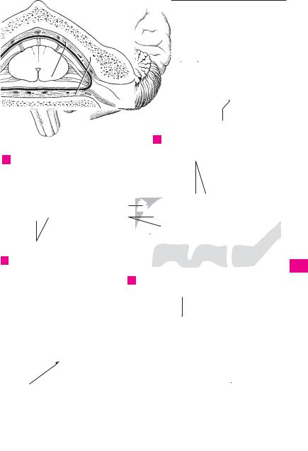
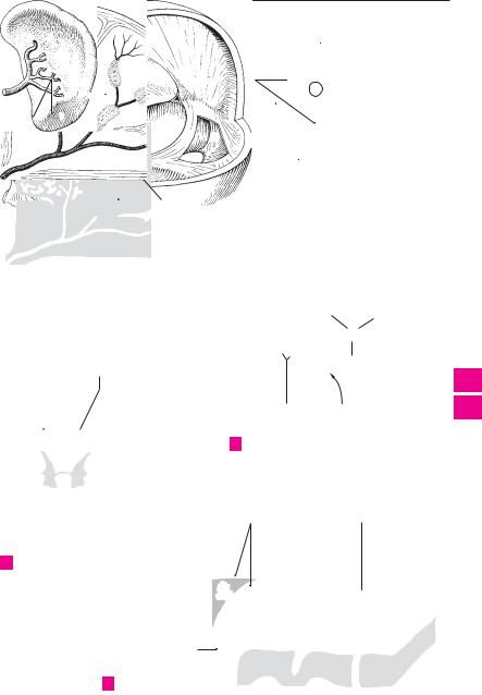
6flows into the subarachnoid space through openings in the fourth ventricle.

7 4 Subarachnoid cisterns. Cisternae subarachnoideae. Expansions of the subarachnoid

space containing cerebrospinal fluid.

8

5Cerebellomedullary cistern (cisterna magna).

Cisterna cerebellomedullaris (magna). Space

9between the cerebellum and medulla oblongata filled with cerebrospinal fluid. It com-

10municates with the fourth ventricle by a median aperture. It is accessible through the foramen magnum. B

116 Cisterna of lateral fossa of cerebrum. Cisterna fossae lateralis cerebri. Space between the in-

12sula, temporal, frontal and parietal lobes. It is filled with cerebrospinal fluid and is accessible through the lateral sulcus. It contains branches

13of the middle cerebral and insular arteries. C

7 Chiasmatic cistern. Cisterna chiasmatica. En-

14larged space around the optic chiasma filled with cerebrospinal fluid. B

158 Interpeduncular cistern. Cisterna interpeduncularis. Space situated behind the chiasmatic cistern and bordered laterally by the

16temporal lobe and the cerebral crura. It is filled

with cerebrospinal fluid and contains the

17oculomotor nerve, branches of the basilar artery, the origin of the superior cerebellar

artery and the posterior cerebral artery. B

18

9 Ambient cistern. Cisterna ambiens. Enlarged cerebrospinal fluid-filled space lateral to the

19cerebral crus. It contains the posterior cerebral artery, superior cerebellar artery, basal vein

20(Rosenthal’s) and the trochlear nerve. F

10Cisterna pericallosa. Space filled with cerebrospinal fluid along the corpus callosum. F

2111 Pontocerebellar cistern. Cisterna pontocerebellaris. Expanded space in the cerebellopon-

22tine angle filled with cerebrospinal fluid. It communicates with the 4th ventricle by a

23lateral aperture. E

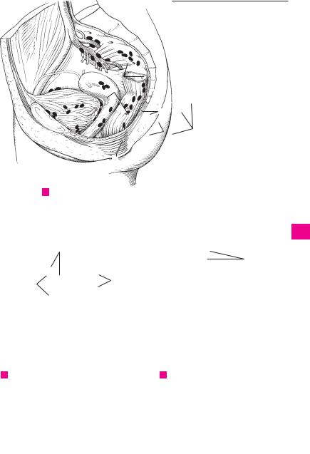
12 Arachnoid granulations. Granulationes arachnoideae. Avascular, villous-like outpock-

24etings of the subarachnoid space into the sagittal sinus and diploic veins. They are more pro-

25nounced after the tenth year of life and are concerned in the excretion of cerebrospinal fluid. D

Meninges 271

13

14

A Spinal meninges

6

C Cerebrum, lateral view

7


8


5

B Sagittal section with choroid plexus

12

12

2

1

D Meninges

10

1

2

3

4

5

6

7

8

9

10

11

12

13

14

15

16

17

18

19

20

21

11


22


23


9

24


Pontocerebellar cistern


System of cisterns in sagittal plane


E


F


25