Материал: Атлас Ханц фениш

|
272 |
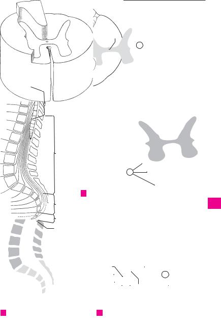
Spinal cord |
|
|
|
|
|
|
|
|
|
|
|
|
|
|||
|
|
1 |
Cranial pia mater. Pia mater cranialis (en- |
14 |
Cervical enlargement. Intumescentia cervi- |
|||||||||||||
1 |
||||||||||||||||||
|
|
cephali). Delicate meninx bearing blood vessels |
|
calis. Enlargement of the spinal cord from C3 to |
||||||||||||||
|
|
|
and covering the surface of the brain as well as |
|
T2 owing to the larger supply region for the |
|||||||||||||
2 |
|
|
extending into its sulci. |
|
|
|
|
arms. D |
|
|
|
|
|
|
||||
|
2 |
Tela choroidea of fourth ventricle. Tela 15 |
Lumbosacral |
enlargement. |
Intumescentia |
|||||||||||||
|
||||||||||||||||||
|
|
|
choroidea ventriculi quarti. Thin membrane of |
|
lumbosacralis. Expansion of the spinal cord |
|||||||||||||
3 |
|
|
|
|||||||||||||||
|
|
pia mater and ependyma in lower part of roof |
|
from T9−10 to L1−2 caused by the greater |
||||||||||||||
|
|
|
of fourth ventricle. It is attached laterally to the |
|
supply region for the lower limbs. D |
|
|
|||||||||||
4 |
|
|
tenia and exhibits lateral and median aper- |
16 |
Conus medullaris. Tapered termination of the |
|||||||||||||
|
|
|
tures. B |
|
|
|
|
|
|
|
spinal cord at the level of L1−2 where it be- |
|||||||
|
|
|
|
|
|
|
|
|
|
|||||||||
5 |
3 |
Choroid |
plexus |
of |
fourth |
ventricle. Plexus |
|
comes continuous with the filum terminale. D |
||||||||||
|
|
choroideus ventriculi quarti. Paired garland- |
17 |
Filum terminale (spinale). Thin terminal pro- |
||||||||||||||
|
|
|
||||||||||||||||
|
|
|
like, ependyma-covered villous projections |
|
longation of spinal cord attached inferiorly to |
|||||||||||||
6 |
|
|
which extend into both lateral apertures. B |
|
the posterior surface of the coccyx. D E |
|
||||||||||||
|
|
4 Tela choroidea |
of |
third |
ventricle. |
Tela |
18 |
Terminal ventricle. Ventriculus terminalis. En- |
||||||||||
|
|
|||||||||||||||||
7 |
|
|
choroidea ventriculi |
tertii. Thin, |
ependyma- |
|
largement of the central canal at the end of the |
|||||||||||
|
|
covered membrane of pia mater between right |
|
conus medullaris. E |
|
|
|
|||||||||||
|
|
|
|
|
|
|
||||||||||||
|
|
|
and left teniae of thalamus. C |
|
|
19 |
Anterior median fissure. Fissura mediana |
|||||||||||
8 |
|
|
|
|
||||||||||||||
5 |
Choroid plexus of third ventricle. Plexus |
|||||||||||||||||
|
anterior. Deep longitudinal fissure along the |
|||||||||||||||||
|
|
|
choroideus ventricul tertii. Paired, highly |
|
anterior aspect of the spinal cord. F |
|
|
|||||||||||
|
|
|
|
|
|
|||||||||||||
9 |
|
|
vascularized villous formations projecting from |
20 |
Posterior |
median |
sulcus. Sulcus |
medianus |
||||||||||
|
|
the thin roof into the third ventricle and con- |
||||||||||||||||
|
|
|
|
posterior. Median longitudinal groove between |
||||||||||||||
|
|
|
tinuing anteriorly through the interventricular |
|
the right and left posterior funiculi. F |
|
||||||||||||
|
|
|
|
|
||||||||||||||
10 |
|
|
foramina into the choroid plexuses of the |
|
|
|||||||||||||
|
|
21 |
Posterior |
median |
septum. |
Septum |
medi- |
|||||||||||
|
6 |
lateral ventricles. C |
|
ventricle. Plexus |
|
anum posterius. |
Thickening |
of |
the |
sub- |
||||||||
|
|
|
||||||||||||||||
11 |
Choroid |
plexus |
of |
lateral |
|
arachnoid connective tissue within the poste- |
||||||||||||
|
|
choroideus ventriculi lateralis. Villous, highly |
|
|||||||||||||||
|
|
|
|
rior median sulcus, less in the cervical region, |
||||||||||||||
|
|
|
vascularized garland invaginated into |
the |
|
|||||||||||||
|
|
|
|
more in the thoracic segment. F |
|
|
||||||||||||
12 |
|
|
|
|
|
|||||||||||||
|
|
lateral ventricle through the choroid fissure. It |
|
|
|
|||||||||||||
|
|
22 |
Anterolateral |
sulcus. Sulcus |
anterolateralis. |
|||||||||||||
|
|
|
extends from the interventricular foramen to |
|||||||||||||||
|
|
|
|
Shallow furrow occasionally present at the exit |
||||||||||||||
|
|
|
the inferior horn. C |
|
|
|
|
|
||||||||||
13 |
|
|
|
|
|
|
|
of the ventral root fibers. F |
|
|
|
|||||||
7 |
Choroid |
glomus. |
Glomus |
choroideum. |
|
|
|
|
||||||||||
|
23 Posterolateral sulcus. Sulcus posterolateralis. |
|||||||||||||||||
|
|
|
Enlargement of the choroid plexus in the region |
|||||||||||||||
14 |
|
|
|
Longitudinal groove external to the boundary |
||||||||||||||
|
|
of the collateral trigone at the root of the infe- |
|
|||||||||||||||
|
|
|
between the lateral and posterior funiculi. It |
|||||||||||||||
|
|
|
rior horn. C |
|
|
|
|
|
|
|||||||||
|
|
|
|
|
|
|
|
|
marks the |
site |
of |
entry of the dorsal spinal |
||||||
15 |
|
8 Spinal pia mater. Pia mater spinalis. Vascu- |
|
nerve roots. F |
|
|
|
|
|
|||||||||
|
|
larized |
connective tissue membrane firmly |
|
|
|
|
|
|
|||||||||
|
|
|
24 Posterior intermediate sulcus. Sulcus inter- |
|||||||||||||||
|
|
|
united to the surface of the spinal cord. A |
|
||||||||||||||
16 |
|
|
|
|
medius posterior. Shallow longitudinal fissure |
|||||||||||||
9 |
Denticulate ligament. Lig. denticulatum. Fron- |
|
||||||||||||||||
|
on both sides of the median sulcus. Externally it |
|||||||||||||||||
|
|
|
tally situated connective tissue membrane con- |
|
marks the boundary between the funiculi |
|||||||||||||
17 |
|
|
necting the spinal cord with the spinal dura |
|
gracilis and cuneatus. F |
|
|
|
||||||||||
|
|
|
mater. It has bow shaped recesses at the level of |
|
|
|
|
|
|
|
|
|||||||
|
|
|
the spinal nerve roots. A |
|
|
|
|
|
|
|
|
|
|
|
||||
1810 Intermediate cervical septum. Septum cervicale intermedium. Connective tissue partition
19in the cervical segment of the spinal cord between the gracilis and cuneatus fasciculi extending from the pia mater to the depths of the
20posterior funiculus. A F
11 Internal filum terminale. Filum terminale in-
21ternum (piale). Filamentous, caudal extension of the spinal cord and pia mater contained in the external terminal ligament. D E
2212 CENTRAL NERVOUS SYSTEM. Systema nervosum centrale. It comprises the brain and spinal
23cord.
13SPINAL CORD. Medulla spinalis. Consists of the myelin-rich white matter and the myelin-poor
24gray matter. It extends from the caudal end of the medulla oblongata, near the exit of the first
25spinal nerves, to the beginning of the filum terminale at L1−2. A D
 11;17
11;17 11;17
11;17 10
10 21
21 19
19

|
274 |
Spinal cord |
|
|
|
|
|||
|
|
|
Funiculi of spinal cord. Funiculi medullae spi- |
14 |
White matter. Substantia alba. Consists of my- |
||||
1 |
|
1 |
|||||||
|
|
|
nalis. Three columns of white matter seg- |
|
elinated nerves and is organized into three |
||||
|
|
|
|
mented by the posterior and anterior horns and |
|
cords (funiculi) which contain the nerve path- |
|||
2 |
|
|
|
their root fibers. |
|
|
ways. A |
|
|
|
|
2 |
Anterior funiculus. Funiculus anterior. Con- |
15 |
Substantia gelatinosa centralis. A narrow zone |
||||
3 |
|
|
|
duction bundle located between the anterior |
|
around the central canal with processes from |
|||
|
|
|
median fissure and the anterior horn with its |
|
ependymal cells. |
||||
|
|
|
|
|
|||||
4 |
|
|
|
root fibers. A |
|
16 |
GRAY COLUMNS. Columnae griseae. Three |
||
|
|
|
|
|
|||||
|
3 |
Lateral funiculus. Funiculus lateralis. Conduc- |
|
ridge-like columns of gray matter. B |
|||||
|
|
|
|
tion bundle located lateral to the gray matter |
17 |
Anterior column. Columna anterior. It is com- |
|||
5 |
|
|
|
and between the posterior and anterior spinal |
|
prised predominantly of motor neurons (ante- |
|||
|
|
|
|
nerve roots. A |
|
|
rior horn cells). B |
||
6 |
|
4 |
Posterior funiculus. Funiculus posterior. Post- |
18 |
Anterior horn. Cornu anterius. Hook-shaped |
||||
|
|
|
erior column situated between the posterior |
|
structure seen in transverse section of the spi- |
||||
|
|
|
|
|
|||||
|
|
|
|
horn with its root fibers and the posterior me- |
|
nal cord. D |
|
||
7 |
|
|
|
dian septum. A |
|
19 |
Anterolateral nucleus. Nucleus anterolater- |
||
|
|
|
5 Segments of spinal cord. Segmenta medullae |
|
alis. Situated anterolaterally in the anterior |
||||
8 |
|
|
|
spinalis. Here, the spinal cord segments are de- |
|
horn, it is localized in segments C4−8 and L2− |
|||
|
|
|
|
fined as regions where root fibers pass through |
|
S1 and innervates the muscles of the limbs. D |
|||
9 |
|
|
|
a specific intervertebral foramen. The boundar- |
20 |
Anteromedial |
nucleus. Nucleus anterome- |
||
|
|
|
ies are not determinable in the isolated spinal |
||||||
|
|
|
|
dialis. From its anteromedial position in the |
|||||
|
|
|
|
cord. |
|
|
|||
|
|
|
|
|
|
anterior horn, it extends the entire length of the |
|||
10 |
|
6 |
Cervical segment. Segmenta cervicalia [1−8] = |
|
|||||
|
|
spinal cord. D |
|
||||||
|
|
|
|
pars cervicalis. Eight cervical segments repre- |
21 |
Posterolateral nucleus. Nucleus posterolater- |
|||
|
|
|
|
sent the seven cervical vertebrae because the |
|||||
11 |
|
|
|
|
alis. It lies posterior to the anterolateral nucleus |
||||
|
|
|
root fibers of segments 1−7 exit above the |
|
|||||
|
|
|
|
in segments C5−T1 and L2−S2 and innervates |
|||||
|
|
|
|
vertebrae of the same number. Root fibers of |
|
||||
|
|
|
|
|
the muscles of the limbs. D |
||||
12 |
|
|
|
the 8th cervical segment, on the other hand, exit |
|
||||
|
|
|
22 |
Retroposterolateral nucleus. Nucleus retro- |
|||||
|
|
|
below C7. The cervical portion of the spinal |
||||||
|
|
|
|
|
posterolateralis. It lies posterior to the post- |
||||
|
|
|
|
cord extends from the atlas to the middle of C7. |
|
||||
13 |
|
|
|
|
erolateral nucleus in segments C8−T1 and S1−3. |
||||
|
|
|
C |
|
|
||||
|
|
7 |
Thoracic segment. Segmenta thoracica [1−12] |
|
D |
|
|||
14 |
|
23 |
Posteromedial |
nucleus. Nucleus posterome- |
|||||
|
|
|
= pars thoracica. The 12 segments comprising |
|
dialis. From the vicinity of the white matter, it |
||||
|
|
|
|
this group extend from the middle of C7 to the |
|
||||
|
|
|
|
|
extends over segments T1−L3 and probably in- |
||||
15 |
|
|
|
middle of T11. C |
|
|
|||
|
|
|
|
|
nervates the trunk musculature. D |
||||
|
8 |
Lumbar segment. Segmental lumbaria [1−5] = |
|
||||||
|
|
24 |
Central nucleus. Nucleus centralis. A less |
||||||
|
|
|
|
pars lumbaris. Comprised of five segments; it |
|||||
|
|
|
|
||||||
16 |
|
|
|
|
prominent group in several cervical and lumbar |
||||
|
|
|
extends from the middle of the body of T11 to |
|
|||||
|
|
|
|
the upper border of the body of L1. C |
|
|
segments. D |
|
|
|
|
|
|
|
25 Nucleus of accessory nerve. Nucleus nervi ac- |
||||
17 |
9 |
Sacral segment. Segmenta sacralia [1−5] |
= |
||||||
|
cessorii (nuc. accessorius). It lies in segments |
||||||||
|
|
|
|
pars sacralis. These five sacral segments |
lie |
|
|||
|
|
|
|
|
C1−6 in the area of the anterolateral nucleus |
||||
|
|
|
|
posterior to the body of L1. C |
|
|
|||
18 |
|
|
|
|
|
and provides the root fibers of the spinal por- |
|||
10 |
Coccygeal segment. Segmenta coccygea [1−3] |
|
|||||||
|
tion of the accessory nerve. D |
||||||||
|
|
|
|
= pars coccygea. Three quite small segments. C |
26 |
Nucleus of phrenic nerve. Nucleus nervi |
|||
19 |
|
|
|
||||||
11 |
SPINAL CORD SECTIONS. Sectiones medullae |
||||||||
|
phrenici (nuc. phrenicus). It lies in the middle |
||||||||
20 |
|
|
|
spinalis. These serve mostly as a foundation for |
|
of the anterior horn and extends from seg- |
|||
|
|
|
description of the following parts. |
|
|
ments C4−C7. D |
|
||
12 Central canal. Canalis centralis. Obliterated re-
21mains of the embryonic neural tube lumen. It is usually located in the central intermediate substance (gray matter). A D
2213 Gray matter. Substantia grisea. In transverse section, it is seen as an H-shaped column
23(columna grisea = gray column) consisting primarily of multipolar ganglion cells and enclosed by white matter. Sections of the spinal
24cord reveal that the ”horns” (cornua) which correspond to the gray column S are charac-
25teristically different in the individual segments. A
Spinal cord 275
|
|
4 |
|
1 |
|
|
|
|
|
|
|
|
|
|
|
|
|
|
2 |
|
|
12 |
|
|
|
|
|
|
|
3 |
|
3 |
||
13 |
|
|||
|
|
14 |
|
|
|
|
|
||
|
|
|
4 |
|
|
|
|
|
|
|
|
13 |
|
|
|
|
|
|
|
|
|
|
5 |
|
|
|
2 |
|
|
|
|
|
|
|
|
|
|
|
|
|
|
|
|
6 |
|
|
|
|
|
|
|
|
|
|
|
|
|
|
7 |
|
|
|
|
|
|
|
|
|
|
6 |
|
|
|
8 |
|
|
|
|
|
|
|
Spinal cord, schematic |
|
9 |
|
A |
|
|
|
|
|
|||
|
|
|
|
10 |
|
|
|
|
|
|
|
|
|
|
|
|
|
|
11 |
|
|
|
|
|
|
|
|
|
|
7 |
|
|
|
12 |
|
|
|
|
|
|
|
13 |
|
16 |
17 |
|
|
14 |
B |
Gray matter of spinal cord, |
15 |
|
||
|
three-dimensional |
|
8 |
|
|
|
|
|
|
|
|
|
|
|
|
16 |
|
|
|
|
|
|
|
|
|
|
|
|
|
|
9 |
|
|
|
|
|
|
|
|
|
|
|
|
17 |
|
|
|
|
|
|
|
|
|
|
|
|
|
|
|
|
|
|
|
|
|
|
|
|
|
|
|
|
10 |
|
|
|
|
|
|
|
|
|
|
|
|
18 |
|
|
|
|
|
|
|
|
|
|
|
|
|
|
|
|
|
|
|
|
|
|
|
|
|
|
|
|
|
|
|
|
|
|
|
|
|
|
|
|
|
19 |
|
|
|
|
|
|
|
|
|
|
|
|
|
|
|
|
|
|
|
|
|
|
|
|
|
|
|
|
|
|
|
|
|
|
|
|
|
|
|
|
|
20 |
|
26 (24) |
|
|
|
|
|
|
|
|
|
|
|
|
|
|
|
|
|
|
|
|
|
|
|
|
||
|
|
|
|
|
|
|
|
12 |
|
|
21 |
||
|
|
|
|
|
|
|
|
|
|||||
|
22 |
|
|
|
|
|
|
18 |
|
|
|
||
|
|
|
|
||||||||||
|
|
|
22 |
||||||||||
|
|
|
|
|
|
|
|||||||
|
21 |
|
|
|
|
|
|
|
|
|
|
|
|
|
|
|
|
|
|
|
|
23 |
|
|
|
|
|
|
|
|
|
|
|
|
|
|
23 |
||||
|
|
|
|
|
|
|
|
|
|
|
|
|
|
|
19 |
20 |
25 |
||||||||||
|
|
||||||||||||
|
|
||||||||||||
|
|
|
|
|
|
|
|
|
|
|
|
|
|
|
|
|
|
|
|
|
|
|
|
|
|
|
24 |
C Segments of spinal cord |
D Nuclei of spinal cord in anterior horn |
|
|||||||||||
|
|||||||||||||
25
|
|
276 |
Spinal cord |
|
|
|
|
|
|
|
|
||
|
|
|
Posterior column. Columna posterior. It is |
|
18 Anterior white commissure. Commissura alba |
||||||||
1 |
|
|
1 |
|
|||||||||
|
|||||||||||||
|
|
|
composed primarily of sensory neurons. B |
|
|
anterior. White matter with fibers crossing be- |
|||||||
|
|
|
2 Posterior horn. Cornu posterius. Hook-shaped |
|
|
tween the central intermediate gray matter and |
|||||||
2 |
|
|
|
|
the anterior median commissure. C |
||||||||
|
|
|
structure seen in transverse section of the spi- |
|
|
||||||||
|
|
|
|
nal cord. A |
|
|
|
|
18 a Posterior |
white |
commissure. |
Commissura |
|
3 |
|
|
3 |
Apex. Apical cap of posterior horn consisting of |
|
|
alba posterior. Individual fibers crossing into |
||||||
|
|
|
large nerve cells ventral to the substantia gelat- |
|
|
the posterior gray commissure. |
|
||||||
4 |
|
|
|
inosa. A C |
|
|
|
|
19 Anterior funiculus. Funiculus anterior. Mass of |
||||
|
|
4 |
Head. Caput. Thickened middle part of poste- |
|
|
white matter between anterior root fibers, |
|||||||
|
|
|
|
rior horn in the lower cervical and thoracic spi- |
|
|
anterior horn and anterior median fissure. A C |
||||||
|
|
|
|
|
20 |
Anterior |
fasciculi |
proprii. Fasciculi proprii |
|||||
5 |
|
|
|
nal cord. A |
|
|
|
|
|||||
|
|
5 Cervix. Thinner segment of posterior horn be- |
|
|
anteriores. Lying directly on the gray matter, |
||||||||
|
|
|
|
|
|||||||||
|
|
|
|
|
these bundles comprise longer |
and shorter |
|||||||
6 |
|
|
|
tween the head and base. A |
|
|
|
||||||
|
|
|
|
|
|
fibers involved in connecting individual sem- |
|||||||
|
|
6 |
Base. Basis. Broadened attachment of the post- |
|
|
||||||||
|
|
|
|
|
gents of the spinal cord with one another. Re- |
||||||||
|
|
|
|
erior horn to the middle part of the gray matter. |
|
|
|||||||
7 |
|
|
|
|
|
flex apparatus. C |
|
|
|||||
|
|
|
A |
|
|
|
|
|
|
|
|||
|
|
|
|
|
|
|
21 |
Sulcomarginal fasciculus. Fasciculus sulcom- |
|||||
|
|
|
7 Substantia gelatinosa. Mobile, slightly glassy |
|
|||||||||
|
|
|
|
|
arginalis. Fibers of the reflex apparatus located |
||||||||
8 |
|
|
|
substance above the apex of the posterior horn. |
|
|
|||||||
|
|
|
|
|
at the anterior median fissure. |
|
|||||||
|
|
|
|
It consists primarily of glia and small ganglion |
|
22 Anterior corticospinal (pyramidal) tract. Trac- |
|||||||
|
|
|
|
cells. A C |
|
|
|
|
|||||
9 |
|
|
|
|
|
|
|
|
tus corticospinalis (pyramidalis) anterior. Un- |
||||
|
|
8 Secondary |
visceral |
substance. |
Substantia |
|
|
||||||
|
|
|
|
|
crossed portion of pyramidal tract lateral to the |
||||||||
|
|
|
|
visceralis secundaria. Small field of autonomic |
|
|
anterior median fissure. C |
|
|||||
10 |
|
|
|
ganglion cells anterior to the central interme- |
|
23 |
Vestibulospinal tract. Tractus |
vestibulospi- |
|||||
|
|
|
|
diate substance. A |
|
|
|
|
nalis. Fibers in the anterior funiculus for im- |
||||
|
|
|
9 Lateral column. Columna lateralis. Gray matter |
|
|
||||||||
11 |
|
|
|
|
pulses from the vestibular organ. C |
||||||||
|
|
|
between the anterior and posterior horns. B |
|
|
||||||||
|
|
|
|
|
24 |
Reticulospinal tract. Tractus reticulospinalis. |
|||||||
|
|
|
10 Lateral horn. Cornu laterale. Lateral promi- |
|
|||||||||
12 |
|
|
|
|
Arising from the reticular formation of the |
||||||||
|
|
|
nence of gray matter. A |
|
|
|
brain stem, it forms a nondefinable tract in the |
||||||
|
|
|
11 |
Interomediolateral |
(autonomic) |
column. |
|
|
middle of the anterior funiculus and ends in the |
||||
13 |
|
|
|
Columna |
intermediolateralis (autonomica). |
|
|
anterior horn. C |
|
|
|||
|
|
|
|
Structure seen as the lateral horn in transverse |
|
25 |
Anterior spinothalamic tract. Tractus sphino- |
||||||
14 |
|
|
|
section. It |
contains |
cells of the sympathetic |
|
|
thalamicus anterior. Fibers ascending to the |
||||
|
|
|
nervous system and extends from T1−L2. A B |
|
|
thalamus for pressure and tactile sensation. C |
|||||||
|
|
|
|
|
|
||||||||
15 |
|
|
12 Central intermediate gray matter. Substantia |
|
26 |
Lateral funiculus. Funiculus lateralis. It lies be- |
|||||||
|
|
|
[grisea] intermedia centralis. Ganglion cells at |
|
|
tween the anterior and posterior horns together |
|||||||
|
|
|
|
the central canal. A C |
|
|
|
with their root fibers. A C |
|
||||
|
|
|
|
|
|
|
|
||||||
16 |
13 |
Lateral intermediate gray matter. Substantia |
|
27 |
Lateral fasciculi proprii. Fasciculi proprii later- |
||||||||
|
|
|
|
[grisea] intermedia lateralis. Part of the sympa- |
|
|
ales. Shorter fibers on the gray matter for con- |
||||||
|
|
|
|
thetic nervous system in the lateral horn. It ex- |
|
|
nection with individual spinal cord segments. C |
||||||
17 |
|
|
|
|
|
||||||||
|
|
|
tends from T1−L2. A C |
|
|
28 |
Lateral corticospinal (pyramidal) tract. Trac- |
||||||
|
|
14 |
Thoracic column. Columna thoracica (nuc. |
|
|
tus corticospinalis [pyramidalis] lateralis. Sit- |
|||||||
18 |
|
|
|
thoracicus) [[Stilling-Clarke]]. It lies at the base |
|
|
uated in front of the posterior horn. It transmits |
||||||
|
|
|
|
of the posterior horn and usually extends from |
|
|
conscious motor impulses. C |
|
|||||
19 |
|
|
|
C8−L2. It belongs partly to the posterior |
|
29 |
Rubrospinal tract. Tractus |
rubrospinalis |
|||||
|
|
|
spinocerebellar tract. A C |
|
|
|
[[Monakow]]. It passes from the red nucleus to |
||||||
|
|
|
15 Sacral parasympathetic nuclei. Nuclei para- |
|
|
the anterior horn cells and lies in front of the |
|||||||
20 |
|
|
|
sympathici sacrales. Cells of the sacral para- |
|
|
lateral corticospinal tract. C |
|
|||||
|
|
|
|
symphathetic nervous system in segments S2− |
|
30 |
Bulboreticulospinal tract. Tractus bulboreticu- |
||||||
21 |
|
|
|
4 situated between the anterior and posterior |
|
|
lospinalis. A tract the existence of which is con- |
||||||
|
|
|
horns. |
|
|
|
|
|
troversial in man. |
|
|
||
|
|
|
|
|
|
|
|
|
|
|
|||
22 |
|
|
16 Reticular formation. Formatio reticularis. Net- |
|
31 |
Pontoreticulospinal tract. Tractus pontoreticu- |
|||||||
|
|
|
like mixture of gray and white matter in the |
|
|
lospinalis. Likewise, a controversial tract in man. |
|||||||
|
|
|
|
angle between the lateral and posterior horns. |
|
32 Tectospinal tract. Tractus tectospinalis. Fibers |
|||||||
|
|
|
|
A C |
|
|
|
|
|||||
23 |
|
|
|
|
|
|
|
|
in the anterolateral region of the anterior |
||||
|
|
16 a |
Anterior/posterior gray commissure. Com- |
|
|
||||||||
|
|
|
|
funiculus from the tectal lamina. They cross |
|||||||||
24 |
|
|
|
missura grisea anterior/posterior. Gray matter |
|
|
into the brain stem and terminate in the ante- |
||||||
|
|
|
situated in front of and behind the central |
|
|
rior horns. C |
|
|
|||||
|
|
|
|
canal. C |
|
|
|
|
33 Olivospinal tract. Tractus olivospinalis. Present |
||||
25 |
|
17 |
WHITE MATTER. Substantia alba. It consists pri- |
|
|
only in the cervical cord, its fibers pass from the |
|||||||
|
|
|
|
marily of myelinated nerve fibers. |
|
|
|
olive region to the anterior root fibers. C |
|||||
|
|
|
|
|
|
|
|
|
|
|
|
|
|