Материал: Атлас Ханц фениш

|
180 |
Peritoneum |
|
|
|
|
|
|||||
|
|
|
1 |
Intersigmoid recess. Recessus intersig- |
18 |
Lateral umbilical fold [epigastric fold]. Plica |
||||||
1 |
|
|||||||||||
|
|
|
moideus. Peritoneal recess left of and below the |
|
umbilicalis lateralis [[plica epigastrica]]. Peri- |
|||||||
|
|
|
|
root of the sigmoid mesocolon. A |
|
|
toneal fold produced by the inferior epigastric |
|||||
2 |
|
2 |
Superior ileocecal recess. Recessus ileocaecalis |
|
artery. A B |
|||||||
|
|
|
|
superior. Peritoneal recess above the opening |
19 Lateral inguinal fossa. Fossa inguinalis later- |
|||||||
3 |
|
|
|
of the ileum into the cecum. A |
|
|
alis. Depression lateral to the lateral umbilical |
|||||
|
|
3 Vascular cecal fold. Plica caecalis vascularis. |
|
fold corresponding to the deep inguinal ring. B |
||||||||
|
|
|
|
|||||||||
4 |
|
|
|
Peritoneal fold in front of the superior ileocecal |
20 |
Transverse vesical fold. Plica vesicalis trans- |
||||||
|
|
|
recess; it contains a branch of the ileocolic |
|
versa. Peritoneal fold extending transversely |
|||||||
|
|
|
|
artery. A |
|
|
|
|
|
|
over the moderately filled bladder. It disap- |
|
5 |
|
|
4 Inferior ileocecal recess. Recessus ileocaecalis |
|
pears when the bladder is full. A |
|||||||
|
|
21 Paravesical fossa. Fossa paravesicalis. Shallow |
||||||||||
|
|
|
inferior. Peritoneal recess below the opening of |
|||||||||
|
|
|
|
|||||||||
6 |
|
|
|
the ileum into the cecum. A |
|
|
depression lateral to the bladder. It is bounded |
|||||
|
5 |
Ileocecal fold. Plica ileocaecalis. Peritoneal fold |
|
laterally by the ductus deferens. B |
||||||||
|
|
|
|
in front of inferior ileocecal recess that extends |
22 Urogenital peritoneum. Peritoneum urogeni- |
|||||||
7 |
|
|
|
inferiorly up to the appendix. A |
|
|
tale. Peritoneum of the reproductive organs. |
|||||
|
|
6 Retrocecal recess. Recessus retrocaecalis. Peri- |
23 |
Vaginal process of peritoneum. [[Processus |
||||||||
|
|
|
||||||||||
|
|
|
|
toneal fold often present on the right side of the |
|
vaginalis peritonei]]. Finger-like diverticulum |
||||||
8 |
|
|
|
|
||||||||
|
|
|
body behind the cecum or ascending colon. A |
|
of the peritoneum that extends through the in- |
|||||||
|
7 |
Cecal folds. Plicae |
caecales. Peritoneal folds |
|
guinal canal accompanying the descent of the |
|||||||
9 |
|
testis. |
||||||||||
|
|
|
outside the cecum. They correspond to the |
|
||||||||
|
|
|
24 Broad ligament of uterus. Lig. latum uteri. Per- |
|||||||||
|
|
|
|
semilunar folds in the colon. A |
|
|||||||
|
8 |
Paracolic |
sulci. Sulci |
paracolici. |
Occasional |
|
itoneal duplication between the uterus and |
|||||
10 |
|
|||||||||||
|
lateral pelvic wall for transmission of vessels |
|||||||||||
|
|
|
pockets left of the descending colon. A |
|
||||||||
|
|
|
|
|
and nerves. A |
|||||||
|
|
|
9 Subphrenic recess. Recessus subphrenici. Peri- |
|
||||||||
11 |
|
|
25 |
Mesometrium. Portion of the broad ligament |
||||||||
|
|
|
toneal recess between the diaphragm and both |
|||||||||
|
|
|
|
lobes of the liver, right and left of the falciform |
|
passing to the uterus. A |
||||||
|
|
|
|
|
||||||||
12 |
|
|
|
ligament; it is bounded posterosuperiorly by |
26 |
Mesosalpinx. Mesentery of uterine tube. A |
||||||
|
|
|
the coronary ligament. C |
|
27 |
Mesovarium. Mesentery of ovary. A |
||||||
|
|
|
|
|
||||||||
|
10 |
Subhepatic |
recess. |
Recessus |
subhepatici. |
28 |
Suspensory ligament of ovary. Lig. suspen- |
|||||
13 |
||||||||||||
|
|
|
Recess between the liver and transverse colon |
|
sorium ovarii. Ligament derived from the |
|||||||
|
|
|
|
or adjoining viscera. C |
|
|
|
cranial gonadal fold; it suspends the ovary su- |
||||
14 |
11 |
Hepatorenal |
recess. Recessus hepatorenalis. |
|
periorly and contains the ovarian vessels. A |
|||||||
29 Ovarian fossa. Fossa ovarica. Depression at the |
||||||||||||
|
|
|
|
Portion of the subhepatic recess bounded by |
||||||||
|
|
|
|
|||||||||
|
|
|
|
the kidney and suprarenal gland. C |
|
origin of the internal and external iliac arteries. |
||||||
15 |
|
|
|
|
||||||||
12 |
Anterior |
parietal |
peritoneum. |
Peritoneum |
30 |
Rectouterine fold. Plica rectouterina. Peri- |
||||||
|
|
|
|
parietale anterius. |
|
|
|
|
toneal fold between the rectum and uterus. A |
|||
16 |
|
|
|
|
|
|
|
|||||
13 |
Median umbilical fold. Plica umbilicalis medi- |
31 Rectouterine pouch [[pouch of Douglas]]. Ex- |
||||||||||
|
|
|
|
ana [[plica umbilicalis media]]. Fold passing |
|
cavatio rectouterina. Deepest part of abdominal |
||||||
|
|
|
|
|
||||||||
17 |
|
|
|
from the apex of the urinary bladder to the |
|
cavity between the rectum, uterus and the two |
||||||
|
|
|
umbilicus. It contains the remains of the ura- |
|
rectouterine folds. A |
|||||||
|
|
|
|
|
||||||||
|
|
|
|
chus. A B |
|
|
|
|
|
32 Vesicouterine pouch. Excavatio vesicouterina. |
||
18 |
|
|
|
|
|
|
|
|
||||
14 |
Supravesical fossa. Fossa supravesicalis. Shal- |
|
Peritoneal space between the uterus and blad- |
|||||||||
|
|
|
|
low depression in front of the urinary bladder |
|
der. A |
||||||
|
|
|
|
|
||||||||
19 |
|
|
|
between the median and medial umbilical |
33 |
Rectovesical pouch. Excavatio rectovesicalis. |
||||||
|
|
|
|
folds. B |
|
|
|
|
|
|
Deepest part of the abdominal cavity between |
|
|
|
15 Medial umbilical fold. Plica umbilicalis medi- |
|
the rectum and bladder in the male. |
||||||||
20 |
|
|||||||||||
|
|
|
alis [[plica umbilicalis lateralis]]. Fold corre- |
|
|
|||||||
|
|
|
|
sponding to the obliterated umbilical artery. It |
|
|
||||||
21 |
|
|
|
is located in the anterior abdominal wall be- |
|
|
||||||
|
|
|
|
tween the median umbilical (obliterated ura- |
|
|
||||||
22 |
|
|
|
chus) and lateral umbilical (inferior epigastric |
|
|
||||||
|
|
|
artery) folds. A B |
|
|
|
|
|
||||
16 Medial inguinal fossa. Fossa inguinalis medi-
23alis. Depression lying opposite the external inguinal ring between the medial and lateral ubilical folds. B
2417 Inguinal trigone. Trigonum inguinale. Triangular area between the lateral margin of the rec-
25tus abdominis, inguinal ligament and lateral umbilical fold (inferior epigastric artery). B
Peritoneum 181
13 |
20 |
15 |
|
18 |
|
|
|
|||||||||||||
32 |
|
|
|
|
|
|
|
|
|
|
|
|
||||||||
|
|
|
|
|
|
|
|
|
|
|
|
|||||||||
|
|
|
|
|
|
|
|
|
|
|
|
|||||||||
|
|
|
|
|
|
|
|
|
|
|
|
|||||||||
32 |
|
|
|
|
|
|
|
|
|
|
|
|
|
|
|
|
|
24 |
||
|
|
|
|
|
|
|
|
|
|
|
|
|
|
|
|
|
||||
|
|
|
|
|
|
|
|
|
|
|
|
|
|
|
|
|
||||
26 |
|
|
|
|
|
|
|
|
|
|
|
|
|
|
|
|
|
|
||
|
|
|
|
|
|
|
|
|
|
|
|
|
|
|
6 |
|||||
|
|
|
|
|
|
|
|
|
|
|
|
|
|
|
||||||
25 |
|
|
|
|
|
|
|
|
|
|
|
|
|
|
|
|
||||
|
|
|
|
|
|
|
|
|
|
|
|
|
|
|
7 |
|||||
27 |
|
|
|
|
|
|
|
|
|
|
|
|
|
|
|
|||||
|
|
|
|
|
|
|
|
|
|
|
|
|
|
|
|
|
|
|
|
|
28 |
|
|
|
|
|
|
|
|
|
|
|
|
30 |
|
|
|
4 |
|
||
|
|
|
|
|
|
|
|
|
|
|
|
|
|
|
|
|||||
|
|
|
|
|
|
|
|
|
|
|
|
|
|
|
||||||
|
|
|
|
|
|
|
|
|
|
|
|
|||||||||
1 |
|
|
|
|
|
|
|
|
|
|
|
|
|
|
|
|
|
5 |
|
|
|
|
|
|
|
|
|
|
|
|
|
|
|
|
|
||||||
|
|
|
|
|
|
|
|
|
|
|
|
|
|
|
|
|
||||
8 |
|
|
|
|
|
|
|
|
|
|
|
|
|
|
|
|
|
|
|
|
|
|
|
|
|
|
|
|
|
|
|
|
|
|
|
|
|
|
|
|
|
|
|
|
|
|
|
|
|
|
|
|
|
|
||||||||
31 |
|
|
|
|
28 3 |
2 |
|
|
||||||||||||
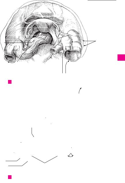
AView of the female true pelvis (pelvis minor) from above
9
10
|
|
|
|
|
13 |
|
|
|
|
|
|
|
|
|
|
|
|
|
|
|
|||
|
|
|
|
15 |
|
|
|
|
|
||
|
|
15 |
|
|
|
|
|
|
|
|
|
|
|
|
|
|
|
|
|
|
|
|
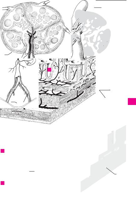
Liver recess |
|
|
|
|
|
|
|
|
|
|
C |
|
17 |
|
|
|
|
|
|
|
|
18 |
|
|
|
|
|
|
|
|
|
|
|
|
||
19 |
|
|
|
|
|
|
|
|
|
||
|
|
||||||||||
|
|
|
|
|
|
17 |
|
|
|||
|
|||||||||||
|
|
||||||||||
16 |
|
|
|
|
|
|
|
|
|
|
|
21 |
14 |
|
B Anterior abdominal wall, posterior view
1
2
3
4
5
6
7
8
9
10
11
12
13
14
15
16
17
18
19
20
21
22
23
24
25
|
254 |
|
Lymphatic system |
|
|
|
|
|
|
|
||
|
|
|
|
|
|
16 |
Right lymphatic duct (right thoracic duct). |
|||||
1 |
|
1 |
|
LYMPHATIC SYSTEM. |
Systema lymphaticum. |
|
||||||
|
||||||||||||
|
2 |
Lymphatic vessels. Vasa lymphatica. |
|
|
|
Ductus lymphaticus dexter (ductus thoracicus |
||||||
|
|
|
|
|
dexter). It is formed by the union of the right |
|||||||
|
|
|
|
|
|
|
|
|
|
|
||
2 |
|
3 |
Lymphatic capillary. Vas lymphocapillare. Any |
|
|
jugular, subclavian |
and bronchomediastinal |
|||||
|
|
|
|
of the vessels of the lymphatic system that |
|
|
trunks. It may be absent. B |
|||||
3 |
|
|
|
form closed networks and have permeable |
|
17 |
Thoracic duct. Ductus thoracicus. Arises from |
|||||
|
|
|
walls. C |
|
|
|
|
|
the cisterna chyli a short distance below the di- |
|||
|
|
4 |
Lymphatic capillary network. Rete lympho- |
|
|
aphragm, courses upward behind the aorta and |
||||||
4 |
|
|
|
opens into the venous angle, i. e., the angle be- |
||||||||
|
|
|
capillare. Network of lymphocapillary veins. C |
|
|
|||||||
|
|
5 |
Lymphatic vessel. Vas lymphaticum. Any of the |
|
|
tween the left internal jugular and subclavian |
||||||
5 |
|
|
|
veins. B |
|
|||||||
|
|
|
valvular lymphatic vessels that communicate |
|
18 |
Arch of thoracic duct. Arcus ductus thoracici. |
||||||
|
|
|
|
with the lymphocapillary vessels. Their thin |
|
|
Arch formed by the thoracic duct before enter- |
|||||
6 |
|
|
|
walls are sparsely lined with smooth muscles. C |
|
|
||||||
|
|
|
|
|
ing the venous angle. B |
|||||||
|
6 |
Lymphatic plexus. Plexus lymphaticus. Net- |
|
|
||||||||
|
|
|
19 |
Cervical part. Pars |
cervicalis. Short cervical |
|||||||
7 |
|
|
|
work of lymphatic vessels lying deeper than |
|
|
segment in front of C7. B |
|||||
|
|
|
the lymphocapillary vessels. In the outer layers |
|
|
|||||||
|
|
|
|
|
20 |
Thoracic part. Pars thoracica. It begins at the |
||||||
|
|
|
|
of the skin, it lies within and directly below the |
|
|||||||
|
|
|
|
|
|
aortic hiatus and ends at the upper margin of |
||||||
8 |
|
|
|
corium. C |
|
|
|
|
|
|||
|
|
|
|
|
|
|
|
T1. B |
|
|||
|
|
|
7 Superficial lymphatic vessel. Vas lymphaticum |
|
|
|
||||||
|
|
|
|
21 |
Abdominal part. Pars abdominalis. Very short |
|||||||
9 |
|
|
|
superficiale. It is situated superficially on the |
|
|||||||
|
|
|
|
|
segment in front of L1. B |
|||||||
|
|
|
|
fascia of the limbs. |
|
|
|
|
22 Cisterna chyli. Variable dilatation at the origin |
|||
10 |
|
8 |
Deep lymphatic vessel. Vas lymphaticum pro- |
|
||||||||
|
|
|
of the thoracic duct. It receives the lumbar and |
|||||||||
|
|
|
|
fundum. It lies beneath the fascia of the limbs |
|
|
intestinal trunks. B |
|
||||
11 |
|
|
|
and often, but not always, accompanies blood |
|
23 Lymph node. Nodus lymphaticus (Lym- |
||||||
|
|
|
vessels. |
|
|
|
|
|||||
|
|
|
|
|
|
|
|
phonodus). Lymphoreticular filtering organ, 1− |
||||
|
|
|
|
|
|
|
|
|
|
|
||
12 |
|
9 |
Lymphatic trunks. Trunci lymphatici. Five |
|
|
25 mm in diameter, within the lymphatic ves- |
||||||
|
|
|
main lymphatic branches of the lymph-vascu- |
|
|
sels. Since lymph must usually traverse two |
||||||
|
|
|
|
lar system. |
|
|
|
|
|
lymph nodes before arriving in the blood |
||
|
|
|
|
|
|
|
|
|
||||
13 |
10 |
Right/left lumbar trunk. Truncus lumbaris |
|
|
stream at the venous angle, there is double pro- |
|||||||
|
|
tection against the invasion of pathogens or |
||||||||||
|
|
|
|
dexter/sinister. Main |
branch |
which |
brings |
|
|
|||
|
|
|
|
|
|
tumor cells into the blood stream. A |
||||||
14 |
|
|
|
lymph to the cisterna chyli from the legs, pelvic |
|
|
||||||
|
|
|
|
24 Afferent lymphatic vessels. Vas lymphaticum |
||||||||
|
|
|
viscera, urogenital system and parts |
of the |
|
|||||||
|
|
|
|
|
|
afferens. Any of the vessels that carry lymph to |
||||||
|
|
|
|
abdominal wall and the abdominal viscera. B |
|
|
||||||
15 |
|
|
|
|
|
a lymph node; located on the convex surface of |
||||||
|
11 |
Intestinal trunks. Trunci intestinales. Main |
|
|
||||||||
|
|
|
the node. A |
|
||||||||
16 |
|
|
|
conduits which transport lymph to the cisterna |
|
25 Efferent lymphatic vessel. Vas lymphaticum |
||||||
|
|
|
chyli from the supply region of the superior and |
|
||||||||
|
|
|
|
|
efferens. Any of the vessels that carry lymph |
|||||||
|
|
|
|
inferior mesenteric arteries. B |
|
|
|
|
away from a lymph node; located on the hilum |
|||
|
|
|
|
|
|
|
|
|
|
|
||
17 |
|
12 |
Right/left bronchomediastinal trunk. Truncus |
|
|
of the node. A |
|
|||||
|
|
|
|
bronchomediastinalis dexter/sinistra. It col- |
|
26 |
Cortex. Part of the lymphoreticular tissue pro- |
|||||
18 |
|
|
|
lects lymph from the heart, lungs and medi- |
|
|
ximal to the capsule. A |
|||||
|
|
|
astinum. On the left |
side it |
opens into the |
|
27 Medulla. Lymphoreticular tissue between cor- |
|||||
|
|
|
|
thoracic duct, on the right side, the right lym- |
|
|||||||
|
|
|
|
|
|
tex and hilum. A |
|
|||||
19 |
|
|
|
phatic duct. Often, however, both may open |
|
|
|
|||||
|
|
|
|
28 |
Hilum. Somewhat retracted area where blood |
|||||||
|
|
|
|
directly into the subclavian veins. B |
|
|
||||||
20 |
|
13 |
Right/left subclavian trunk. Truncus sub- |
|
|
vessels enter and where blood and lymphatic |
||||||
|
|
|
vessels exit. A |
|
||||||||
|
|
|
|
clavius dexter/sinister. Arises from the arm, ac- |
|
29 |
Lymphatic nodule. |
Nodulus lymphaticus |
||||
|
|
|
|
companies the subclavian vein and usually |
|
|||||||
21 |
|
|
|
|
|
(lymphonodulus). Spherical condensation of |
||||||
|
|
|
opens on the right side into the right lymphatic |
|
|
|||||||
|
|
|
|
duct and on the left side into the angle between |
|
|
lymphoreticular tissue predominantly occupy- |
|||||
|
|
|
|
|
|
ing the cortex. It exhibits a lighter central area |
||||||
22 |
|
|
|
the left subclavian vein and internal jugular |
|
|
||||||
|
|
|
|
|
(“reaction center”). A |
|
||||||
|
|
|
vein. B |
|
|
|
|
|
|
|||
|
|
|
|
|
|
|
|
|
|
|
||
23 |
|
14 |
Right/left jugular trunk. Truncus jugularis |
|
|
|
|
|||||
|
|
|
|
dexter/sinister. Accompanies the internal jugu- |
|
|
|
|
||||
|
|
|
|
lar vein and passes to the angle between the in- |
|
|
|
|
||||
24 |
|
|
|
ternal jugular and subclavian veins (venous |
|
|
|
|
||||
|
|
|
|
angle). B |
|
|
|
|
|
|
|
|
25 |
|
15 |
Lymphatic ducts. Ductus lymphatici. The main |
|
|
|
|
|||||
|
|
|
|
drainage ducts of the lymphatic system. |
|
|
|
|
|
|||
|
|
|
|
|
|
|
|
|
|
|
|
|
|
256 |
Lymphatic system |
|
|
|
|
|
|
|
|
|
|
|
|||||
|
|
|
REGIONAL LYMPH NODES. Nodi lymphatici re- |
|
15 |
Submental lymph nodes. Nodi lymphatici sub- |
||||||||||||
1 |
|
1 |
|
|||||||||||||||
|
||||||||||||||||||
|
|
|
gionales. |
|
|
|
|
|
|
|
mentales. Nodes between the anterior bellies |
|||||||
|
|
|
2 Head and neck. Caput et collum. |
|
|
|
of the digastric muscles. Afferents: middle of |
|||||||||||
2 |
|
|
|
|
|
lower lip, floor of mouth and tip of tongue. |
||||||||||||
|
|
3 Occipital lymph nodes. Nodi lymphatici occipi- |
|
|
||||||||||||||
|
|
|
|
|
Efferents: deep cervical and submental lymph |
|||||||||||||
3 |
|
|
|
tales. One to three lymph nodes lying close to |
|
|
nodes. B |
|
|
|
|
|
||||||
|
|
|
the margin of the trapezius. Afferents: scalp, |
|
16 |
Submandibular lymph nodes. Nodi lymphatici |
||||||||||||
|
|
|
|
deep cervical muscles. Efferents: deep cervical |
|
|
submandibulares. |
Nodes |
between |
the |
||||||||
4 |
|
|
|
lymph nodes. A |
|
|
|
|
|
|
||||||||
|
|
|
|
|
|
|
|
|
mandible and submandibular gland that serve |
|||||||||
|
|
|
|
|
|
|
|
|
|
|
|
|||||||
|
|
|
4 Mastoid [retroauricular] lymph nodes. Nodi |
|
|
as first and second filter stations. Direct affer- |
||||||||||||
5 |
|
|
|
lymphatici mastoidei [[retroauriculares]]. Usu- |
|
|
ent area: inner canthus of eye, cheek, side of |
|||||||||||
|
|
|
ally two nodes on the mastoid process. Affer- |
|
|
nose, upper lip, lateral lower lip, gingiva and |
||||||||||||
|
|
|
|
|
|
|||||||||||||
|
|
|
|
ents: posterior surface of pinna, posterior wall |
|
|
anterior lateral margin of tongue. Indirect affer- |
|||||||||||
6 |
|
|
|
of external acoustic meatus and corresponding |
|
|
ents: facial and submental lymph nodes. Effer- |
|||||||||||
|
|
|
|
parts of scalp. Efferents: deep cervical lymph |
|
|
ents: deep cervical lymph nodes. B |
|
||||||||||
7 |
|
|
|
nodes. A |
|
|
|
|
|
|
17 |
Anterior cervical lymph nodes. Nodi lymphat- |
||||||
|
|
|
|
|
|
|
|
|
|
|
||||||||
|
|
|
5 Superficial partodi lymph nodes. Nodi lym- |
|
|
ici cervicales anteriores. |
|
|
|
|||||||||
8 |
|
|
|
phatici parotidei superficiales. They lie on the |
|
18 |
Superficial (anterior jugular) lymph nodes. |
|||||||||||
|
|
|
parotid fascia in front of the tragus. Afferents: |
|
|
Nodi lymphatici superficiales (jugulares anteri- |
||||||||||||
|
|
|
|
|
|
|||||||||||||
|
|
|
|
junction of temporal region and anterior sur- |
|
|
ores). Nodes on the internal jugular vein. Affer- |
|||||||||||
9 |
|
|
|
face of pinna. Efferents: deep cervical lymph |
|
|
ent region: skin of anterior side of neck. Effer- |
|||||||||||
|
|
|
|
nodes. A |
|
|
|
|
|
|
|
ents: bilateral deep cervical lymph nodes. A |
||||||
10 |
|
6 |
Deep parotid lymph nodes. Nodi lymphatici |
|
19 |
Deep lymph nodes. Nodi lymphatici profundi. |
||||||||||||
|
|
|
|
parotidei profundi. Group beneath the parotid |
|
|
Anterior group. |
|
|
|
|
|||||||
11 |
|
|
|
fascia. |
Afferents: |
tympanic |
cavity, |
external |
|
19 a |
Infrahyoid lymph nodes. Nodi lymphatici in- |
|||||||
|
|
|
acoustic meatus, frontotemporal region, eye- |
|
||||||||||||||
|
|
|
|
lids, root of nose, and sometimes the posterior |
|
|
frahyoidei. They lie in the midline below the |
|||||||||||
|
|
|
|
|
|
body of the hyoid bone. Afferent areas: larny- |
||||||||||||
12 |
|
|
|
floor of the nose and nasopharyngeal cavity. |
|
|
||||||||||||
|
|
|
|
|
geal vestibule, piriform recess and adjacent hy- |
|||||||||||||
|
|
|
Efferents: deep cervical lymph nodes. A |
|
|
|||||||||||||
|
|
|
|
|
|
popharynx. |
Efferents: deep |
cervical |
lymph |
|||||||||
|
|
|
|
Preauricular |
lymph |
nodes. Nodi lymphatici |
|
|
||||||||||
|
7 |
|
|
|||||||||||||||
13 |
|
|
nodes. B |
|
|
|
|
|
||||||||||
|
|
|
prae-auriculares. Group located in front of the |
|
20 |
Prelaryngeal |
lymph |
nodes. |
Nodi |
lymphatici |
||||||||
|
|
|
|
|
||||||||||||||
|
|
|
|
pinna. A |
|
|
|
|
|
|
||||||||
14 |
|
|
|
|
|
|
|
|
|
|
praelaryngeales3. Nodes on |
the |
cricothyroid |
|||||
|
|
|
|
|
|
|
|
|
|
|
|
|||||||
|
8 |
Infra-auricular lymph nodes. Nodi lymphat- |
|
|
ligament. Afferent area: lower half of larynx. |
|||||||||||||
|
|
|
|
|||||||||||||||
|
|
|
|
ici infra-auriculares. Group beneath the pinna. |
|
|
Efferents: deep cervical lymph nodes. B |
|
||||||||||
15 |
|
|
|
A |
|
|
|
|
|
|
|
21 |
Thyroid lymph nodes. Nodi lymphatici thyroidei. |
|||||
|
|
9 |
Intraglandular lymph nodes. Nodi lymphatici |
|
|
Nodes on the thyroid gland. Efferents: as in 20. |
||||||||||||
16 |
|
|
|
intraglandulares. |
Group situated |
directly |
|
|
B |
|
|
|
|
|
||||
|
|
|
|
within the parotid. A |
|
|
|
|
22 |
Pretracheal lymph nodes. Nodi lymphatici pre- |
||||||||
17 |
|
10 |
Facial lymph nodes. Nodi lymphatici faciales. |
|
|
tracheales. Nodes in front of the trachea. Affer- |
||||||||||||
|
|
|
ent regions: trachea and larynx. Efferents: deep |
|||||||||||||||
|
|
|
|
Variable lymph nodes that receive lymph from |
|
|
||||||||||||
18 |
|
|
|
the eyelids, nose and the rest of the face and |
|
|
cervical lymph nodes. B |
|
|
|
||||||||
|
|
|
buccal |
mucosa. |
Efferents: |
submandibular |
|
23 |
Paratracheal lymph nodes. Nodi lymphatici par- |
|||||||||
|
|
|
|
lymph nodes. The vessels accompany the facial |
|
|||||||||||||
|
|
|
|
|
|
atracheales. Nodes beside the trachea. Actions |
||||||||||||
|
|
|
|
artery. |
|
|
|
|
|
|
|
|
||||||
19 |
|
|
|
|
|
|
|
|
|
|
|
similar to those of 22. B |
|
|
|
|||
|
11 |
Buccinator |
node. |
[Nodus |
buccinatorius]. |
|
|
|
|
|
||||||||
|
|
|
23 a Retropharyngeal lymph nodes. Nodi lymphatici |
|||||||||||||||
20 |
|
|
|
Lymph node situated deep within the buccina- |
|
|
retropharyngeales. Deep cervical lymph nodes |
|||||||||||
|
|
|
|
tor muscle. A |
|
|
|
|
|
|
|
in front of the arch of the atlas. See p. 258.13 |
||||||
21 |
|
12 |
Nasolabial node. [Nodus nasolabialis]. Lymph |
|
|
|
|
|
|
|
|
|||||||
|
|
|
|
node located below the nasolabial fold. A |
|
|
|
|
|
|
|
|
||||||
22 |
|
13 |
Malar node. [Nodus malaris]. Superficial |
|
|
|
|
|
|
|
|
|||||||
|
|
|
|
lymph node of the cheek. |
|
|
|
|
|
|
|
|
|
|
||||
23 |
|
14 Mandibular |
node. |
[Nodus |
mandibularis]. |
|
|
|
|
|
|
|
|
|||||
|
|
|
|
Lymph node located on the mandible. A |
|
|
|
|
|
|
|
|
||||||
24 |
|
14 a |
Lingual lymph nodes. Nodi lymphatici lingu- |
|
|
|
|
|
|
|
|
|||||||
|
|
|
ales. Nodes located on the hyoglossus muscle. |
|
|
|
|
|
|
|
|
|||||||
|
|
|
|
They drain lymph from the lower surface and |
|
|
|
|
|
|
|
|
||||||
25 |
|
|
|
lateral margin to tongue as well as the medial |
|
|
|
|
|
|
|
|
||||||
|
|
|
|
anterior two-thirds of its dorsal surface. |
|
|
|
|
|
|
|
|
||||||
|
|
|
|
|
|
|
|
|
|
|
|
|
|
|
|
|
|
|