Материал: Атлас Ханц фениш

|
160 |
Urogenital system |
|
|
|
|
|
|
||||||
|
|
|
|
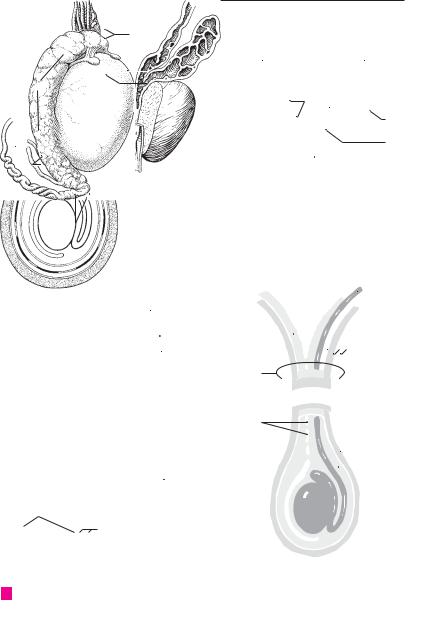
Epididymis. Lying on the posteromedial surface |
19 |
Ejaculatory duct. Ductus ejaculatorius. Sper- |
||||||||
1 |
|
1 |
|
|||||||||||
|
|
|
|
of the testis, it serves as a storage receptacle for |
|
matic duct formed by the union of the ductus |
||||||||
|
|
|
|
|
sperm. A |
|
|
|
|
|
deferens and the duct of the seminal vesicle. It |
|||
2 |
|
2 |
|
Head of epididymis. Caput epididymidis. It is |
|
traverses the prostate and empties into the |
||||||||
|
|
|
|
|
occupied by the efferent ductules. A |
|
prostatic urethra. B |
|
||||||
|
|
|
|
|
20 |
Seminal |
vesicle. |
Vesicula seminalis. Er- |
||||||
3 |
|
3 |
|
Body |
of |
epididymis. |
Corpus |
epididymidis. |
||||||
|
|
|
roneously designated as a receptacle for sperm, |
|||||||||||
|
|
|
|
Middle segment of the epididymis consisting of |
|
|||||||||
|
|
|
|
|
|
this organ is a vesicular gland which consists of a |
||||||||
|
|
|
|
|
the convolutions of the duct of the epididymis. A |
|
||||||||
4 |
|
|
|
|
|
coiled tube, about 12 |
cm in length. B C |
|||||||
|
4 |
Tail of epididymis. Cauda epididymidis. Infe- |
|
|||||||||||
|
|
21 Tunica adventitia. Connective tissue covering |
||||||||||||
|
|
|
|
|
rior, terminal portion of the epididymis con- |
|||||||||
5 |
|
|
|
|
|
of the seminal vesicle. C |
||||||||
|
|
|
|
sisting of the convolutions of the duct of the |
|
|||||||||
|
|
|
|
22 |
Tunica muscularis. Muscular layer in the wall of |
|||||||||
|
|
|
|
|
epididymis. A |
|
|
|
||||||
6 |
|
5 |
|
Lobules (cones) of epididymis. Lobuli coni |
|
the seminal vesicle. C |
||||||||
|
|
23 |
Tunica mucosa. Multilocular mucous mem- |
|||||||||||
|
|
|
|
|
epididymidis. Wedge-shaped lobules in the |
|||||||||
|
|
|
|
|
head of the epididymis separated by connective |
|
brane of the seminal vesicle lined by a simple |
|||||||
|
|
|
|
|
|
|||||||||
|
|
|
|
|
|
|||||||||
7 |
|
|
|
|
tissue and formed by one or two efferent duc- |
|
secretory epithelium. C |
|||||||
|
|
|
|
|
tules. A |
|
|
|
|
24 |
Excretory duct. Ductus excretorius. Efferent |
|||
|
|
|
|
|
|
|
|
|
||||||
8 |
6 |
|
Duct |
of |
epididymis. |
Ductus |
epididymidis. |
|
duct of the seminal vesicle. It unites with the |
|||||
|
|
ductus deferens to form the ejaculatory duct. B |
||||||||||||
|
|
|
|
|
Coiled duct, 5−6 |
meters long, beginning at the |
|
|||||||
|
|
|
|
|
25 |
Spermatic cord. [[Funiculus spermaticus]]. It |
||||||||
|
|
|
|
|
end of the head of the epididymis where it re- |
|||||||||
9 |
|
|
|
|
||||||||||
|
|
|
|
ceives the efferent ductules. It terminates at the |
|
consists of the ductus deferens, accompanying |
||||||||
|
|
|
|
|
end of the tail where it is continuous with the |
|
vessels, nerves and connective tissue, together |
|||||||
10 |
|
|
|
|
ductus deferens. A |
|
|
|
with its coverings. D |
|
||||
7 |
|
Aberrant ductules. Ductuli aberrantes. Blind |
26 |
Tunicae funiculi spermatici. Coverings of the |
||||||||||
|
|
|||||||||||||
|
|
|||||||||||||
|
|
|
|
|
branches of the efferent ductules and duct of the |
|
spermatic cord and the testis, described below. |
|||||||
11 |
|
|
|
|
|
|||||||||
|
|
|
|
epididymis representing vestiges of the caudal |
|
D |
|
|
||||||
|
|
|
|
|
mesonephric tubules. |
|
|
27 |
External spermatic fascia. Fascie spermatica |
|||||
12 |
|
|
|
|
|
|
||||||||
8 |
|
[Ductulus aberrans superior]. Superior aber- |
|
externa. Outer covering of the spermatic cord, |
||||||||||
|
|
|
|
|
rant ductule in the head of the epididymis. |
|
which is continuous with the fascia of the exter- |
|||||||
|
|
|
|
|
|
|||||||||
13 |
9 |
|
[Ductulus aberrans inferior]. Inferior aber- |
|
nal oblique m. of the abdomen. It also envelops |
|||||||||
|
|
the testis together with its remaining coverings. |
||||||||||||
|
|
|
|
|
rant ductule in the tail of the epididymis. A |
|
D |
|
|
|||||
|
|
|
|
|
|
|
|
|||||||
14 |
10 |
|
Appendix testis. Vesicular appendage superior |
|
|
|
||||||||
|
28 |
M. cremaster. Elevator of the testis. It is derived |
||||||||||||
|
|
|
|
to the testis (vestige of the paramesonephric |
||||||||||
|
|
|
|
|
|
mainly |
from internal abdominal oblique |
|||||||
|
|
|
|
|
duct). A |
|
|
|
|
|
||||
15 |
|
|
|
|
|
|
|
|
|
muscle. D |
|
|||
11 |
|
[Appendix |
epididymidis]. Appendix of epid- |
|
|
|||||||||
|
29 Cremasteric fascia. Fascia cremasterica. Con- |
|||||||||||||
|
|
|
|
|
idymis. Pedunculate appendage at the head of |
|
nective tissue on and between the cremaster |
|||||||
16 |
|
|
|
|
the epididymis (vestige of the mesonephros). A |
|
||||||||
|
|
|
|
|
muscle fibers. D |
|
||||||||
|
|
12 Paradidymis. Bilateral blind ductules superior |
30 Internal spermatic fascia. Fascia spermatica in- |
|||||||||||
|
||||||||||||||
17 |
|
|
|
|
to the head of the epididymis and in front of the |
|
terna [tunica vaginalis communis]]. The finger- |
|||||||
|
|
|
|
spermatic |
cord |
(remnant of |
mesonephric |
|
||||||
|
|
|
|
|
|
like inner covering of the spermatic cord, which |
||||||||
|
|
|
|
|
tubules). A |
|
|
|
|
|
||||
|
|
|
|
|
|
|
|
|
|
is derived from the transverse fascia. It lies |
||||
18 |
|
|
|
|
|
|
|
|
|
|||||
|
|
|
|
|
|
|
|
|
|
|
||||
13 |
|
Ductus deferens. Spermatic duct, about 60 cm |
|
beneath the cremaster muscle and surrounds |
||||||||||
|
|
|
|
|
long, between the epididymis and the seminal |
|
the testis, epididymis and ductus deferens to- |
|||||||
19 |
|
|
|
|
vesicle. It is initially coiled and then becomes |
|
gether with blood vessels and nerves. D |
|||||||
|
|
|
|
|
straight. A B D E |
|
|
|
31 |
Vestige of vaginal process. [Vestigium proces- |
||||
|
|
|
|
|
|
|
|
|||||||
20 |
14 |
Ampulla of ductus deferens. Ampulla ductus |
|
sus vaginalis]. Remnant of the not completely |
||||||||||
|
|
|
|
deferentis. Oval enlargement of ductus deferens |
|
obliterated embryological vaginal process of the |
||||||||
|
|
|
|
|
just prior to joining the duct of the seminal ves- |
|
peritoneum. D |
|
||||||
21 |
|
|
|
|
icle. B |
|
|
|
|
|
|
|
|
|
|
15 |
Diverticula of ampulla. Diverticula ampullae. |
|
|
|
|
||||||||
|
|
|
|
|
||||||||||
22 |
|
|
|
|
Lateral sacculations in the wall of the ampulla of |
|
|
|
|
|||||
|
|
|
|
the ductus deferens. B |
|
|
|
|
|
|
||||
|
|
|
|
|
|
|
|
|
|
|
||||
|
16 |
Tunica adventitia. Connective tissue covering |
|
|
|
|
||||||||
23 |
|
|
|
|
||||||||||
|
|
|
|
of the ductus deferens. E |
|
|
|
|
|
|
||||
24 |
|
17 Tunica muscularis. Relatively very thick muscle |
|
|
|
|
||||||||
|
|
|
|
layer of the ductus deferens. E |
|
|
|
|
|
|||||
18 Tunica mucosa. Mucous membrane of ductus
25deferens lined by pseudostratified, stereociliated, columnar epithelium. E
Urogenital system 161
|
|
|
|
12 |
|
|
|
|
|
|
|
|
|
|
|
|
|
|
|
|
|
|
|
|
|
|
|
|
|
|
|
|
1 |
||
5 |
|
|
|
|
|
|
|
|
|
|
|
|
|
|
|
|
|
|
|
|
|
|
|
|
|
|
|
|
2 |
||||||
2 |
|
|
|
|
|
|
|
|
|
|
|
|
|
|
|
|
|
|
|
|
|
|
|
|
|
|
|
|
|
|
|||||
5 |
|
|
|
|
|
|
|
|
|
|
|
|
13 |
|
|
|
|
|
|
|
13 |
|
|
|
|
|
|
|
|
|
|||||
|
|
|
|
|
|
|
11 |
|
|
|
|
|
|
|
|
|
|
|
|
|
|
|
|
|
|
|
|
|
3 |
||||||
|
|
|
|
|
|
|
|
|
|
|
|
|
|
|
|
|
|
|
|
|
|
|
|
|
|
|
|
|
|
|
|
|
|||
|
|
|
|
|
|
|
|
|
|
|
|
|
|
|
|
|
|
|
|
|
|
|
|
|
|
|
|
|
|
|
|||||
1 |
10 |
|
|
|
|
|
|
|
|
|
|
|
|
|
|
|
|
|
|
|
|
|
|
|
|
|
|
||||||||
|
|
|
15 |
|
|
|
|
|
|
|
|
|
|
|
4 |
||||||||||||||||||||
|
|
|
|
|
|
|
|
|
|
|
|
|
|
|
|
|
|
|
|
|
|
|
|||||||||||||
3 |
|
|
|
|
|
|
|
|
14 |
|
|
|
|
|
|
|
|
|
|
|
|
|
|||||||||||||
|
|
|
|
|
|
|
|
|
|
|
|
|
|
|
|
|
|
|
|
|
|||||||||||||||
|
|
|
|
|
|
|
|
|
|
|
|
20 |
|
|
|
|
|
|
|
|
|
|
|
|
|
|
|
|
|
|
|
20 |
|
5 |
|
|
|
|
|
|
|
|
|
|
|
|
|
|
|
|
|
|
|
|
|
|
|
|
|
|
|
|
|
|
|
|
|
|
|
|
|
|
|
|
13 |
|
|
|
|
|
|
|
|
|
|
|
|
|
|
|
|
|
|
|
|
|
|
|
|
|
|
|
|
|
24 |
|
6 |
|
|
|
|
|
|
|
|
|
|
|
|
|
|
|
|
|
|
|
|
|
|
|
|
|
|
|
|
|
|
|
|
19 |
|
||
9 |
|
|
|
|
|
|
|
|
|
|
|
|
|
|
|
|
|
|
|
|
|
|
|
|
|
|
|
|
|
|
|
||||
|
|
|
|
|
|
|
|
|
|
|
|
|
|
|
|
|
|
|
|
|
|
|
|
|
|
|
|
|
|
|
|||||
|
|
|
|
|
|
|
|
|
|
|
|
|
|
|
|
|
|
|
|
|
|
|
|
|
|
|
|
|
|
|
|
||||
|
|
|
|
|
|
|
|
|
|
|
|
|
|
|
|
|
|
|
|
|
|
|
|
|
|
|
|
|
|
|
7 |
||||
|
|
|
|
|
|
|
|
|
|
|
|
|
|
|
|
|
|
|
|
|
|
|
|
|
|
|
|
|
|
|
|
|
|
|
|
|
|
|
|
|
4 |
|
|
|
|
|
|
|
|
|
|
|
|
|
|
|
|
|
|
|
|
|
|
|
|
|
|
|
|
|
|
|
|
|
|
|
|
|
|
|
|
|
|
|
|
|
|
|
|
|
|
|
|
|
|
|
|
|
|
|
|
|
|
|
|
|
|
|
|
|
|
|
|
|
|
|
|
|
|
|
|
|
|
|
|
|
|
|
|
|
|
|
|
|
|
|
|
|
|
|
|
8 |
|
|
|
|
|
|
|
|
|
|
|
|
|
|
|
|
|
|
|
|
|
|
|
|
|
|
|
|
|
|
|
|
|
|
|
||
|
|
|
|
|
|
|
|
|
|
|
|
|
|
|
|
|
|
|
|
|
|
|
|
|
|
|
|
|
|
|
|
|
|
|
|
|
|
|
|
|
|
|
|
|
|
|
|
|
|
|
|
|
|
|
|
|
|
|
|
|
|
|
|
|
|
|
|
|
|
|
|
|
|
|
|
|
|
|
|
|
|
|
|
|
|
|
|
|
|
|
|
|
|
|
|
|
|
|
|
|
|
|
|
|
|
|
|
6 |
|
|
|
|
|
|
|
|
|
|
|
|
|
|
|
|
|
|
|
|
|
|
|
|
|
|
|
|
|
|
|
9 |
|||
|
|
|
|
|
|
|
|
|
|
|
|
|
|
|
|
|
|
|
|
|
|
|
|
|
|
|
|
|
|
|
|
||||
|
Testicle and epididymis |
|
Postate and seminal vesicle, |
|
|
|
|
|
|
|
|
|
|
|
|
||||||||||||||||||||
A |
B |
|
|
|
|
|
|
|
|
|
|
|
10 |
||||||||||||||||||||||
|
|
|
|
|
|
|
|
|
|
|
|
|
opened, frontal view |
|
|
|
|
|
|
|
|
|
|
|
|
||||||||||
|
|
|
|
|
|
|
|
|
|
|
|
|
|
|
|
|
|
|
|
|
|
|
|
||||||||||||
|
|
|
|
|
|
|
|
|
|
|
|
|
|
|
|
|
|
|
|
|
|
|
|
|
|
|
|
|
|
|
|
|
|
|
11 |
|
|
|
|
|
|
|
|
|
|
|
|
|
|
|
|
|
|
|
|
|
|
|
|
|
|
|
|
|
|
|
|
13 |
|
|
|
|
|
|
|
|
|
|
|
|
|
|
|
|
|
|
|
|
|
|
|
|
|
|
|
|
|
|
|
|
|
|
|
|
|
||
|
|
|
|
|
|
|
|
|
|
|
21 |
176.23 |
|
|
|
|
12 |
||||||||||||||||||
|
|
|
|
|
|
|
|
|
|
|
|
|
|
|
|
|
|
|
|
|
|
||||||||||||||
|
|
|
|
|
|
|
|
|
|
|
|
|
|
|
|
|
|
|
|
|
|
|
|
||||||||||||
|
|
|
|
|
|
|
|
|
|
|
|
|
|
|
|
|
|
|
|
|
|
|
|
|
|
|
|
|
|
|
|
|
|
|
|
|
|
|
|
|
|
|
|
|
|
|
|
|
|
|
|
|
|
|
|
|
|
|
|
|
|
|
|
|
|
|
|||||
|
|
|
|
|
|
|
|
|
|
|
|
86.28 |
|
|
|
|
|
|
|
|
|
|
|
|
|
|
|
|
|
|
|
|
|
13 |
|
|
|
|
|
|
|
|
|
|
|
22 |
|
|
|
|
|
|
|
|
|
|
|
|
|
|
|
|
|
|
|
|
|
|
|||
|
|
|
|
|
|
|
|
|
|
|
|
|
|
|
|
|
|
|
|
|
|
|
|
|
|
||||||||||
|
|
|
|
|
|
|
|
|
|
|
|
|
|
|
|
|
|
|
|
|
|
|
|
|
|
|
|
||||||||
|
|
|
|
|
|
|
|
|
|
|
|
|
|
|
|
|
|
|
|
|
|
|
26 |
|
|
14 |
|||||||||
|
|
|
|
|
|
|
|
|
23 |
25 |
|
|
|
|
|
|
|
|
|
|
|
|
|
|
|
|
|||||||||
|
|
|
|
|
|
|
|
|
|
|
|
|
|
|
|
|
|
|
|
||||||||||||||||
|
|
|
|
|
|
|
|
|
|
|
|
|
|
|
|
|
|
|
|
|
|
|
|
||||||||||||
|
|
|
|
|
|
|
|
|
|
|
|
|
|
|
|
|
|
|
|
|
|
|
|
|
|
|
|
|
|
|
|
|
|
||
|
|
|
|
|
|
|
|
|
|
|
|
|
|
|
|
|
|
|
|
|
|
|
|
|
|
|
|
|
|
|
|
|
15 |
||
|
|
|
|
|
|
|
|
|
|
|
|
27 |
|
|
|
|
|
|
|
|
|
|
|
|
|
|
|||||||||
|
|
|
|
|
|
|
|
|
|
|
|
|
|
|
|
|
|
|
|
|
|
|
|
|
|
||||||||||
|
|
|
|
|
|
|
|
|
|
|
|
|
|
|
|
|
|
|
|
|
|
|
|
|
16 |
||||||||||
|
|
|
|
|
|
|
|
|
|
|
|
|
|
|
|
|
|
|
|
|
|
|
|
|
|
|
|
|
|
|
|
|
|
|
|
|
Seminal vesicle, histological section |
|
|
|
|
|
|
|
|
|
|
|
|
|
|
|
|
|
|
|
28 |
|
|
|
|||||||||||
|
|
|
|
|
|
|
|
|
|
|
|
|
|
|
|
|
|
|
|
||||||||||||||||
C |
31 |
|
|
|
|
|
|
|
|
|
|
|
|
|
|
|
|
17 |
|||||||||||||||||
|
|
|
|
|
|
|
|
|
|
|
|
|
|
|
|
||||||||||||||||||||
|
|
|
|
|
|
|
|
|
|
|
|
|
|
|
|
|
|
|
|
|
|
|
|
|
|
|
|
|
|
|
|
|
|||
|
|
|
|
|
|
|
|
|
|
|
|
|
|
|
|
|
|
|
|
|
|
|
|
|
|
|
|
|
|
|
|
|
|
|
|
|
|
|
|
|
|
|
|
|
|
|
|
|
|
|
|
|
|
|
|
|
|
|
|
|
|
|
|
|
|
|
|
|
|
|
|
|
|
|
|
|
|
|
|
|
|
|
|
|
|
|
|
|
|
|
|
|
|
|
|
|
|
|
|
29 |
|
|
18 |
||||
|
|
|
|
|
|
|
|
|
|
|
|
|
|
|
|
|
|
|
|
|
|
|
|
|
|
|
|
|
|||||||
|
|
|
|
|
|
|
|
|
|
|
|
|
|
|
|
|
|
|
|
|
|
|
|
|
|
|
|
|
30 |
|
|
|
|||
|
|
16 |
19 |
|
|
|
|
|
|
|
|
|
|
|
20 |
17 |
|
|
|
|
|
|
|
|
|
21 |
|
18 |
|
|
|
|
|
|
22
23
ESperm duct (ductus deferens), cross section
D |
Coverings of the spermatic |
24 |
|
cord and the testis |
|
|
|
|
|
|
|
|
|
25 |
|
|
|

|
162 |
Urogenital system |
|
|
|
||||
|
|
|
Tunica vaginalis testis. Double-layered serous |
19 |
Capsule of prostate. Capsula prostatica. Pro- |
||||
1 |
|
1 |
|||||||
|
|
|
covering of the testis, a remnant of the vaginal |
|
vided with smooth muscle fibers, it is firmly |
||||
|
|
|
|
process of the peritoneum. A |
|
|
fused to the prostate. D |
||
2 |
|
2 |
Parietal layer. |
Lamina parietalis |
[[peri- |
20 |
Parenchyma. Glandular component of the pros- |
||
|
|
|
|
orchium]]. External layer of the serous tunica |
|
tate. |
|||
3 |
|
|
|
vaginalis testis. A |
|
|
21 |
Prostatic ductules. Ductuli prostatici. 15−30 |
|
|
3 |
Visceral layer. Lamina visceralis [[eporchium]]. |
|
glandular excretory ductules which open into |
|||||
|
|
|
|||||||
4 |
|
|
|
Layer of the tunica vaginalis testis that attaches |
|
the prostatic urethra. C |
|||
|
|
|
superiorly to the testis. A |
|
22 |
Substantia muscularis. Smooth muscle situated |
|||
|
|
4 |
Superior ligament of epididymis. Lig. epid- |
|
between the glandular alveoli. C |
||||
5 |
|
|
|
idymidis superius. Reflected fold of the tunica |
23 |
M. puboprostaticus. Tracts of smooth muscle |
|||
|
|
|
vaginalis testis located superiorly at the head of |
|
contained within the puboprostatic (pubovesi- |
||||
|
|
|
|
|
|||||
6 |
|
|
|
the epididymis. A |
|
|
|
cal) ligament extending from the pubic symphy- |
|
|
5 |
Inferior ligament of epididymis. Lig. epididy- |
|
sis to the prostate. |
|||||
|
|
|
|
midis inferius. Reflected fold of the tunica vagi- |
24 |
Bulbourethral [[Cowper’s]] gland. Glandula |
|||
|
|
|
|
nalis testis situated inferiorly at the tail of the |
|
bulbourethralis [[Cowper’s]]. Pea-sized mucous |
|||
7 |
|
|
|
|
|||||
|
|
|
epididymis. A |
|
|
|
gland located in the urogenital diaphragm. E |
||
|
|
|
|
|
|
|
|||
|
6 |
Sinus of epididymis. Sinus epididymidis. Ser- |
25 |
Duct of bulbourethral gland. Ductus gl. bul- |
|||||
8 |
|||||||||
|
|
|
ous cleft between the testis and epididymis. It is |
|
bourethralis. Excretory duct of the bulbourethral |
||||
|
|
|
|
accessible laterally and is bordered above and |
|
gland, 3−4 cm long. E |
|||
9 |
|
|
|
below by the superior and inferior ligaments of |
26 |
EXTERNAL MALE GENITALIA. Organa genitalia |
|||
|
|
|
|
the epididymis. A |
|
|
|
masculina externa. E |
|
|
7 |
Descent of testis. [[Descensus testis]]. Down- |
27 |
Penis. Male copulatory organ consisting of |
|||||
10 |
|||||||||
|
|
|
ward migration of the fetal testis during the last |
|
cavernous bodies and the urethra. E |
||||
|
|
|
|
weeks of pregnancy. It descends from the peri- |
28 |
Root of penis. Radix penis. Portion of the penis |
|||
11 |
|
|
|
toneal cavity into the scrotal sac via the inguinal |
|
attached to the pubis. E |
|||
|
|
|
|
canal. |
|
|
29 |
Body (shaft) of penis. Corpus penis. Portion of |
|
|
8 |
[[Gubernaculum testis]]. Fetal connective tissue |
|||||||
12 |
|
the penis situated between the root and the |
|||||||
|
|
|
band which arises from the caudal gonadal fold |
|
glans. E |
||||
|
|
|
|
and guides the testis during its descent. |
|
30 |
Crus penis. Cavernous body attached to the infe- |
||
13 |
|
|
|
|
|||||
9 |
Genitoinguinal ligament. [[Lig. genitoingui- |
||||||||
|
rior ramus of the pubis. E |
||||||||
|
|
|
|
nale]]. Embryonic |
precursor of the |
guber- |
31 |
Dorsum penis. Flattened upper surface of penis. |
|
|
|
|
|
||||||
14 |
|
|
|
naculum testis. |
|
|
32 |
Urethral surface. Facies urethralis. Underside of |
|
|
10 Prostate. Prostata (glandula prostatica). Chest- |
||||||||
|
|
|
penis. It bears the urethra within the corpus |
||||||
|
|
|
|
nut-sizedorganconsistingof30−50tubulo-alve- |
|
||||
|
|
|
|
|
spongiosum. E |
||||
15olarglands.Situatedbelowtheurinarybladder,it is penetrated by the urethra. B C D
1611 Base of prostate. Basis prostatae. Part of the prostate fused with the urinary bladder. B
17 |
12 |
Apex of prostate. Apex prostatae. Portion of |
|
|
prostate directed downward and forward and |
||
|
|
containing the urethra. B |
|
18 |
13 |
Anterior surface. Facies anterior. Surface of the |
|
|
|
prostate facing the symphysis. B D |
|
|
|
||
19 |
14 |
Posterior surface. Facies posterior. Surface of |
|
|
|
the prostate facing the rectum. B |
|
|
15 |
Inferolateral surface. Facies inferolateralis. Sur- |
|
20 |
|||
|
face of the prostate directed downward and |
||
|
|
lateral. D |
|
|
|
||
21 |
16 |
Right/left lobe. Lobus (dexter/sinister). Part of |
|
|
|
the prostate arising from the caudal anlage. B D |
|
|
17 Isthmus of prostate. Isthmus prostatae. Median |
||
22 |
|||
|
part of the prostate located in front of the urethra |
||
|
|
and connecting the right and left lobes. It is |
|
23 |
|
devoid of glands and possesses a fibromuscular |
|
|
|
stroma. D |
|
|
18 |
Middle lobe. [Lobus medius]. Prostatic lobe sit- |
|
24 |
|||
|
uated between the ejaculatory duct and the |
||
|
|
urethra. It tends to undergo hormone-induced |
|
25 |
|
hypertrophy in the elderly, thus closing the |
|
|
|
urethral canal like a valve. B D |
|
Urogenital system 163
|
3 |
|
|
|
|
|
|
|
|
|
11 |
|
|
||
|
|
|
|
|
|
|
|
|
|
|
|
|
|
||
|
|
|
|
|
|
|
|
|
|
|
|
|
|
|
|
|
|
|
|
4 |
|
|
|
|
|
|
|
|
|
||
|
|
18 |
|
||||||||||||
|
|
|
|
|
|
|
|
||||||||
|
|
|
|
|
|
|
|
|
|
||||||
6 |
|
|
|
|
2 |
1 |
13 |
|
|
|
|
|
|
||
|
|
|
|
|
|
|
|
||||||||
|
|
|
|
|
|
|
|||||||||
|
|
|
3 |
|
|
|
|
|
|
||||||
|
|
|
|
|
|
|
|
|
|
||||||
5 |
|
|
|
14 |
|
||||
16 |
|
|
||
|
|
|
||
|
12 |
|
|
|
ARight testicle with epididymis and investing layers, lateral view
21
21
C Prostate, histologic view
13 |
|
|
|
15 |
|
|
|
17 |
|
|
19 |
|
|||
16 |
16 |
|
|
18 |
|
|
|
D Prostate, horizontal section
B Prostate, sagittal section
29 |
27 |
|
32 |
25
30 |
|
|
|
30 |
|
|
|||
28 |
|
|
28 |
|
24
E Penis from below
1
2
3
4
5
6
7
8
9
10
11
12
13
14
15
16
17
18
19
20
21
22
23
24
25

|
164 |
Urogenital system |
|
|
|
|||
|
|
|
Glans penis. Expanded end of the corpus spon- |
22 |
Preputial glands. Gll. preputiales. Sebaceous |
|||
1 |
|
1 |
||||||
|
|
|
giosum penis. A D |
|
|
glands, mainly on the corona of the glans. |
||
2 |
|
2 |
Corona glandis. Raised posterior margin of the |
23 |
Male urethra. Urethra masculina. D |
|||
|
|
|
glans. A D |
|
24 |
Prostatic part of urethra. Pars prostatica. Por- |
||
|
|
3 |
Septum glandis. Median partition in the glans. |
|
tion of male urethra passing through the pros- |
|||
3 |
|
|
|
C |
|
|
tate. D |
|
|
|
4 |
Collum glandis. Neck of glans. Constricted por- |
25 |
Urethral crest. Crista urethralis. Mucosal fold in |
|||
4 |
|
|
|
tion behind the corona. A |
|
the dorsal wall of the prostatic urethra continu- |
||
|
|
5 Prepuce (foreskin) of penis. Preputium penis. |
|
ous with the uvula of the urinary bladder. D |
||||
|
|
|
|
|||||
|
|
|
26 |
Colliculus seminalis. Elevated portion (veru- |
||||
5 |
|
|
|
Double layer of skin over the glans. A |
||||
|
6 |
Frenulum of prepuce. Frenulum preputii. Re- |
|
montanum) of the urethral crest containing the |
||||
|
|
|
openings of the ejaculatory duct. D |
|||||
6 |
|
|
|
flected fold passing from the prepuce to the un- |
|
|||
|
|
|
27 |
Prostatic utricle. Utriculus prostaticus. Blind |
||||
|
|
|
derside of the glans. A |
|
||||
|
|
7 |
Raphe penis. Seam on the skin on the underside |
|
sac in the colliculus seminalis measuring up to |
|||
|
|
|
1 cm in length and representing a rudiment of |
|||||
7 |
|
|||||||
|
|
|
of the penis that forms during development. B |
|
||||
|
|
|
|
the paramesonephric duct. D |
||||
|
8 |
Corpus cavernosum |
penis. Cavernous body |
|
||||
|
28 |
Prostatic sinus. Sinus prostaticus. Furrow on |
||||||
8 |
|
|
|
divided into two halves by the septum of the |
||||
|
|
|
|
both sides of the colliculus seminalis containing |
||||
|
|
|
|
penis. A B D |
|
|
the openings of the prostatic ductules. D |
|
|
|
|
|
|
|
|||
9 |
|
|
9 Corpus spongiosum penis.Cavernousbodysur- |
|
||||
|
|
29 |
Membranous part of urethra. Pars mem- |
|||||
|
|
|
rounding the urethra. A B D |
|||||
|
|
|
|
|
branacea. Portion of the male urethra passing |
|||
|
|
|
|
|
|
|
||
10 |
10 |
Bulb of penis. Bulbus penis. Posterior thickened |
|
through the urogenital diaphragm. D |
||||
|
|
|
|
end of the corpus spongiosum. D |
30 Spongy part of urethra. Pars spongiosa. Portion |
|||
|
|
11 Tunica albuginea of corpora cavernosa.Tunica |
|
of male urethra surrounded by the corpus spon- |
||||
11 |
|
|||||||
|
|
|
albuginea corporum cavernosorum. Tough con- |
|
giosum. D |
|||
|
|
|
|
nective tissue covering of the corpora cavernosa. |
31 |
Navicular fossa of urethra. Fossa navicularis |
||
12 |
|
|
|
B |
|
|||
|
|
|
|
|
urethrae. Oval dilatation of the male urethra |
|||
|
|
|
|
|
|
|||
|
12 |
Tunica albuginea of corpus spongiosum. |
|
before its external opening. A D |
||||
|
|
|||||||
13 |
|
|
|
Tunica albuginea corporis spongiosi. Less firm |
32 |
Valve of navicular fossa. [Valvulafossaenavic- |
||
|
|
|
connective tissue covering of the corpus spon- |
|||||
|
|
|
|
|
ularis]. Mucosal fold on the upper wall of the |
|||
|
|
|
|
giosum. B |
|
|
navicular fossa. |
|
14 |
|
|
|
|
|
|||
13 |
Septum of penis. Septum penis [[septum pec- |
33 |
External urethral orifice. Ostium urethrae ex- |
|||||
|
|
|
|
tiniforme]]. Pectinate partition between the |
|
ternum. D |
||
15 |
|
|
|
right and left corpus cavernosum. Gaps are pre- |
34 |
Urethral lacunae. Lacunae urethrales. Numer- |
||
|
|
|
sent. B |
|
||||
|
|
|
|
|
|
ous outpocketings in the urethral mucosa with |
||
|
14 |
Trabeculae corporum cavernosorum. Connec- |
|
|||||
16 |
|
the openings of the urethral glands. D |
||||||
|
|
|
tivetissuetractswithinthecorporacavernosain- |
35 Urethral glands. Gll. urethrales. Small mucous |
||||
|
|
|
|
|||||
|
|
|
|
terspersed with smooth muscle. A B D |
||||
17 |
|
|
|
|
glands opening into the urethral lacunae. |
|||
15 |
Trabeculae corporis |
spongiosi. Connective |
|
|||||
36 |
Ductus (canales) paraurethrales. Inconstant |
|||||||
|
|
|
|
tissue tracts within the corpus spongiosum in- |
||||
|
|
|
|
|
paraurethralductsthatdraintheurethralglands. |
|||
18 |
|
|
|
terspersed with smooth muscle. B |
|
|||
|
|
|
|
They open in the vicinity of the external urethral |
||||
16 |
Cavernae corporum cavernosorum. Wide- |
|
||||||
|
|
orifice. |
||||||
|
|
|||||||
19 |
|
|
|
meshed, blood-filled spaces within the corpora |
|
|
||
|
|
|
cavernosa. B D |
|
|
|
||
|
|
|
|
|
|
|
||
|
|
|
|
|
|
|
|
|
17 Cavernae corporis spongiosi Blood-filled,
20finely meshed spongy network within the corpus spongiosum. A B
2118 Helicine arteries. Arteriae helicinae. Coiled branches of the deep artery of the penis. D
2219 Cavernous veins. Vena cavernosae. Dilated veins in the cavernous bodies.
2320 Superficial fascia of penis. Fascia penis superficialis. Delicate subcutaneous fascia with individual smooth muscle fibers, continuous with
24the tunica dartos of the scrotum. B
21 Deep fascia of penis. Fascia penis profunda.
25Deeper, somewhat tougher fascia surrounding the three cavernous bodies. B