Материал: Атлас Ханц фениш

|
150 |
Respiratory system |
|
|
|
|
|
||
|
|
|
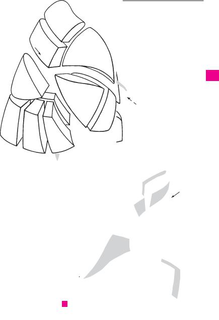
BRONCHOPULMONARY |
SEGMENTS. Segmenta |
19 |
Superior lingular segment (S IV). Segmen- |
|||
1 |
|
1 |
|||||||
|
|
|
bronchopulmonalia. Lung segments supplied by |
|
tum lingulare superius. It lies predominantly |
||||
|
|
|
|
individual bronchi and arteries and separated by |
|
on the inferior lingular segment. B |
|||
2 |
|
|
|
veins and connective tissue septa. A B |
20 |
Inferior lingular segment (S V). Segmentum |
|||
|
|
|
2 Upper lobe of right lung. (According to Dor- |
|
lingulare inferius. It lies between the superior |
||||
3 |
|
|
|
land’s lobus superior pulmonis dextri). Pulmo |
|
lingular segment and the oblique fissure. B |
|||
|
|
|
dexter, lobus superior. A |
|
|
21 Lower lobe of left lung. (Lobus inferior pul- |
|||
|
|
|
|
|
|
||||
|
|
|
3 Apical segment of upper lobe of right lung. |
||||||
4 |
|
|
|
monis sinistri). Pulmo sinister, lobus inferior |
|||||
|
|
|
Segmentum apicale (S |
I). It is inserted like a |
|
B |
|
||
|
|
|
|
wedge between the anterior and posterior seg- |
22 |
Superior segment (S VI). |
Segmentum su- |
||
5 |
|
|
|
ments. A |
|
|
|||
|
|
|
|
|
|
perius. Apical portion of lower lobe situated |
|||
|
4 |
Posterior segment of upper lobe of right |
|
||||||
|
|
|
posterosuperiorly near the vertebral column. |
||||||
|
|
|
|
lung. Segmentum posterius (S II). It lies be- |
|
B |
|
||
6 |
|
|
|
|
|
||||
|
|
|
tween the apical segment and the lower lobe. A |
23 |
Subapical segment. [[Segmentum subapi- |
||||
|
5 |
Anterior segment of upper lobe of right |
|||||||
|
|
cale]]. Accessory segment occasionally pre- |
|||||||
7 |
|
||||||||
|
|
|
lung. Segmentum anterius (S III). It lies be- |
|
sent below the superior segment of the lower |
||||
|
|
|
|
tween the apical segment and the middle lobe. A |
|
lobe. |
|
||
|
|
|
|
|
|
||||
8 |
6 |
Middle lobe of right lung. (Lobus medius pul- |
24 |
Medial basal (cardiac) segment (S VII). Seg- |
|||||
|
|
|
|
monis dextri). Pulmo dexter, lobus medius. A |
|
mentum basale mediale (cardiacum). It is |
|||
|
|
|
|
|
|||||
9 |
7 |
Lateral segment of middle lobe. Segmentum |
|
often an inseparable part |
of the anterior |
||||
|
|
|
laterale (S IV). It occupies the dorsal portion |
|
basal segment. B |
|
|||
|
|
|
|
|
|
||||
|
|
|
|
of the middle lobe and does not |
reach the |
25 |
Anterior basal segment (S |
VIII). Segmentum |
|
10 |
|
|
|
||||||
|
|
|
hilum. A |
|
|
||||
|
|
|
|
|
|
basale anterius. It lies between the oblique |
|||
|
8 |
Medial segment of middle lobe. Segmentum |
|
fissure and the lateral basal segment. B |
|||||
11 |
|
|
|
mediale (S V). It forms the medial and dia- |
26 |
Lateral basal segment (S |
IX). Segmentum |
||
|
|
|
|
phragmatic surfaces of the middle lobe. A |
|
basale laterale. It lies between the anterior |
|||
|
|
|
|
|
|
|
|
||
12 |
9 |
Lower lobe of right lung. (Lobus inferior pul- |
|
and posterior basal segments. B |
|||||
|
|
|
monis dextri). Pulmo dexter, lobus inferior. A |
27 |
Posterior basal segment (S X). Segmentum |
||||
|
|
|
|
||||||
|
10 |
Superior segment. |
Segmentum |
superius |
|||||
|
|
basale posterius. It lies beside the vertebral |
|||||||
13 |
|
||||||||
|
|
|
(S VI). Posterosuperiorly situated apical por- |
|
column below the superior segment of the |
||||
|
|
|
|
tion of lower lobe. A |
|
|
|
lower lobe. B |
|
1411 Subapical segment. [[Segmentum subapicale]]. An accessory segment occasionally present below the superior segment.
1512 Medial basal (cardiac) segment (S VII). Segmentum basale mediale (cardiacum). It does
16not reach the lateral surface of the lung and is only seen from the medial and inferior sur-
17faces. A
13Anterior basal segment (S VIII). Segmentum basale anterius. It lies between the middle
18 |
lobe and diaphragm. A |
14 Lateral basal segment (S IX). Segmentum
19basale laterale. It lies between the posterior and anterior basal segments. A
2015 Posterior basal segment (S X). Segmentum basale posterius. Located between the vertebral column and lateral basal segment. A
2116 Upper lobe of left lung. (Lobus superior pulmonis sinistri). Pulmo sinister, lobus superior.
22B
17 Apicoposterior segment (S I + II). Segmen-
23tum apicoposterius. It comprises two wedgeshaped segments (apical and posterior) which lie between the oblique fissure and the
24anterior segment of the upper lobe. B
18 Anterior segment (S III) of upper lobe. Seg-
25mentum anterius. It lies between the superior lingular and apicoposterior segments. B
Respiratory system 151
I
|
|
|
|
|
|
3 |
|
|
|
|
|
|
|
|
||
148.26 |
|
II |
|
|
|
III |
|
|
|
|
|
|
|
|||
|
|
|
|
|
|
|
|
|
|
|
||||||
|
|
|
|
|
|
|
|
|
|
|
|
|
|
|
|
|
|
|
|
|
|
|
4 |
|
|
|
|
|
|
|
|
|
|
|
|
|
|
|
|
5 |
|
|
|
|
|
|
|
|||
|
|
|
|
|
|
|
|
|
|
2 |
|
|
|
|
||
|
|
|
VI |
|
|
|
|
|
|
|
|
|
|
|
|
|
|
|
|
|
|
|
|
|
|
|
|
|
|
|
|
|
|
|
|
10 |
|
|
|
|
|
IV |
|
|
|
|
|
|
|
|
|
|
|
|
|
|
|
|
|
|
|
|
|
|
|||
|
|
|
|
|
|
|
|
|
|
|
|
|
|
|
|
|
9 |
|
|
|
12 |
|
|
|
|
148.27 |
|
|
|
|
|||
|
|
|
|
|
|
|
|
|
|
|
|
|||||
|
|
|
|
|
|
7 |
V |
|
|
|
|
|||||
|
|
|
|
|
|
|
|
|
|
|
||||||
|
|
|
|
|
|
|
|
|
|
|
|
|
|
|
||
|
|
|
X |
|
|
|
VII |
6 |
|
|
|
|
||||
|
|
|
|
|
|
|
|
|
8 |
|
|
|
|
|
|
|
|
|
|
|
|
|
|
|
|
|
|
|
|
|
|
||
|
|
|
|
|
|
|
|
|
|
|
|
|
|
|
|
|
|
|
15 |
IX |
|
VIII |
|
|
|
|
|
|
|
||||
|
|
|
|
|
|
|
|
|
|
|
||||||
|
|
|
|
|
|
|
|
|
|
|
|
|
||||
|
|
|
|
14 |
|
13 |
|
|
|
|
|
|
|
|
|
|
|
|
|
|
|
|
|
|
|
|
|
|
|
|
|
||
|
|
|
|
|
|
|
|
|
|
|
|
|
|
|
|
|
|
|
|
|
|
|
|
|
|
|
|
|
|
I |
|
|
|
|
|
|
|
|
|
|
|
|
|
|
|
|
|
|
|
|
A |
Segments |
|
|
|
|
|
|
17 |
|
|
|
|||||
|
|
|
|
|
|
|
|
|
|
|||||||
|
of the right lung |
|
|
|
|
|
|
|
|
|
|
|||||
|
|
|
|
|
|
|
|
|
|
|
|
|
|
|||
|
|
|
|
|
|
|
|
|
|
|
|
|
II |
|
|
|
|
|
|
|
|
|
|
|
|
|
|
|
|
|
|
|
|
|
|
|
|
|
|
|
|
|
III |
|
|
|
17 |
148.26 |
|
|
|
|
|
|
|
|
|
|
|
18 |
|
|
|
|
|
||
|
|
|
|
|
|
|
|
|
|
|
|
|
|
|
|
|
|
|
|
|
|
|
|
19 |
|
|
|
|
|
|
|
||
|
|
|
|
|
|
|
|
|
|
|
|
|
|
|||
|
|
|
|
|
|
|
|
|
IV |
|
|
|
|
|
|
|
|
|
|
|
|
|
|
|
|
|
|
|
|
|
VI |
|
|
|
|
|
|
|
|
16 |
|
|
|
|
|
|
||||
|
|
|
|
|
|
|
|
|
|
|
|
|
|
|
||
|
|
|
|
|
|
|
|
|
|
|
|
|
|
22 |
|
|
|
|
|
|
|
|
|
|
|
|
|
|
|
|
|
|
|
|
|
|
|
|
|
|
|
|
|
|
|
|
|
|
|
|
|
|
|
|
|
|
|
|
|
V |
|
|
|
|
21 |
||
|
|
|
|
|
|
|
20 |
|
|
VII |
|
|
|
|
||
|
|
|
|
|
|
|
|
|
|
|
|
|
||||
|
|
|
|
|
|
|
|
|
|
|
|
|
|
|
|
|
|
|
|
|
|
|
|
|
|
|
24 |
|
|
|
|
||
|
|
|
|
|
|
|
|
|
|
|
|
VIII |
IX |
X |
|
|
|
|
|
|
|
|
|
|
|
|
25 |
26 |
27 |
|
|
||
|
|
|
|
|
|
148.22 |
||||||||||
|
|
|
|
|
|
|
|
|
|
|
|
|
||||
|
|
|
|
|
|
|
|
|
|
|
|
|
|
|
|
|
BSegments
of the left lung
1
2
3
4
5
6
7
8
9
10
11
12
13
14
15
16
17
18
19
20
21
22
23
24
25

|
152 |
Respiratory system |
|
|
|
|
|
|
||||
|
|
|
Bronchioles. |
Bronchioli. |
Noncartilaginous |
16 |
Mediastinal part of parietal pleura (mediastinal |
|||||
1 |
|
1 |
||||||||||
|
|
|
segments of the respiratory tree directly fol- |
|
pleura). Pars mediastinalis. Portion of the |
|||||||
|
|
|
|
lowing the bronchi. They are lined initially by |
|
parietal pleura lining the mediastinum. B C |
||||||
2 |
|
|
|
pseudostratified, |
ciliated, |
columnar |
17 |
Costal part of parietal pleura (costal pleura). Pars |
||||
|
|
|
|
epithelium which is subsequently replaced by |
|
costalis. It lines the ribs. B C |
|
|||||
|
|
|
|
simple cuboidal epithelium. A |
|
|
|
|||||
3 |
|
|
|
|
18 |
Diaphragmatic part of parietal pleura (diaphrag- |
||||||
|
2 |
Respiratory bronchioles. Bronchioli respira- |
||||||||||
|
|
|
matic pleura). Pars diaphragmatica. It covers |
|||||||||
|
|
|
|
torii. Last bronchiolar segment the wall of |
|
|||||||
4 |
|
|
|
|
the diaphragm. B |
|
|
|||||
|
|
|
which already consists partially of alveoli. A |
19 Pleural recesses. Recessus pleurales. Fissure- |
||||||||
|
|
3 |
Alveolar ducts. Ductuli alveolares. Terminal |
|||||||||
|
|
|
shaped spaces formed by the parietal pleura |
|||||||||
5 |
|
|
|
branches of the respiratory bronchioles the |
|
for reception of the lungs during inspiration. |
||||||
|
|
|
|
walls of which contain only alveoli. A |
20 |
Costodiaphragmatic recess. Recessus cos- |
||||||
|
4 |
Alveolar sacs. Sacculi alveolares. Blind, ex- |
||||||||||
6 |
|
todiaphragmaticus. Pleural recess |
between |
|||||||||
|
|
|
|
panded ends of the alveolar ducts. A |
|
the descending sides of the diaphragm and |
||||||
|
|
|
5 Pulmonary alveoli. Alveoli pulmonis. Small- |
|
the lateral wall of the thorax. B |
|
||||||
7 |
|
|
|
|
||||||||
|
|
21 |
Costomediastinal recess. Recessus costome- |
|||||||||
|
|
|
est outpocketings, 0.1−0.9 mm. in diameter, |
|||||||||
|
|
|
|
|||||||||
|
|
|
|
the thin walls of which permit the exchange |
|
diastinalis. Anterior pleural space between |
||||||
8 |
|
|
|
|
||||||||
|
|
|
of gases. A |
|
|
|
|
|
the costal and mediastinal pleura; it is more |
|||
|
6 |
THORACIC |
CAVITY. |
|
Cavitas |
thoracis |
|
extensive on the left than on the right. C |
||||
|
|
|
||||||||||
9 |
|
22 |
Phrenicomediastinal |
recess. |
Recessus |
|||||||
|
|
|
(thoracica). Internal thoracic space enclosed |
|||||||||
|
|
|
|
by the ribs and limited inferiorly by the dia- |
|
phrenicomediastinalis. Pleural recess situated |
||||||
10 |
|
|
|
phragm. B C |
|
|
|
|
|
dorsally between the diaphragm and the me- |
||
|
|
|
|
|
|
|
|
|
diastinum. |
|
|
|
7Pleuropulmonary regions. Regiones pleuro-
|
|
pulmonales. Regions connecting the pleura |
23 Pulmonary ligament. Lig. pulmonale. Double |
||||||||||
11 |
|
||||||||||||
|
|
fold extending from the right and left sides of |
|||||||||||
|
and lungs. |
|
|
|
|
|
|
|
|
||||
|
8 |
Endothoracic |
fascia. |
Fascia |
endothoracica. |
|
the hilum, connecting the visceral and medi- |
||||||
|
|
||||||||||||
12 |
|
astinal pleura. Between both folds the lung |
|||||||||||
|
Displaceable layer of loose connective tissue |
|
abuts against the mediastinal connective |
||||||||||
|
|
between the parietal pleura and chest wall. B |
|
tissue free of pleura. B. See also p. 149 B D |
|||||||||
13 |
|
|
|||||||||||
9 |
Suprapleural membrane. Membrana supra- |
24 |
Mediastinum. Thoracic area between both |
||||||||||
|
|
pleuralis [[Sibson]]. Thickened portion of the |
|
pleural sacs. It extends from the anterior sur- |
|||||||||
|
|
|
|||||||||||
14 |
|
endothoracic |
fascia |
in the |
region |
of |
the |
|
face of the vertebral column to the posterior |
||||
|
pleural cupola. B |
|
|
|
|
|
|
|
surface of the sternum and from the upper |
||||
|
|
|
|
|
|
|
|
|
|||||
|
10 |
Phrenicopleural fascia. Fascia phrenico- |
|
thoracic aperture to the diaphragm. B |
|||||||||
15 |
|
||||||||||||
25 Superior mediastinum. Mediastinum su- |
|||||||||||||
|
pleuralis. Portion of the endothoracic fascia |
||||||||||||
|
|
which connects the parietal pleura with the |
|
perius. Portion of the mediastinum above the |
|||||||||
16 |
|
diaphragm. B |
|
|
|
|
|
|
|
|
heart. It contains the arch of the aorta to- |
||
|
11 |
Pleural cavity. Cavitas pleuralis. Capillary fis- |
|
gether with its branches, as well as the bra- |
|||||||||
|
|
chiocephalic veins, superior vena cava, tra- |
|||||||||||
17 |
|
sure-like |
space |
between |
the parietal |
and |
|
||||||
|
|
chea, esophagus, vagus nerves, thoracic duct, |
|||||||||||
|
visceral pleura containing a small amount of |
|
|||||||||||
|
|
|
thymus, etc. B |
||||||||||
|
|
serous fluid. B C |
|
|
|
|
|
|
|
||||
18 |
|
|
|
|
|
|
|
26 |
Inferior mediastinum. Mediastinum inferius. |
||||
12 |
Pleura. |
Serous |
membrane |
consisting |
of |
||||||||
|
Collective term for the following three divi- |
||||||||||||
|
|
simple squamous epithelium and underlying |
|
||||||||||
|
|
|
sions. |
||||||||||
|
|
|
|||||||||||
19 |
|
connective tissue. It comprises two portions |
|
||||||||||
|
27 Anterior mediastinum. Mediastinum an- |
||||||||||||
|
|
(visceral and parietal pleura) which become |
|||||||||||
|
|
continuous at the hilum. The visceral (pulmo- |
|
terius. Area between the pericardium and |
|||||||||
20 |
|
|
|||||||||||
|
nary) pleura covers the lungs whereas the |
|
sternum. C |
||||||||||
|
|
parietal pleura lines the chest wall, dia- |
28 |
Middle mediastinum. Mediastinum medium. |
|||||||||
21 |
|
phragm and mediastinum. B |
|
|
|
|
Area occupied by the heart, pericardium and |
||||||
|
13 |
Cupula (dome) of pleura. Cupula pleurae. It |
|
phrenic nerves with their accompanying ves- |
|||||||||
|
|
||||||||||||
|
|
sels. C |
|||||||||||
22 |
|
covers the apex of the lung at the superior |
|
||||||||||
|
29 |
Posterior mediastinum. Mediastinum post- |
|||||||||||
|
thoracic aperture and forms the boundary be- |
||||||||||||
|
|
||||||||||||
|
|
tween the neck and thorax. B |
|
|
|
erius. Area between the pericardium and the |
|||||||
23 |
|
|
|
|
|||||||||
14 |
Visceral (pulmonary) pleura. Pleura |
viscer- |
|
vertebral column. It contains the esophagus, |
|||||||||
|
|
alis (pulmonalis). Portion |
of |
the pleura |
that |
|
vagus nerves, descending aorta, thoracic duct |
||||||
|
|
|
and the azygos and hemiazygos veins. C |
||||||||||
24 |
|
envelops the lung and passes into the inter- |
|
||||||||||
|
|
|
|||||||||||
|
|
lobar spaces. B C |
|
|
|
|
|
|
|
|
|||
|
|
|
|
|
|
|
|
|
|
||||
2515 Parietal pleura. Pleura parietalis. Serous lining of the space in which the lungs lodge. B C
Respiratory system 153
9 |
13 |
25
|
|
|
|
|
14 |
|
|
|
|
|
|
|
|
|
|
|
|
|
|
|
|
|
|
|
|
|
|
|
|
|
|
|
|
|
|
|
|
|
|
|
|
|
|
|
|
|
|
|
15 |
|
|
|
|
|
|
|
|
|
|
|
23 |
|
|
|
|
|
|
|
|
|
|
|
|
|
|
|
|
|
|
|
|
|
|
|
|||
|
|
|
|
|
|
|
|
|
|||||||||||||
1 |
2 |
3 |
|
8 |
|
|
|
|
|
|
|
|
|
|
|
|
|
|
|
|
|
|
24 |
|
|||||||||||||||||||
|
|
|
|||||||||||||||||||
|
|
|
4 |
11 |
|
||||||||||||||||
|
|
|
|
|
|
|
|
|
|
|
|
|
|
|
|
|
|
|
|||
|
|
|
|
|
11 |
|
|
|
|
|
|
|
|
|
|
|
|
|
16 |
|
|
|
|
|
|
|
5 |
|
|
|
|
|
|
|
|
|
|
|
|
|
|
|
|
|
|
|
|
|
|
|
|
|
|
|
|
|
|
|
|
|
|
|
|
|
|
|
|
|
|
|
12 |
10 |
|
|
|
|
|
|
|
|
|||||||
|
|
|
|
|
|
|
|
|
|
|
|
|
|
|
|
|
|
|
|
|
|
|
|
|
|
|
17 |
|
|
|
|
|
|
18 |
|
|
|
|
|
|
|
|
|
|
|
|
|
|
|
|
|
|
|
|
|
|
|
|
|
|
|
|
|
||
|
|
|
|
|
|
|
|
|
|
|
|
|
|
|
|
|
|
||||
|
|
|
|
|
|
|
|
|
|
|
|
|
|||||||||
|
Bronchiole and |
|
|
|
|
|
20 |
|
|
Frontal section |
|||||||||||
|
|
|
|
|
|||||||||||||||||
|
|
|
|
|
|
|
|
|
|
|
|
|
|||||||||
A |
|
|
|
|
|
|
|
|
|
|
B |
||||||||||
|
alveolar ducts |
|
|
|
|
|
|
|
|
|
|
|
|
through both lungs |
|||||||
|
|
|
|
|
|
|
|
|
|
|
|
IX |
||
|
15 |
|
|
|
|
|
|
|
|
|
|
29 |
|
|
|
|
|
|
|
|
|
|
|
|
|
|
|||
|
|
|
|
|
|
|
|
|
|
|
|
|||
|
|
|
|
|
|
|
|
|
|
|
|
|
||
|
|
|
|
|
|
|
|
|
|
|
|
246.21 |
||
|
|
|
|
|
|
|
|
|
|
|
|
|
|
|
|
14 |
|
|
|
|
|
|
|
|
28 |
|
|||
|
|
|
|
|
|
|
|
|
|
|
|
|||
|
11 |
|
|
|
|
|
|
|
|
|
|
|
||
|
|
|
|
|
|
|
|
|
|
|
|
|||
|
16 |
|
|
|
|
|
|
|
|
|
||||
|
|
|
|
|
|
|
|
|
|
|||||
|
17 |
|
|
|
|
|
|
|
27 |
|
||||
|
|
|
|
|
|
|
|
|
||||||
|
|
|
|
|
|
|
|
|
|
|
|
|
||
|
Horizontal section at level |
|
|
|
|
|
||||||||
|
|
|
|
|
|
|||||||||
|
|
|
|
|
|
|||||||||
|
|
|
|
|
|
|||||||||
C |
|
|
21 |
|||||||||||
21 |
||||||||||||||
|
of the ninth thoracic vertebra. |
|||||||||||||
|
|
|
|
|
|
|||||||||
View from below
1
2
3
4
5
6
7
8
9
10
11
12
13
14
15
16
17
18
19
20
21
22
23
24
25

|
154 |
|
Urogenital system |
|
|
|
|
|
|||
|
|
|
|
|
Apparatus urogenitalis |
26 |
Renal cortex. Cortex renalis. About 6 mm |
||||
|
|
1 |
|
|
|||||||
1 |
|
|
UROGENITAL SYSTEM. |
||||||||
|
|
|
|
(systema urogenitale). |
Urinary and genital or- |
|
thick, it consists of glomeruli and predomi- |
||||
|
|
|
|
gans. |
|
nantly convoluted uriniferous tubules. With |
|||||
2 |
|
2 |
URINARY ORGANS. Organa urinaria. |
|
the renal columns, it extends up to the wall of |
||||||
|
|
|
3 KIDNEY. Ren (nephros). A B F |
|
the renal pelvis. F |
|
|
||||
3 |
|
|
27 |
Convoluted part (cortical labyrinth). Pars |
|||||||
|
4 |
Lateral margin. Margo lateralis. Convex lateral |
|||||||||
|
|
convoluta. Cortical region consisting of glomer- |
|||||||||
4 |
|
|
|
border of kidney. A |
|
uli and convoluted uriniferous tubules. F |
|||||
|
5 |
Medial margin. Margo medialis. Border of the |
28 |
Radiating part (medullary rays). Pars radi- |
|||||||
|
|
|
|
kidney which becomes concave at the hilum. A |
|
ata. Collecting tubules coursing radially into |
|||||
5 |
|
6 |
Renal hilum. Hilum renale. Site of entry and |
|
the cortex from the medulla. F |
|
|
||||
|
|
|
|
|
|
||||||
|
|
|
|
exit of renal blood vessels and ureter. A |
29 |
Cortical lobules. Lobuli corticales. Areas |
|||||
6 |
|
7 |
Renal sinus. Sinus renalis. Very concave cavity |
|
delimited by interlobular arteries. |
|
|||||
|
|
|
at the medial border for the renal hilum. B D |
29 a |
Medullary rays. Radii medullares. Consisting |
||||||
|
|
|
|
||||||||
|
|
|
8 Anterior surface. Facies anterior. Curved ante- |
|
of pale collecting tubules which project into the |
||||||
|
|
|
|
||||||||
7 |
|
|
|
||||||||
|
|
|
rior surface of the kidney. A D |
|
cortex. F |
|
|
|
|||
|
9 |
Posterior surface. Facies posterior. Nearly flat |
30 |
Renal medulla. Medulla renalis. Medullary |
|||||||
8 |
|
tissue in the shape of renal pyramids and con- |
|||||||||
|
|
|
posterior surface of the kidney. D |
|
|||||||
|
|
10 Upper pole of kidney. Extremitas superior. A B |
|
sisting of the straight portions of the urinifer- |
|||||||
|
|
|
ous tubules and the collecting ducts. F |
||||||||
9 |
|
||||||||||
11 |
Lower pole of kidney. Extremitas inferior. B |
|
|||||||||
31 |
Renal pyramids. Pyramides renales. Six to 20 |
||||||||||
|
|
12 Renal fascia. Fascia renalis. Fibrous sheath that |
|
pyramidal areas separated by renal columns. |
|||||||
10 |
|
||||||||||
|
|
|
separates the adipose capsule from the per- |
|
They form the medullary substance. F |
||||||
|
|
|
|
irenal fat. D |
32 |
Base of pyramid. Basis pyramidis. It lies at the |
|||||
|
|
|
|
||||||||
11 |
13 |
Pararenal fatty body. Corpus adiposum para- |
|
corticomedullary border. F |
|
|
|||||
|
|
|
|
renale. Fat pad between the posterior layer of |
33 |
Renal papillae. Papillae renales. Rounded apical |
|||||
|
|
|
|
||||||||
|
|
|
|
the renal fascia and the transversalis fascia. D |
|
portion of the renal pyramid projecting into the |
|||||
12 |
|
|
|
|
|||||||
|
14 Fatty capsule. Capsula adiposa. Fatty capsule of |
|
renal calyx. F |
|
|
||||||
|
|
|
|
|
|||||||
|
|
|
|
the kidney, more prominent posteriorly and |
34 |
Area cribrosa. Surface of renal papillae with |
|||||
13 |
|
|
|
||||||||
|
|
|
medially. D |
|
sieve-like perforations created by the openings |
||||||
|
|
15 Fibrous capsule. Capsula fibrosa. Tough organ |
|
of the uriniferous tubules. F |
|
|
|||||
|
|
|
|
||||||||
14 |
|
|
|
capsule fused with the surface of the kidney, |
35 |
Papillary foramina. Foramina papillaria. Holes |
|||||
|
|
|
|
but removable. D F |
|
in the area cribrosa produced by the openings |
|||||
|
|
|
|
|
|||||||
15 |
16 |
Renal segments. Segmenta renalia. Five seg- |
|
of the uriniferous tubules. |
|
|
|||||
|
Renal columns. Columnae renales. Cortical |
||||||||||
|
|
|
|
ments of the kidney corresponding to the blood |
36 |
||||||
|
|
|
|
supply regions of the branches of the renal |
|
substance which extends toward the hilum be- |
|||||
16 |
|
|
|
artery. |
|
tween the renal pyramids. F |
|
|
|||
|
17 |
Superior segment. Segm. superius. Upper seg- |
37 |
Renal corpuscle. Corpusculum renale. Com- |
|||||||
|
|||||||||||
17 |
|
|
|
ment extending up to the posterior surface. A B |
|
posed of a glomerulus and its capsule; it lies in |
|||||
|
|
18 Upper anterior segment. Segm. anterius su- |
|
the convoluted part of the cortex. E |
|
||||||
|
|
|
|||||||||
18 |
|
|
|
perius. A |
38 |
Glomerulus. Capillary tuft within a renal cor- |
|||||
19 |
Lower anterior segment. Segm. anterius in- |
|
puscle. E |
|
|
|
|||||
|
|
|
|
ferius. A |
39 |
Glomerular [[Bowman’s]] capsule. Capsula |
|||||
19 |
|
|
|
||||||||
|
20 Inferior segment. Segm. inferius. It reaches as |
|
glomerularis [[Bowman’s]]. The capsule around |
||||||||
|
|
|
|
far as the posterior and anterior surfaces. A B |
|
a capillary |
tuft (glomerulus) of |
a |
renal cor- |
||
20 |
|
|
|
|
puscle. It |
is continuous with |
a |
convoluted |
|||
21 |
Posterior segment. Segm. posterius. B |
|
|||||||||
|
tubule. E |
|
|
|
|||||||
|
|
|
|
|
|
|
|
|
|
|
|
22 Renal (uriniferous) tubule (nephron). Tubulus
21renalis. Tubular system representing the structural unit of the kidney in which filtration and
22selective reabsorption take place. C
23Convoluted uriniferous tubules. [[Tubuli renales contorti]]. Tortuous parts of the renal
23 |
tubules. C |
24Straight segments of renal tubules. [[Tubuli
24 renales recti]]. C
25Renal lobes. Lobi renales. Still preserved in the
25newborn, they correspond to renal pyramids with cortical caps.