Материал: Атлас Ханц фениш

|
130 |
Digestive system |
|
|
|
|||
|
|
|
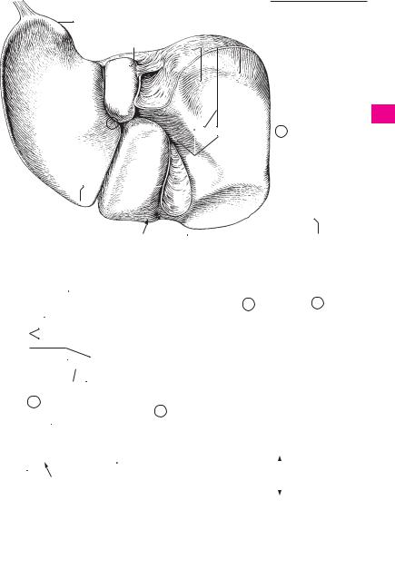
Visceral surface. Facies visceralis. Posteroinfe- |
19 |
Anterior segment of liver. Segmentum an- |
|||
1 |
|
1 |
||||||
|
|
|
rior, partially concave surface of liver facing the |
|
terius. C D |
|
||
2 |
|
|
|
viscera. |
20 |
Posterior segment of liver. Segmentum post- |
||
|
|
|
Fossa of gallbladder. Fossa vesicae biliaris. |
|||||
|
2 |
|
erius. C D |
|
||||
|
|
|
|
Fossa on the visceral surface of the liver that |
21 Left lobe of liver. Lobus hepatis sinister. Its right |
|||
|
|
|
|
lodges the gallbladder. A |
||||
3 |
|
|
|
|
border corresponds to a line connecting the in- |
|||
|
3 |
Fissure for round ligament. Fissura ligamenti |
|
|||||
|
|
|
ferior vena cava and fundus of gallbladder. A B C |
|||||
4 |
|
|
|
teretis. Fissure on the visceral surface of the liver |
22 |
Medial segment. Segmentum mediale. C D |
||
|
|
|
that lodges the ligamentum teres hepatis. A |
|||||
|
|
|
23 |
Quadrate part. Pars quadrata. Part belonging |
||||
|
|
4 |
Round ligament of liver. Lig. teres hepatis. The |
|||||
|
|
|
to the quadrate lobe. D |
|||||
5 |
|
|||||||
|
|
|
connective tissue remains of the umbilical vein. |
|
||||
|
|
|
24 |
Lateral segment. Segmentum laterale. C D |
||||
|
|
|
|
B |
||||
|
|
|
|
25 Quadrate lobe. Lobus quadratus. Liver lobe sit- |
||||
6 |
5 |
Ligamentum venosum. [[Arantii]]. Venous liga- |
||||||
|
uated between the gallbladder, round ligament |
|||||||
|
|
|
ment of liver, the fibrous remains of the ductus |
|
||||
|
|
|
|
venosus. B |
|
and hilum. A B |
||
7 |
|
|
|
|
||||
|
|
|
26 |
Caudate lobe. Lobus caudatus. Liver lobe sit- |
||||
6 |
Hilum of liver. Porta hepatis. Fissure between |
|||||||
|
|
uated between the inferior vena cava, hilum and |
||||||
|
|
|
|
the caudate and quadrate lobes in which run the |
|
|||
8 |
|
|
|
hepatic artery proper, portal vein and hepatic |
|
ligamentum venosum. A B |
||
|
|
|
27 |
Papillary process. Processus papillaris. Portion |
||||
|
|
|
|
duct. A B |
||||
|
7 |
Tuber omentale. Bulge on the visceral surface |
|
of caudate lobe projecting caudally. A B |
||||
9 |
|
|||||||
28 |
Caudate |
process. Processus caudatus. |
||||||
|
|
|
of the left lobe to the left of the ligamentum ve- |
|||||
|
|
|
|
|||||
|
|
|
|
nosum. A B |
|
Parenchymal connection between the caudate |
||
10 |
|
|
|
|
||||
8 |
Esophageal impression. Impressio oe- |
|
and right lobes cranial to the hilum. A B |
|||||
|
|
|
|
sophagea. Groove on the left lobe where the |
29 |
Tunica serosa. Peritoneal covering containing |
||
11 |
|
|
|
liver is indented by the esophagus. A |
|
simple squamous epithelium. |
||
|
9 |
Gastric impression. Impressio gastrica. Im- |
30 |
Tela subserosa. Connective tissue layer beneath |
||||
|
||||||||
12 |
|
|
|
pression caused by contact of the stomach with |
|
the serosa. |
|
|
|
|
|
the visceral surface of the left lobe of the liver. A |
31 |
Tunica fibrosa. Immobile connective tissue |
|||
|
|
|
|
|||||
|
10 |
Duodenal impression. Impressio duodenalis. |
|
capsule of liver; thick, especially in the bare area |
||||
13 |
|
|||||||
|
|
|
Impression caused by contact of the duodenum |
|
not covered by peritoneum. |
|||
|
|
|
|
with the right side of the visceral surface of the |
32 Ligament of vena cava. [[Lig. venae cavae]]. |
|||
|
|
|
|
liver next to the neck of the gallbladder. A B |
||||
14 |
|
|
|
|
Connective tissue bridging over the inferior |
|||
|
|
|
|
|
||||
|
11 |
Colic impression. Impressio colica. Impression |
|
vena cava. |
|
|||
|
|
|
||||||
|
|
|
|
caused by contact of the colon with the visceral |
|
|
|
|
15surface of the right lobe of liver to the right of the gallbladder. A
1612 Renal impression. Impressio renalis. Impression caused by contact of the right kidney with the visceral surface of the right lobe. It overlaps
17the bare area. A
18 |
13 |
Suprarenal |
impression. Impressio |
su- |
|
prarenalis. Impression caused by contact of the |
|||
|
|
right suprarenal gland with the bare area on the |
||
19 |
|
right side near the inferior vena cava. A |
|
|
|
14 |
[Appendix fibrosa hepatis]. Connective |
tissue |
|
|
||||
20 |
band occasionally present at the upper end of |
|
the left lobe of the liver. A |
||
|
||
|
|
15 Inferior margin. Margo inferior. Border be-
21tween the diaphragmatic and visceral surfaces of the liver. A
2216 Incisura lig. teretis. Notch for round ligament at the lower border of the liver. A B
2317 Lobes of liver. Lobi hepatis. The four macroscopically visible lobes of the liver, which are described below.
2418 Right lobe of liver. Lobus hepatis dexter. Its border to the left lobe corresponds to the line
25connecting the inferior vena cava and fundus of the gallbladder. A B C
Digestive system 131
14
8 |
32 |
28 |
13 |
926
7 |
|
|
|
|
|
|
|
|
|
12 |
|||||
|
|
|
|
|
|
|
|
|
|
||||||
21 |
|
|
|
|
|
|
|
|
|
|
|||||
|
|
27 |
|
18 |
|||||||||||
|
|
|
|
|
|
|
|
|
|
|
|
|
|
|
|
|
|
|
|
|
|
|
|
|
|
|
|
|
|
||
|
|
|
|
|
|
|
|
|
6 |
|
|
|
|||
3 |
|
|
|
|
|
|
|
|
10 |
|
|||||
|
|
|
25 |
|
|
|
|||||||||
|
|
|
|
|
|
||||||||||
|
|
|
|
|
|
|
|
|
|
|
|
||||
15 |
|
|
|
|
|
|
|
|
|
2 |
|
||||
|
|
|
|
|
|
|
|
|
|
|
|
|
|
|
|
|
|
|
|
|
|
|
|
|
|
|
|
|
|
|
11 |
A |
Liver from behind and below |
|
|
|
|
|
|
|
|
||||||
|
|
|
|
|
|
|
|
|
|||||||
|
16 |
|
|
|
|
|
|
|
|
15 |
|||||
|
|
|
|
|
|
|
|
|
15 |
|
|
|
|
||
246.21 |
|
|
|
|
|
|
|
|
|
|
|
||||
|
|
|
|
|
|
|
|
|
|
|
|
|
|
18 |
21 |
|
|
|
|
|
|
|
|
|
|
|
|
|
|
||
|
|
|
|
|
|
|
|
|
|
|
|
|
|
||
5 |
|
|
|
|
|
|
|
|
|
|
|
|
|
24 |
|
|
|
|
|
|
|
|
20 |
19 |
22 |
||||||
|
|
|
|
|
|
|
|||||||||
26 |
|
|
|
|
|
|
|||||||||
7 |
|
|
|
|
|
|
|
|
|
|
|
|
|
|
|
28 |
|
|
|
|
|
|
|
|
|
|
|
|
|
|
|
27 |
|
|
|
|
|
|
|
|
|
|
|
|
|
|
|
|
|
|
|
|
|
|
|
|
|
|
|
|
|
||
6 |
|
|
|
|
|
|
|
|
|
|
|
|
|
|
|
|
|
|
|
|
|
|
|
|
|
|
|
|
|
||
21 |
|
|
|
|
|
|
|
|
|
|
Liver segments, |
|
|||
|
|
|
|
18 |
|
|
|
C |
|
||||||
|
|
|
|
|
|
|
|
|
|
|
anterior view |
|
|||
|
|
|
|
|
|
|
|
|
|
|
|
|
|
||
4 |
|
|
|
10 |
|||
|
|||
|
25 |
||
1
2
3
4
5
6
7
8
9
10
11
12
13
14
15
16
17
18
19
20
|
|
|
|
24 |
|
|
|
|
||
|
|
|
|
|
|
|
21 |
|||
|
|
16 |
|
22 |
20 |
|||||
|
|
|
|
|
|
|
|
|
||
178.22 |
134.1 |
|
|
23 |
|
|
22 |
|||
|
|
|
|
|
|
|
|
|
||
|
Hepatic porta |
|
|
|
|
|
|
|
||
|
|
|
|
|
|
|
||||
|
|
|
|
|
|
|
|
|
||
B |
|
|
|
|
19 |
|
23 |
|||
|
|
|
|
|
|
|
|
|||
|
|
|
|
|
|
|
|
|
|
|
|
|
|
|
|
|
|
|
|
|
|
|
|
|
|
|
|
|
|
|
|
24 |
|
|
|
|
|
|
Liver segments, |
|
|
|
|
|
|
|
|
|
D |
|
|
|
|
|
|
|
|
|
|
|
25 |
||||
|
|
|
|
|
|
posterior view |
|
|
|
|
|
|
|
|
|
|
|
|
|
|
|

132 |
Digestive system |
||
|
|
1 Hepatic lobules. Lobuli hepatis. They measure |
|
1 |
|
||
|
|
1−2 mm in size. A |
|
|
|
|
|
2Perivascular fibrous capsule. Capsula fibrosa
2perivascularis. Connective tissue sheath accompanying the liver vessels and biliary ducts until
3
their terminal branches. A
3 Interlobular arteries. Arteriae interlobulares. Branches of the hepatic artery proper between
4the liver lobules. A
4 Interlobular veins. Venae interlobulares.
5Branches of the portal veins between the liver lobules. A
6 |
5 Central veins. Venae centrales. Efferent veins in |
|
the center of the liver lobule. A |
||
|
6 Interlobular ductules. Ductuli interlobulares.
7Biliary drainage channels between the liver lobules. A
87 Ductuli biliferi. Biliary drainage channels which connect the interlobular ductules with the right and left hepatic ducts.
98 Common hepatic duct. Ductus hepaticus communis. Duct between the cystic duct and the
10 junction of right and left hepatic ducts. B
|
9 Right hepatic duct. Ductus hepaticus dexter. |
11 |
Duct arising from the right lobe of liver. B |
|
10 Anterior branch. Ramus anterior. Anterior branch of the right hepatic duct. B
1211 Posterior branch. Ramus posterior. Posterior branch of the right hepatic duct. B
1312 Left hepatic duct. Ductus hepaticus sinister. Duct arising from the left lobe of the liver. B
1413 Lateral branch. Ramus lateralis. Lateral branch of the left hepatic duct. B
1514 Medial branch. Ramus medialis. Medial branch of the left hepatic duct. B
15 Right duct of caudate lobe. Ductus lobi caudati
16dexter. Branch coming from the right half of the caudate lobe and usually leading into right he-
17patic duct. B
16Left duct of caudate lobe. Ductus lobi caudati sinister. Branch arising from the left half of the
18caudate lobe and emptying usually into the left hepatic duct. B
19
20
21
22
23
24
25
Digestive system 133
2
 5 4 3 6
A Two liver lobules
15 16
11
13
14
10 |
9 |
12 |
|
|
8 |
BBranches of the hepatic duct, anterior view
1
2
3
4
5
6
7
8
9
10
11
12
13
14
15
16
17
18
19
20
21
22
23
24
25

134 Digestive and respiratory system
1 |
|
1 |
GALLBLADDER. Vesica biliaris (fellea). 8− |
17 |
|
RESPIRATORY SYSTEM. |
Apparatus |
respira- |
|||||
|
|
|
12 cm long, pear-shaped reservoir for bile. A |
|
|
torius (systema respiratorium). |
|
||||||
2 |
|
2 |
Fundus of gallbladder. Fundus vesicae biliaris. |
18 |
EXTERNAL NOSE. Nasus externus. E |
|
|||||||
|
|
|
Caudally directed rounded end of gallbladder. A |
19 |
Root of nose. Radix nasi (nasalis). Upper por- |
||||||||
|
|
|
3 Body of gallbladder. Corpus ves. biliaris. Por- |
|
tion of the nose which lies between the two or- |
||||||||
3 |
|
|
|
tion of gallbladder between |
the fundus and |
|
bits. D E |
|
|
|
|
||
|
|
|
neck. A |
|
20 Dorsum (bridge) of nose. Dorsum nasi. E |
||||||||
|
|
|
|
|
|||||||||
|
|
|
4 Neck of gallbladder. Collum |
vesicae biliaris. |
|||||||||
4 |
|
|
21 Tip of nose. Apex nasi. E |
|
|||||||||
|
|
|
The portion of the gallbladder continuous with |
|
|||||||||
|
|
|
22 |
Alae (wings) of |
nose. Alae nasi. Winglike |
||||||||
|
|
|
|
the cystic duct. A |
|
||||||||
|
|
|
|
|
|
structures forming the lateral borders of the |
|||||||
5 |
|
|
|
|
|
||||||||
5 |
Tunica serosa. Peritoneal lining of the gallblad- |
|
|||||||||||
|
nares. E |
|
|
|
|
||||||||
|
|
|
|
der. B |
|
23 |
Nasal cartilages. Cartilagines nasi (nasalis). |
||||||
|
|
|
|
|
|||||||||
6 |
6 |
Tela subserosa. Connective tissue underlying |
|||||||||||
|
Pieces of cartilage which form the non-osseous |
||||||||||||
|
|
|
|
the peritoneum. B |
|
|
supporting skeleton of the nose. C |
|
|||||
7 |
|
|
7 Tunica muscularis. Muscle layer in the wall of |
24 |
Lateral nasal cartilage. [Cartilago nasi later- |
||||||||
|
|
|
the gallbladder. B |
|
|||||||||
|
|
|
|
|
|
alis]. No longer classified as independent left |
|||||||
8 |
|
8 |
Tunica mucosa. Mucous membrane of gall- |
|
and right plates of cartilage, but as part of the |
||||||||
|
|
|
bladder with simple columnar epithelium |
|
nasal septum with which each is partially |
||||||||
|
|
|
|
comprised of tall prismatic cells. B |
|
fused. C |
|
|
|
|
|||
9 |
|
9 |
Plicae mucosae. Mucosal folds that project |
25 |
Greater alar cartilage. Cartilago alaris major. |
||||||||
|
|
|
|
into the lumen, thus producing a loculate relief |
|
Hook-shaped cartilage surrounding the nares |
|||||||
10 |
|
|
|
pattern. A B |
|
|
and forming the tip of the nose. C |
|
|||||
|
10 |
Cystic duct. Ductus cysticus. Duct that drains |
26 |
Medial crus. Crus mediale. The part of the |
|||||||||
|
|
|
|
the gallbladder. It joins the common hepatic |
|
greater alar cartilage that forms the anterior |
|||||||
11 |
|
|
|
duct to form the common bile duct (ductus |
|
and lower part of the nasal septum. C D |
|||||||
|
|
|
|
choledochus). A |
|
27 |
Lateral crus. Crus laterale. The part of the |
||||||
12 |
|
11 |
Spiral fold. Plica spiralis. A spiral elevation in |
|
greater alar |
cartilage |
that curves |
laterally |
|||||
|
|
|
the neck of the gallbladder and the cystic duct. |
|
around the nares. C |
|
|
||||||
|
|
|
|
|
|
|
|||||||
13 |
|
|
|
A |
|
28 |
Lesser alar cartilages. Cartilagines alares |
||||||
|
12 |
Bile duct. Ductus choledochus (biliaris). Duct |
|
minores. Single, small plates of cartilage which |
|||||||||
|
|
|
|
formed by the union of the cystic duct and |
|
supplement the greater alar cartilage. C |
|||||||
14 |
|
|
|
common hepatic duct; it passes into the greater |
29 |
Accessory |
nasal |
cartilages. Cartilagines |
|||||
|
|
|
duodenal papilla. A |
|
|||||||||
|
|
|
|
|
|
nasales accessoriae. Small pieces of cartilage |
|||||||
15 |
|
|
13 Sphincter muscle of bile duct. M. sphincter |
|
occasionally present between the cartilaginous |
||||||||
|
|
|
ductus choledochi. Thickened, annular muscle |
|
nasal septum and greater alar cartilage that |
||||||||
|
|
|
|
that forms sphincter directly before the hepa- |
|
supplement the cartilaginous nasal skeleton. |
|||||||
16 |
|
|
|
topancreatic ampulla. A |
|
30 |
Cartilaginous nasal septum. Cartilago septi |
||||||
|
|
14 |
Hepatopancreatic ampulla. Ampulla hepato- |
|
nasi. Large independent piece of cartilage in the |
||||||||
17 |
|
|
|
pancreatica. Dilatation in the wall of the |
|
wall of the nasal septum between the perpen- |
|||||||
|
|
|
duodenum immediately after the opening of |
|
dicular plate of the ethmoid and the vomer. D |
||||||||
18 |
|
|
|
the pancreatic duct into the bile duct. A |
30 a Lateral process. Processus lateralis. Dorsolateral |
||||||||
|
15 |
M. sphincter ampullae hepatopancreaticae |
|
cartilaginous ridge of the nasal septum fused |
|||||||||
|
|
|
|
(sphincter ampullae). Thickened annular |
|
above with the lateral nasal cartilage. D |
|||||||
19 |
|
|
|
sheath of muscle that invests the ampulla |
31 |
Posterior process. Processus posterior (sphe- |
|||||||
|
|
|
(sphincter of Oddi). A |
|
|||||||||
|
|
|
|
|
|
noidalis). Variably long process between the |
|||||||
20 |
|
|
16 Biliary glands. Gll. biliares. Mucous glands in |
|
vomer and the perpendicular plate; it can ex- |
||||||||
|
|
|
the wall of the biliary excretory ducts. |
|
tend as far as the sphenoid. D |
|
|||||||
|
|
|
|
|
|
32 |
Vomeronasal cartilage. Cartilago vomer- |
||||||
21 |
|
|
|
|
|
|
onasalis [[Jacobson’s cartilage]]. Narrow strip of |
||||||
|
|
|
|
|
|
|
cartilage on the lower end of the nasal septum |
||||||
22 |
|
|
|
|
|
|
between the cartilaginous nasal septum and |
||||||
|
|
|
|
|
|
the vomer. D |
|
|
|
|
|||
23 |
|
|
|
|
|
33 Mobile part of nasal septum. [[Pars mobilis |
|||||||
|
|
|
|
|
|
septi nasi]]. Anterioinferior, very mobile part of |
|||||||
|
|
|
|
|
|
|
the nasal septum which contains the medial |
||||||
24 |
|
|
|
|
|
|
crus of the greater alar cartilage. |
|
|||||
|
|
|
|
|
|
|
|
|
|
|
|
||
25 |
|
|
|
|
|
|
|
|
|
|
|
|
|
|
|
|
|
|
|
|
|
|
|
|
|
|
|
|
|
|
|
|
|
|
|
|
|
|
|
|
|