Материал: Атлас Ханц фениш

|
120 |
Digestive system |
|
|
|
|
|||
|
|
|
DIGESTIVE TRACT (ALIMENTARY CANAL). 20 |
Angular |
notch. Incisura angularis. Radio- |
||||
1 |
|
1 |
|||||||
|
|
|
[[Canalis alimentarius]]. |
|
|
graphically evident notch at the deepest point of |
|||
2 |
|
2 |
ESOPHAGUS. Oesophagus. 23−26 cm long pas- |
|
the lesser curvature. D |
||||
|
|
|
|
||||||
|
|
|
sage extending from below the cricoid cartilage |
21 |
Cardiac part of stomach. Pars cardiaca. The |
||||
|
|
|
|
at the level of C6 to the cardiac part of the |
|
portion of the stomach into which the |
|||
3 |
|
|
|
stomach. A B |
|
|
esophagus opens. D |
||
|
3 |
Cervical part. Pars cervicalis. The segment of |
22 |
Cardiac |
orifice. Ostium cardiacum (cardia). |
||||
|
|
||||||||
4 |
|
|
|
the esophagus situated in front of the cervical |
|
Opening of the esophagus into the cardiac |
|||
|
|
|
vertebral column (from C6−T1). A |
|
|
stomach. D |
|||
|
|
|
4 Thoracic part. Pars thoracica. The portion of |
23 Fundic stomach. Fundus gastricus (ventricu- |
|||||
|
|
|
|||||||
5 |
|
|
|
the esophagus extending from T1 to the pas- |
|||||
|
|
|
|
laris). Fundus (dome) of the stomach located |
|||||
|
|
|
|
sageway through the diaphragm (about T11). A |
|
beneath the diaphragm. D |
|||
|
|
|
5 Abdominal part. Pars abdominalis. The short |
|
|||||
6 |
|
|
23 a Fornix of stomach. Fornix gastricus (ventricu- |
||||||
|
|
|
esophageal segment between the diaphragm |
||||||
|
|
|
|
and the stomach. A |
|
|
laris). Forms the upper boundary of the lower |
||
|
|
|
|
|
|
||||
|
|
|
|
|
|
diaphragm. |
|||
7 |
|
|
|
|
|
||||
6 |
Tunica adventitia. Loose connective tissue in- |
|
|||||||
24 |
Cardiac notch. Incisura cardiaca. Acute angle |
||||||||
|
|
|
|
vestment which binds the esophagus to the |
|||||
|
|
|
|
|
between the esophagus and the stomach wall. D |
||||
8 |
|
|
|
surrounding tissues and permits its movement. |
|
||||
|
|
|
|
|
|
||||
|
|
|
C |
|
25 |
Body of stomach. Corpus gastricum (ventricu- |
|||
|
|
|
|
|
|||||
|
|
|
7 Tunica muscularis. Two layers of muscle, inner |
|
lare). Portion of stomach situated between the |
||||
9 |
|
|
|
||||||
|
|
|
circular and outer longitudinal, occupying the |
|
cardia/fundus and pylorus. D |
||||
|
|
|
|
esophageal wall and consisting of skeletal |
26 |
Gastric canal. Canalis gastricus (ventricularis). |
|||
|
|
|
|
||||||
10 |
|
|
|
muscle in the upper third, smooth muscle in |
|
Channel formed by the longitudinal mucosal |
|||
|
|
|
the lower third. C |
|
|
||||
|
|
|
|
|
|
folds along the lesser curvature of the stomach. |
|||
|
8 |
Cricoesophageal tendon. Tendo cricoe- |
|
D |
|
||||
11 |
|
|
|||||||
|
|
|
sophageus. Tendon that attaches the longitudi- |
27 Pyloric part of stomach. Pars pyloricum. Distal |
|||||
|
|
|
|
nal muscle fibers of the esophagus to the poste- |
|
segment of the stomach beginning with the |
|||
12 |
|
|
|
rior wall of the cricoid cartilage. B |
|
|
|||
|
|
|
|
|
angular notch and ending with the pylorus. D |
||||
9 |
Bronchoesophageal muscle. M. |
bronchoe- |
|
||||||
|
28 |
Pyloric antrum. Antrum pyloricum. Initial seg- |
|||||||
|
|||||||||
|
|
|
|
sophageus. Smooth muscle fibers from the left |
|||||
13 |
|
|
|
|
ment of the pyloric stomach beginning at the |
||||
|
|
|
main bronchus to the esophagus. B |
|
|
||||
|
|
|
|
|
angular notch. It may be temporarily closed off |
||||
|
10 |
Pleuroesophageal muscle. M. pleuroe- |
|
from the rest of the gastric lumen during the |
|||||
14 |
|
||||||||
|
|
|
sophageus. Smooth muscle tract between the |
|
passage of a peristaltic wave. D |
||||
|
|
|
|
esophagus and the left mediastinal pleura. B |
29 |
Pyloric canal. Canalis pyloricus. Lower termi- |
|||
|
|
|
|
||||||
15 |
11 |
Tela submucosa. Movable layer between the |
|||||||
|
nal segment of the stomach, about 2−3 cm long. |
||||||||
|
|
|
|
muscularis and mucosa consisting |
predomi- |
|
D |
|
|
16 |
|
|
|
nantly of collagenous connective tissue with |
|
|
|||
|
|
|
|
|
|
||||
|
|
|
blood vessels, nerves and glands. C |
|
|
|
|
||
12 Tunica mucosa. Mucous membrane lining of
17the esophagus consisting of stratified, nonkeratinized squamous epithelium, lamina propria (connective tissue) and muscularis mucosae. C
1813 Lamina muscularis mucosae. Distinct layer of smooth muscle between the submucosa and
19lamina propria. C
14Esophageal glands. Gll. esophageae. Discrete mixed glands scattered throughout the submu-
20 |
cosa. C |
|
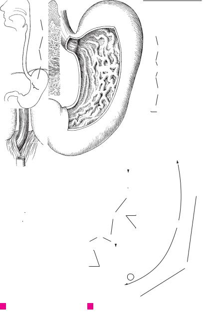
15 STOMACH. Gaster (Ventriculus). It extends from
21the end of the esophagus to the pylorus. A D 16 Anterior surface of stomach. Paries anterior D
2217 Posterior surface of stomach. Paries posterior.
18 Greater curvature of stomach. Curvatura ga-
23strica (ventricularis) major. Large curvature of the stomach profile directed toward the left and downward. D
2419 Lesser curvature of stomach. Curvatura gastrica (ventricularis) minor. Small curvature of
25stomach directed toward the right and upward. D
Digestive system 121
1
2
3
|
|
|
|
|
|
|
|
|
|
|
|
|
|
|
|
3 |
|
|
|
|
|
|
|
|
|
|
|
|
|
|
|
12 |
|
|
|
|
|
|
|
|
|
|
|
|
|
|
|
|
|
|
4 |
||
|
2 |
|
|
|
13 |
|
|
|
|
|
|
|
|||||
|
|
|
|
|
|
|
|
|
|
|
|
||||||
|
|
|
|
|
|
|
|
|
|
|
|
|
|
||||
|
4 |
|
|
|
14 |
|
|
|
|
11 |
|
5 |
|||||
|
|
|
|
|
|
||||||||||||
|
|
|
|
|
|
|
|
|
|
|
|
|
|
|
|
||
|
|
|
|
|
|
|
|
|
|
|
|
|
|
|
6 |
||
|
|
|
|
|
|
|
|
|
|
|
|
|
|
|
|
|
|
|
|
|
|
|
|
|
|
|
|
|
|
|
|
|
|
|
|
|
|
|
|
|
|
|
|
|
|
|
|
|
|
|
|
|
|
|
5 |
|
|
|
|
|
|
|
|
|
7 |
|
7 |
||||
|
|
|
|
|
|
|
|
|
|
|
|
||||||
|
15 |
|
|
|
|
|
|
|
|
||||||||
|
|
|
|
|
|
|
|
|
|
|
|||||||
|
|
|
|
|
|
|
|
|
|
8 |
|||||||
|
|
|
|
|
|
|
|
|
|
|
|
|
|
|
|
|
|
|
|
|
|
|
|
|
|
|
|
|
|
|
|
6 |
|
|
|
|
|
|
|
|
|
|
|
|
|
|
|
|
|
|
|||
|
|
|
|
|
|
|
|
|
|
|
|
|
|
|
9 |
||
|
Esophagus and stomach |
|
Microscopic cross-section |
|
|
||||||||||||
|
|
|
|
||||||||||||||
A |
C |
|
10 |
||||||||||||||
|
|
|
|
|
|
|
|
|
of esoghagus |
|
|||||||
|
|
|
|
|
|
|
|
|
|
|
|||||||
|
|
|
|
|
|
|
|
|
|
|
|
|
|
|
|
|
|
|
|
|
|
|
|
|
8 |
|
|
|
|
|
|
|
|
|
11 |
|
|
|
|
|
|
|
|
|
|
|
|
|
|
|
|
|
|
|
|
|
|
|
|
|
|
|
|
|
|
|
|
||||
|
|
|
|
|
|
|
|
|
|
|
|
|
|
23 |
|
12 |
|
|
|
|
|
|
|
|
|
|
|
|
|
|
|
|
|
||
|
|
|
|
|
|
|
|
|
|
|
|
|
|
24 |
|
||
|
|
|
|
|
|
|
|
|
|
|
|
|
|
|
13 |
||
|
|
|
|
|
|
|
|
|
|
|
|
|
|
|
|
|
|
|
|
|
|
|
|
|
|
|
|
|
|
|
|
|
|
|
|
|
|
|
|
|
|
|
|
|
|
|
|
|
|
16 |
|
14 |
|
|
|
|
|
|
|
|
|
|
|
|
|
|
|
21 |
|
|
|
|
|
|
|
|
|
|
|
|
|
|
|
|
|
|
|
|
|
|
|
|
|
|
|
|
|
22 |
|
|
|
|
|
|
15 |
||
|
|
|
|
|
|
|
|
|
|
|
|
|
|
||||
|
|
|
|
|
|
|
|
|
|
|
|
|
|
|
|
|
|
|
|
|
|
|
|
|
|
|
|
|
|
|
|
|
|
|
16 |
|
|
|
|
|
|
|
|
|
|
|
|
|
|
122.12 |
|
|
|
|
|
|
|
|
|
|
|
|
|
|
|
|
|
|
|||
|
|
|
|
|
9 |
|
|
|
|
|
|
|
17 |
||||
|
|
|
|
|
10 |
|
|
|
19 |
|
26 |
|
|
|
|||
|
|
|
|
|
|
|
|
|
|
||||||||
|
|
|
|
|
|
|
|
|
25 |
|
18 |
||||||
|
|
|
|
|
|
|
|
|
|||||||||
|
|
|
|
|
|
|
|
|
|
|
|
|
|
|
|||
|
|
|
|
|
|
|
|
27 |
|
20 |
|
|
|
|
|||
|
|
|
|
|
|
|
|
|
|
|
|
|
|||||
|
|
|
|
|
|
|
|
|
|
|
|
19 |
|||||
|
|
|
|
|
|
|
|
|
|
|
|
|
|
|
|
|
|
|
|
|
|
|
|
|
|
|
|
|
|
|
|
|
|
|
|
|
|
|
|
|
|
|
|
|
|
|
|
|
|
|
|
|
20 |
|
|
|
|
|
|
|
|
28 |
|
|
|
|
|
|
|
|
|
|
|
|
|
|
|
|
|
|
|
|
|
|
|
|
|
||
|
|
|
|
|
|
|
|
|
|
|
|
|
18 |
21 |
|||
|
|
|
|
|
|
|
|
29 |
|
|
|
|
|
|
|||
|
|
|
|
|
|
|
|
|
|
|
|
|
|
|
|||
|
|
|
|
|
|
|
|
|
|
|
|
|
|
|
|||
|
|
|
|
|
|
|
|
|
|
|
|
|
|
|
|
||
|
|
|
|
|
|
|
|
|
|
|
|
|
|
16 |
|
22 |
|
|
|
|
|
|
|
|
|
|
|
|
|
|
|
|
|
||
|
|
|
|
|
|
|
|
|
|
|
|
|
|
|
|
|
|
|
|
|
|
|
|
|
|
|
|
|
|
|
|
|
|
|
23 |
|
|
|
|
|
|
|
|
|
|
|
|
|
|
|
|
|
|
|
|
|
|
|
|
|
|
|
|
|
|
|
|
|
|
|
|
|
|
|
|
|
|
|
|
|
|
|
|
|
|
|
|
|
24 |
B |
Esophagus from behind |
D Stromach from front right |
|
|
|||||||||||||
|
|
||||||||||||||||
25

|
122 |
Digestive system |
|
|
|
|
|
|||
|
|
|
Pylorus. Distal end of the stomach reinforced by |
20 |
SMALL INTESTINE. Intestinum tenue. It com- |
|||||
1 |
|
1 |
||||||||
|
|
|
a strong band of circular muscle. A |
|
prises the duodenum, jejunum and ileum. |
|||||
2 |
|
2 |
Pyloric opening. Ostium pyloricum. Lumen of |
21 |
Tunica serosa. Peritoneal covering containing |
|||||
|
|
|
the pylorus connecting the stomach with the |
|
simple squamous epithelium. F |
|||||
|
|
|
|
duodenum. A |
22 |
Tela subserosa. Connective tissue underlying |
||||
3 |
|
3 |
Serous membrane. Tunica serosa. Serous, peri- |
|
the serosa. F |
|
|
|
||
|
|
|
|
toneal covering containing simple squamous |
23 |
Tunica muscularis. Two principal muscle layers |
||||
4 |
|
|
|
epithelium. B |
|
of the intestinal wall. F |
|
|
||
|
4 |
Tela subserosa. Connective tissue component |
24 |
Longitudinal |
layer. |
Stratum longitudinale. |
||||
|
|
|||||||||
|
|
|
|
of the serosa underlying the epithelium. B |
|
Outer layer of longitudinally coursing muscle |
||||
|
|
|
|
|
||||||
5 |
|
|
|
|
||||||
5 |
Tunica muscularis. Muscular coat of the |
|
fibers. F |
|
|
|
||||
|
|
|
|
stomach comprised of three types of fibers |
25 |
Circular layer. Stratum circulare. Inner layer of |
||||
6 |
|
|
|
(longitudinal, circular, oblique). A B |
|
circularly coursing muscle fibers. F |
||||
|
6 |
Longitudinal layer. Stratum longitudinale. Ex- |
26 |
Tela sumucosa. Displaceable layer between the |
||||||
|
||||||||||
7 |
|
|
|
ternal layer of longitudinal muscle fibers sit- |
|
muscularis mucosae and the muscularis con- |
||||
|
|
|
uated primarily along the greater and lesser cur- |
|
sisting primarily of collagenous connective |
|||||
|
|
|
|
|
||||||
|
|
|
|
vatures. A B |
|
tissue bearing blood vessels and nerves. F |
||||
8 |
|
|
|
|
||||||
7 |
Circular layer. Stratum circulare. Middle layer |
27 |
Tunica mucosa. Intestinal mucous membrane |
|||||||
|
|
|
|
of circular muscle. A B |
|
consisting of simple columnar epithelium, con- |
||||
9 |
8 |
Pyloric sphincter. M. sphincter pyloricus. Thick |
|
nective tissue (lamina propria) and muscularis |
||||||
|
|
|
|
layer of circular muscle around the pylorus. A |
|
mucosae. |
|
|
|
|
|
|
|
|
28 |
Lamina muscularis mucosae. Layer of smooth |
|||||
10 |
9 |
Oblique fibers. Fibrae obliquae. Inner layer of |
||||||||
|
muscle between the lamina propria and submu- |
|||||||||
|
|
|
|
obliquely coursing fibers of the tunica muscu- |
|
|||||
|
|
|
|
|
cosa. Its action produces folds in the mucosa. F |
|||||
11 |
|
|
|
laris. A B |
|
|||||
|
|
|
29 |
Circular folds [Valves of |
Kerckring]. Plicae |
|||||
10 |
Tela submucosa. Displaceable layer between |
|||||||||
|
|
circulares. Permanent folds of mucosa and sub- |
||||||||
|
|
|
|
the muscularis mucosae and the muscularis; it |
|
|||||
12 |
|
|
|
|
mucosa spanning nearly 2/3 of the intestinal |
|||||
|
|
|
is composed primarily of collagenous connec- |
|
||||||
|
|
|
|
lumen and projecting up to 8 mm high, perpen- |
||||||
|
|
|
|
tive tissue bearing vessels and nerves. B |
|
|||||
|
|
|
|
|
dicular to the intestinal axis. E F |
|||||
13 |
11 |
Tunica mucosa. Gastric mucous membrane |
|
|||||||
30 |
Intestinal villi. Villi intestinales. Fingerlike pro- |
|||||||||
|
|
|
|
consisting of simple columnar epithelium, con- |
|
jections about 0.5−1.5 |
mm in length. F |
|||
|
|
|
|
|
||||||
14 |
|
|
|
nective tissue (lamina propria) and muscularis |
|
|||||
|
|
|
31 |
Intestinal glands. Gll. intestinales. Cryptlike |
||||||
|
|
|
mucosae. B |
|||||||
|
12 |
Gastric folds. Plicae gastricae. Mucosal folds |
|
glands. F |
|
|
|
|||
|
|
|
|
|
||||||
15 |
32 |
Solitary lymphatic follicles. Folliculi lym- |
||||||||
|
|
|
extending primarily in a longitudinal direction. |
|||||||
|
|
|
|
See p. 121 D |
|
phatici solitarii. Solitary lymphatic nodules in |
||||
|
|
|
|
|
the lamina propria (p. 405.42) F |
|||||
16 |
13 |
Lamina muscularis mucosae. Layer of smooth |
|
|||||||
33 |
Aggregated |
lymphatic |
follicles [[Peyer’s |
|||||||
|
|
|
|
muscle between the lamina propria and the |
||||||
|
|
|
|
|
patches]]. Folliculi lymphatici aggregati. Aggre- |
|||||
|
|
|
|
submucosa. Its contraction produces folds in the |
|
|||||
17 |
|
|
|
|
gation of several lymphatic nodules in the |
|||||
|
|
|
mucosa. B |
|
||||||
|
14 |
Gastric (mamillated) areas. Areae gastricae. |
|
ileum. |
|
|
|
|||
|
|
|
|
|
||||||
|
|
|
|
|
|
|||||
18Mamillated areas on the mucosal surface that are bounded by shallow grooves and have a
19diameter of 1−6 mm. B
15Villous folds. Plicae villosae. Epithelial ridges invisible to the naked eye between the openings
20 |
of glands. B C D |
16 Gastric pits (foveolae). Foveolae gastricae.
21Openings of the gastric glands between the villous folds. B C D
2217 Gastric glands proper. Glandula gastrica propria. Tubular glands in the fundus and body of the stomach made up of 4 cell types. B C
2318 Pyloric glands. Glandula pylorica. Mucoid glands in the pyloric part of the stomach con-
24sisting of two cell types. D
19 Lymphatic nodules (follicles). [[Folliculi lym-
25phatici gastrici]]. Small aggregations of lymphatic tissue in the lamina propria. C D
Digestive system 123
|
|
|
|
|
|
|
|
|
|
|
|
|
|
|
|
|
1 |
|
|
|
|
|
|
|
|
|
|
|
14 |
|
|
|
|
2 |
|
|
5 |
|
|
|
|
|
15 |
|
|
|
|
|
|
|
|
3 |
|
|
|
|
|
|
|
|
|
|
|
|
|
|
|
||||
|
|
|
|
|
9 |
16 |
|
|
|
|
|
|
|
|
|||
|
|
|
|
|
|
|
|
|
|
|
|
|
|
|
|||
|
|
|
|
|
|
|
11 |
|
|
|
|
|
|
4 |
|||
|
|
|
|
|
|
|
|
|
|
|
|
|
|
||||
|
|
|
|
|
|
|
|
|
|
|
|
|
|
|
|||
|
|
|
|
|
|
|
7 |
|
|
|
|
|
|
|
|
||
|
|
|
|
|
|
|
17 |
|
|
|
|
|
|
|
|
|
|
|
|
|
|
|
|
|
|
|
|
||||||||
|
8 |
|
|
|
|
|
|
|
|
5 |
|||||||
|
|
|
|
|
|
|
|
|
|
|
|
|
|
|
|||
|
|
|
|
|
|
13 |
|
|
10 |
|
|
|
|
|
|||
|
|
|
|
|
|
|
|
||||||||||
|
|
6 |
|
|
|
|
|
|
|
|
|
|
|
6 |
|||
2 |
|
9 |
|
|
|
9 |
|
|
|
|
|||||||
|
|
|
|
|
|
|
|
|
|||||||||
|
|
|
|
|
|
|
|
||||||||||
|
|
|
7 |
|
|
|
|
|
5 |
7 |
|
|
|
|
7 |
||
|
|
|
|
|
|
|
|
|
|
|
|
|
|
||||
|
|
|
|
|
|
|
|
|
|
|
|
|
4 |
|
|
||
|
|
|
|
|
|
|
|
|
|
|
|
|
|
|
|
|
|
|
1 |
|
|
|
|
|
|
|
|
|
6 |
8 |
|||||
|
|
|
|
|
|
|
|
|
|
|
|
||||||
|
|
|
|
|
|
|
|
|
|
|
|
|
|
3 |
|
|
|
|
|
|
|
|
|
|
|
|
|
||||||||
A |
Stomach musculature |
B |
Stomach wall, overview |
|
|
|||||||||
16 |
15 |
19 |
|
16 |
19 |
|||||||||
|
|
|
|
|
|
|||||||||
|
|
|
|
|
|
|||||||||
|
|
|
|
|
|
|
|
|
|
|
|
|
15 |
|
|
|
|
|
|
|
|
|
|
|
|
|
|
|
|
|
|
|
|
|
|
|
|
|
|
|
|
|
|
|
|
|
|
|
|
|
|
|
|
|
Mucosa of pylorus |
|
|
||
17 |
|
|
|
|
|
|
|
D |
|
|
||||
|
|
|
|
|
|
|
|
18 |
|
|
|
|
||
|
|
|
|
|
|
|
|
|
|
|
|
|
|
|
|
|
|
|
|
|
|
|
|
|
|
|
|
|
|
CTunica mucosa, fundus ventriculi
30
|
|
|
|
|
|
|
|
|
|
|
|
29 |
||
|
|
|
|
|
|
|
|
|
||||||
|
|
|
|
|
|
|
|
31 |
|
|||||
|
|
|
|
|
|
|
|
|
|
|
|
|
|
|
|
|
|
|
|
|
|
|
32 |
|
|||||
|
|
|
|
29 |
28 |
|
26 |
|||||||
|
|
|
|
|
||||||||||
|
|
|
|
|
|
|||||||||
|
|
|
|
|
|
|
25 |
|
|
|
|
|
|
|
|
|
|
|
|
|
|
|
|
|
|
|
|
|
|
|
|
|
|
|
|
24 |
|
|
|
|
|
|
|
|
|
|
|
|
|
|
|
22 |
|
|
|
|
|
|
|
29 |
|
|
21 |
|
|
|
|
|
|
|
||||
|
Intestinal canal |
|
|
|
|
|
|
|
|
|
Intestinal wall, histological section |
|||
E |
|
|
|
|
|
|
|
|
F |
|||||
9
10
11
12
13
14
15
16
17
18
19
20
21
22
23
24
25

|
124 |
Digestive system |
|
|
||||
|
|
|
|
DUODENUM. Initial segment of the small in- |
19 |
Ileocecal valve. Valva ileocaecalis (Valva ilealis). |
||
1 |
|
1 |
|
|||||
|
|
|
|
testine, about 25−30 mm long, between the py- |
|
Two-lipped valve at the entrance of the ileum |
||
2 |
|
|
|
|
lorus and the duodenojejunal flexure. A |
|
into the large intestine. D |
|
|
2 |
|
Superior part. Pars superior. Horizontal begin- |
19 a |
Papilla ilealis. B |
|||
|
|
|
|
|
ning part of the duodenum. A |
20 |
Opening of ileocecal valve. Ostium valvae |
|
3 |
|
3 |
|
Ampulla [[bulbus duodeni]]. Functional dilata- |
|
ilealis. Transverse, slitlike aperture of ileum at |
||
|
|
|
entrance into the large intestine in the cadaver. |
|||||
|
|
|
|
|
tion at the beginning of the duodenum. It is |
|
||
4 |
|
|
|
|
briefly visible in radiograms. A |
|
D |
|
|
4 |
|
Descending part. Pars descendens. Lateral, ver- |
21 |
Frenulum valvae ilealis. Fold formed by the |
|||
|
|
|
|
|
tical segment. A |
|
union of the lips of the ileocecal valve. B D |
|
|
|
|
|
|
|
|||
5 |
|
|
|
|
|
|||
5 |
|
Horizontal (inferior) part. Pars horizontalis |
21 a Ostium papillae ilealis. B |
|||||
|
|
|
|
|
(inferior). Horizontal segment below the head |
22 |
Vermiform appendix. Appendix vermiformis. |
|
6 |
|
|
|
|
of the pancreas. A |
|
Appendage of the cecum, usually 9 cm long, |
|
6 |
Ascending part. Pars ascendens. Segment of |
|
with abundant lymphatic tissue. B C D |
|||||
|
|
|||||||
|
23 |
Ostium appendicis vermiformis. Opening of |
||||||
7 |
|
|
|
|
duodenum lying to the left of the head of the |
|||
|
|
|
|
pancreas and ascending up to the duodenoje- |
|
vermiform appendix into the cecum. D |
||
|
|
|
|
|
junal flexure. A |
24 |
Aggregated lymphatic nodules of ver- |
|
8 |
7 |
|
Superior duodenal flexure. Flexura duodeni |
|
miform appendix. Folliculi lymphatici aggre- |
|||
|
|
|
|
|
superior. Flexure between the superior and |
|
gati appendicis vermiformis. Lymphatic tissue |
|
|
|
|
|
|
|
|||
9 |
|
|
|
|
horizontal parts of the duodenum medial to the |
|
within the wall of the appendix. |
|
|
|
|
|
gallbladder. A |
25 |
COLON. Portion of large intestine extending |
||
|
|
|
|
|
||||
|
|
|
8 Inferior duodenal flexure. Flexura duodeni in- |
|
from the ileocecal valve to the rectum. |
|||
10 |
|
|
|
|||||
|
|
26 |
Ascending colon. Colon ascendens. Segment of |
|||||
|
|
|
|
ferior. Flexure between the descending and |
||||
|
|
|
|
|
horizontal parts of the duodenum. A |
|
large intestine ascending retroperitoneally on |
|
11 |
9 |
|
Duodenojejunal flexure. Flexura duodenoje- |
|
the right side. C |
|||
|
27 |
Right colic flexure. Flexura coli dextra. Flexure |
||||||
|
|
|
|
|
junalis. Flexure between the duodenum and je- |
|||
|
|
|
|
|
||||
|
|
|
|
|
junum. A |
|
between the ascending and transverse colon. C |
|
12 |
|
|
|
|
|
|||
10 |
|
Suspensory muscle of duodenum. M. suspen- |
28 Transverse colon. Colon transversum. Trans- |
|||||
|
|
|||||||
|
|
|
verse segment of large intestine situated in- |
|||||
|
|
|
|
|
sorius duodeni. Bundle of smooth muscle which |
|
||
13 |
|
|
|
|
|
|||
|
|
|
|
|
traperitoneally between the right and left colic |
|||
|
|
|
|
attaches the duodenojejunal flexure to the dia- |
|
|||
|
|
|
|
|
phragm or to the celiac trunk. A |
|
flexures. C |
|
|
|
|
|
|
|
|||
14 |
11 |
|
Longitudinal folds of duodenum. Plicae longi- |
29 Left colic flexure. Flexura coli sinistra. Flexure |
||||
|
|
between the transverse and descending colon |
||||||
|
|
|
|
|
tudinales duodeni. Longitudinal mucosal folds |
|
||
|
|
|
|
|
|
below the left subphrenic space. In its vicinity |
||
15 |
|
|
|
|
on the left side of the posterior wall of the de- |
|
||
|
|
|
|
|
lies Cannon-Böhm’s ring, the boundary be- |
|||
|
|
|
|
scending part of the duodenum produced by the |
|
|||
|
|
|
|
|
pancreatic and bile ducts. A |
|
tween the cranial (vagus nerve) and sacral para- |
|
|
|
|
|
|
|
|||
16 |
|
|
|
|
|
sympathetics. C |
||
12 |
|
Greater duodenal papilla. Papilla duodeni |
|
|||||
|
30 Descending colon. Colon descendens. Segment |
|||||||
|
|
|
|
|
major. Elevation at the end of the longitudinal |
|||
|
|
|
|
|
|
of large intestine extending retroperitoneally on |
||
17 |
|
|
|
|
fold at the openings of the common bile duct |
|
||
|
|
|
|
|
the left side between the left colic flexure and |
|||
|
|
|
|
and pancreatic duct. A |
|
|||
|
|
|
|
|
|
sigmoid colon. C |
||
|
13 |
|
Lesser duodenal papilla. Papilla duodeni |
|
||||
18 |
|
31 |
Sigmoid colon. Colon sigmoideum. Portion of |
|||||
|
|
|
|
minor. Elevation located superior to the greater |
||||
|
|
|
|
|
duodenal papilla, usually at the opening of the |
|
colon lying intraperitoneally between the de- |
|
|
|
|
|
|
|
scending colon and the rectum. C |
||
19 |
|
|
|
|
accessory pancreatic duct. A |
|
||
|
|
|
|
32 |
Semilunar folds of colon. Plicae semilunares |
|||
14 |
|
Duodenal glands [[Brunner’s glands]]. Glan- |
||||||
|
|
|
coli. Crescentic contraction folds between two |
|||||
20 |
|
|
|
|
dulae duodenales. Mucous glands located pre- |
|
haustra comprising all layers of the intestinal |
|
|
|
|
|
dominantly in the submucosa of the duodenum. |
|
|||
|
|
|
|
|
|
wall. C D |
||
|
15 |
JEJUNUM. Middle segment of the small in- |
|
|||||
|
33 |
Haustra (sacculationes) coli. Outpocketings |
||||||
21 |
||||||||
|
|
|
|
testine. It extends about 2.5 meters from the |
||||
|
|
|
|
|
between two semilunar folds. C D |
|||
|
|
|
|
|
duodenojejunal flexure. A C |
|
||
|
|
|
|
|
34 |
Appendices epiploicae (omentales). Serosa- |
||
22 |
16 |
|
ILEUM. Terminal segment of small intestine, |
|||||
|
|
covered appendages containing adipose tissue, |
||||||
|
|
|
|
|
about 3.5 mm long. C |
|
located on the free and omental tenia. C |
|
|
|
|
|
|
|
|||
2317 LARGE INTESTINE. Intestinum crassum. About 1.5−1.8 m long; it extends from the cecum to the anus and consists of tenia, haustra and ap-
24pendices epiploicae.
18 CECUM. Caecum. Initial portion of the large in-
25testine, ca. 7 cm long, located below the opening of the ileum. C D