Материал: Атлас Ханц фениш
Digestive system 115
86
5
4
15
6
15
7
15
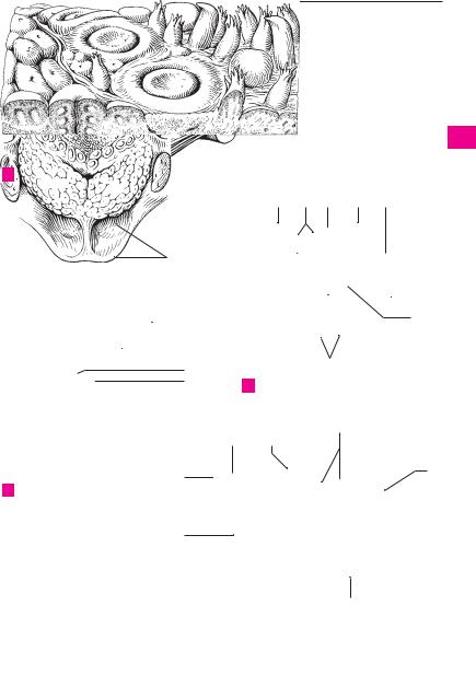
A Surface of tongue, enlarged
10
|
6 |
|
||
112.32 |
|
|
|
|
|
|
|
9 |
|
|
|
|||
|
|
|
|
7 |
|
|
|
||
|
11 |
|||
112.33 |
12 |
|||
14 |
|
|
|
|
|
|
|
|
|
22
B Dorsum of tongue, overview
118.27 
118.22
118.29 
1 23 10 17 25
26 
16 |
|
|
|
24 |
|
|
|
||
|
196.5
19
C Cross-section of tongue
9
116.2214
2
19
20
21
82.12
1
2
3
4
5
6
7
8
9
10
11
12
13
14
15
16
17
18
19
20
21
22
23
|
Tongue muscles |
24 |
D |
||
|
|
|
|
|
25 |
|
|
|

|
116 |
Digestive system |
|
|
|
|
|
|
|
|
|
|
||||
|
|
|
|
Pharynx. Passageway for air and food. 14− |
19 |
M. levator veli palatini. o: Petrous portion of |
||||||||||
1 |
|
1 |
|
|||||||||||||
|
|
|
|
16 cm long, it extends from the fornix to the |
|
temporal bone in front of the lower opening of |
||||||||||
|
|
|
|
|
beginning of the esophagus in front of the 6th |
|
the carotid canal. i: Palatine aponeurosis. It |
|||||||||
2 |
|
|
|
|
cervical vertebra. E |
|
|
passes through the pharyngeal wall above the |
||||||||
|
|
2 |
|
FAUCES. Space between soft palate and base of |
|
superior constrictor muscle and moves the soft |
||||||||||
|
|
|
|
palate backward and upward, thereby taking |
||||||||||||
3 |
|
|
|
|
tongue. E |
|
|
|||||||||
|
|
|
|
|
|
along the dorsomedial part of the auditory tube |
||||||||||
|
3 |
|
Isthmus of fauces. Isthmus faucium. Space be- |
|
||||||||||||
|
|
|
|
cartilage below the pharyngeal opening of the |
||||||||||||
|
|
|
|
|
tween right and left palatoglossal and |
pala- |
|
|||||||||
4 |
|
|
|
|
|
auditory tube. I: Vagus (X) nerve. C |
|
|||||||||
|
|
|
|
topharyngeal arches. |
|
|
|
|||||||||
|
|
|
|
|
20 M. tensor veli palatini. o: Spine of sphenoid, |
|||||||||||
|
|
4 |
|
Soft palate. Palatum molle (velum palatinum). |
||||||||||||
|
|
|
|
scaphoid fossa and anterior (lateral) lip of car- |
||||||||||||
|
|
|
|
|||||||||||||
5 |
|
|
|
|
The dorsal portion projects downward in front |
|
||||||||||
|
|
|
|
|
tilaginous |
auditory |
tube. |
i: After |
looping |
|||||||
|
|
|
|
|
of the posterior pharyngeal wall and assists in |
|
around the pterygoid hamulus, it radiates into |
|||||||||
|
|
|
|
|
swallowing by closing off like a valve the na- |
|
||||||||||
6 |
|
|
|
|
|
the palatine aponeurosis, stiffens the anterior |
||||||||||
|
|
|
|
sopharyngeal space from the oral cavity. A D E |
|
|||||||||||
|
|
|
|
|
(lateral) membranous wall of the auditory tube |
|||||||||||
|
5 |
|
Uvula. Uvula palatina. Conical process project- |
|
||||||||||||
|
|
|
and tenses the soft palate. I: Mandibular nerve. |
|||||||||||||
7 |
|
|
|
|
ing downward from the posterior margin of the |
|
C |
|
|
|
|
|
|
|
||
|
|
|
|
|
soft palate. A D E |
|
21 M. uvulae. o: Palatine aponeurosis. i: Connec- |
|||||||||
|
|
|
|
|
|
|||||||||||
8 |
6 |
|
Palatoglossal arch. Arcus palatoglossus. Mu- |
|
tive tissue of uvulae. I: Vagus nerve. C |
|
||||||||||
|
|
|
|
cosal fold overlying the palatoglossal muscle |
|
|
||||||||||
|
|
|
|
|
22 |
M. |
palatoglossus. |
o: |
Transversus |
linguae |
||||||
|
|
|
|
|
and extending from the palate to the tongue in |
|||||||||||
9 |
|
|
|
|
|
muscle. i: Palatine aponeurosis. A: Elevates the |
||||||||||
|
|
|
|
front of the tonsillar fossa. A |
|
|
||||||||||
|
|
|
|
|
|
base of the tongue, depresses the palate and |
||||||||||
|
7 |
|
Palatopharyngeal arch. Arcus palatopharyn- |
|
narrows the isthmus of fauces. I: Vagus nerve. D |
|||||||||||
10 |
|
|
|
|
geus. Mucosal fold overlying the palatopharyn- |
23 |
M. |
palatopharyngeus |
[[m. |
pharyngopalat- |
||||||
|
|
|
|
geal muscle and extending between the palate |
||||||||||||
|
|
|
|
|
|
inus]]. |
o: |
Palatine |
aponeurosis, pterygoid |
|||||||
|
|
|
|
|
and pharyngeal wall behind the tonsillar fossa. |
|
||||||||||
11 |
|
|
|
|
|
hamulus and medial plate of pterygoid process. |
||||||||||
|
|
|
|
A |
|
|
||||||||||
|
|
|
|
|
|
i: Lateral wall of pharynx and thyroid cartilage. |
||||||||||
|
8 |
|
Salpingopalatine fold. Plica salpingopalatina |
|
||||||||||||
|
|
|
A: It lowers the palate and constricts the isth- |
|||||||||||||
12 |
|
|
|
|
[[plica palatotubalis]]. Fold extending from the |
|
mus of fauces. I: Vagus nerve. D |
|
||||||||
|
|
|
|
|
anterior lip of the auditory tube to the soft pa- |
24 PHARYNGEAL CAVITY. Cavitas pharyngis. Space |
||||||||||
|
|
|
|
|
late in front of the tubal elevation. A |
|
||||||||||
13 |
|
|
|
|
|
|
enclosed by the pharyngeal walls. |
|
||||||||
9 |
|
Palatine tonsil. Tonsilla palatina. Tonsil sit- |
|
|
||||||||||||
|
25 Pharyngeal fornix. Fornix pharyngis. Roof of |
|||||||||||||||
|
|
|
|
|
uated between the palatoglossal and |
pala- |
||||||||||
|
|
|
|
|
|
the pharyngeal cavity beneath the sphenoid |
||||||||||
14 |
|
|
|
|
topharyngeal arches. A |
|
|
|||||||||
|
|
|
|
|
|
bone. E |
|
|
|
|
|
|
||||
|
10 |
Tonsillar pits. Fossulae tonsillae. Pit-like open- |
26 Nasopharynx. Pars nasalis pharyngis. The por- |
|||||||||||||
|
||||||||||||||||
15 |
|
|
|
|
ings of the tonsillar crypts visible on the sur- |
|||||||||||
|
|
|
|
|
tion of the pharyngeal cavity located behind |
|||||||||||
|
|
|
|
face. B |
|
|
||||||||||
|
|
|
|
|
|
|
the choanae. E |
|
|
|
|
|||||
|
11 |
Tonsillar crypts. Cryptae tonsillares. Epithelial |
27 |
|
|
|
Tonsilla |
|||||||||
16 |
Nasopharyngeal tonsil |
(adenoids). |
||||||||||||||
|
|
|
|
invaginations extending into the tonsil |
from |
|||||||||||
|
|
|
|
|
pharyngealis (adenoidea). It lies at the pharyn- |
|||||||||||
|
|
|
|
|
the tonsillar pits. B |
|
|
|||||||||
|
|
|
|
|
|
|
geal fornix. E |
|
|
|
|
|||||
17 |
12 |
|
Capsule of tonsil. Capsula tonsillaris. Fibrous |
|
|
|
|
|
||||||||
|
28 |
Tonsillar pits. Fossulae tonsillares. Openings |
||||||||||||||
|
|
|
|
|
capsule covering the organ. |
|
|
of crypts visible on surface of tonsil. See also |
||||||||
|
|
|
|
|
|
|
||||||||||
18 |
13 |
|
Triangular fold. Plica triangularis. Triangular |
|
p. 116.10. B |
|
|
|
|
|
||||||
|
|
|
|
|
fold emanating from the palatoglossal arch in |
29 |
Tonsillar |
crypts. Cryptae |
tonsillares. Epithelial |
|||||||
|
|
|
|
|
front of the tonsil. A |
|
||||||||||
19 |
|
|
|
|
|
|
invaginations emanating from the tonsillar |
|||||||||
14 |
|
Semilunar fold. Plica semilunaris. Arched fold |
|
|||||||||||||
|
|
pits. See also p. 116.11. B |
|
|
|
|||||||||||
|
|
|
|
|
between the palatoglossal and palatopharyn- |
|
|
|
|
|
|
|
|
|
||
20geal arches. It forms the upper boundary of the tonsillar fossa. A
21 |
15 Tonsillar fossa. Fossa tonsillaris. Recess for the |
|
tonsil bordered by the palatoglossal and pala- |
||
|
topharyngeal arches as well as by the triangular |
|
22 |
and semilunar folds. D |
|
|
16 Supratonsillar fossa. Fossa supratonsillaris. Su- |
|
|
||
23 |
perior portion of tonsillar fossa not occupied by |
|
the tonsil. A |
||
|
||
24 |
17 Muscles of palate and fauces. Musculi palati et |
|
faucium. |
18 Palatine aponeurosis. Aponeurosis palatina. It
25is formed primarily by the tendon of the tensor veli palatini muscle. C
Digestive system 117
8
4
14
5
6
16
13 9
7
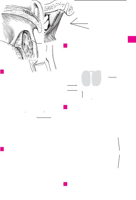
ATonsillar fossa and soft palate
4
22 |
|
|
|
5 |
|
||||
|
|
|
23
15
D Muscles of tonsillar fossa
10;(28)
11;(29)
B Palatine tonsil, microscopic view
19
20
102.2
20 |
|
21 |
|
CNasal cavity from behind and muscles of soft palate
25
26 27
4
5
2 118.8
1
118.12
E Head, sagittal section
1
2
3
4
5
6
7
8
9
10
11
12
13
14
15
16
17
18
19
20
21
22
23
24
25

118 Digestive system
1 Pharyngeal bursa. [Bursa pharyngealis]. Blind 1 pouch in the roof of the pharynx; it is more frequently present in children, less often in
2adults. A
2Pharyngeal opening of auditory tube. Ostium pharyngeum tubae auditivae (auditoriae). Open-
3ing found in the nasopharynx. A
3Torus tubarius. Elevation produced by the dor-
4somedial cartilage of the auditory tube posterior to the tube opening. A
4 Salpingopharyngeal fold. Plica salpingopharyngea.
5 Mucosal fold overlying the salpingopharyngeal muscle and extending obliquely downward from
6the dorsomedial lip of the auditory tube cartilage. A
5 Torus levatorius. Elevation situated in front of
7the dorsomedial lip of the cartilage of the auditory tube and below the tube opening. It overlies
8the levator veli palatini muscle. A
|
6 |
Tubal tonsil. Tonsilla tubaria. Submucosal lym- |
|
9 |
|
phatic tissue near the opening of the auditory |
|
|
tube. |
||
|
|
||
|
7 |
Pharyngeal recess. Recessus pharyngeus |
|
10 |
|||
|
[[Rosenmüller’s]]. Lateral recess of the na- |
||
|
|
sopharyngeal space behind the auditory tube. A |
|
|
|
||
11 |
8 |
Oropharynx. Pars oralis pharyngis. The portion |
|
|
|
of the pharyngeal cavity located behind the oral |
cavity. See p. 117. E
129 Vallecula epiglottica. Fossa between the median and lateral glossoepiglottic folds. B
1310 Median glossoepiglottic fold. Plica glossoepiglottica mediana. Unpaired mucosal fold located in the median plane between the base of
14the tongue and the epiglottis. B
|
11 |
Lateral glossoepiglottic fold. Plica glos- |
|
15 |
|||
|
soepiglottica lateralis. Bilateral mucosal fold be- |
||
|
|
tween the base of the tongue and the epiglottis. B |
|
|
|
||
16 |
12 |
Laryngopharynx. Pars laryngea pharyngis. The |
|
|
|
portion of the pharyngeal cavity situated behind |
the larynx. See p. 117. E
1713 Piriform recess. Recessus piriformis. Channel between the aryepiglottic fold and the thyrohyoid
18membrane or thyroid cartilage. B
14Plica nervi laryngei. Mucosal fold in the piriform recess produced by the underlying internal
19branch of the superior laryngeal nerve and the superior laryngeal artery. B
2015 Pharyngobasilar fascia. Fascia pharyn-
gobasilaris. Membranous wall of the uppermost muscle free portion of the pharynx. It corre-
21 sponds to the thickened tela submucosa. C D E
|
16 |
Submucosa. Tela submucosa. Connective tissue |
|
22 |
|||
|
layer between the mucosa and muscularis. A |
||
|
17 |
Mucosa. Tunica mucosa. Pharyngeal mucous |
|
|
|||
23 |
|
membrane lined by stratified squamous or pseu- |
|
|
dostratified ciliated columnar (nasopharynx) |
||
|
|
||
|
|
epithelium. |
|
24 |
|
||
18 |
Pharyngeal glands. Gll. pharyngis. Small sub- |
||
|
|
epithelial mixed salivary glands. |
|
|
|
||
25 |
19 |
Muscularis of pharynx. Tunica muscularis |
|
|
|
pharyngis. Muscular layer of pharyngeal wall. A |
20Pharyngeal raphe. Raphe pharyngis. Connective tissue band between the right and left pharyngeal muscles, extending posteriorly in the midline. C
21Pterygomandibular raphe. Raphe pterygomandibularis. Tendinous seam between the buccinator muscle and the superior pharyngeal constrictor. It extends from the pterygoid hamulus to the mandible. D
22Superior pharyngeal constrictor muscle. M. constrictor pharyngis superior. Uppermost constrictor of the pharynx consisting of the four parts described below, which attach to the pharyngeal raphe. I: Pharyngeal plexus. C D
23Pterygopharyngeal part. Pars pterygopharynea. o: Medial plate of pterygoid process and pterygoid hamulus. D
24Buccopharyngeal part. Pars buccopharyngea. o: Pterygomandibular raphe. D
25Mylopharyngeal part. Pars mylopharyngea. o: Posterior end of mylohyoid line of mandible. D
26Glossopharyngeal part. Pars glossopharyngea.
o:Intrinsic muscles of tongue. D
27M. stylopharyngeus. o: Styloid process. i: Between superior and middle pharyngeal constrictors radiating into the pharyngeal wall, thyroid cartilage and epiglottis. I: Glossopharyngeal (IX) nerve. C
28M. salpingopharyngeus. o: Dorsomedial lip of auditory tube cartilage and part of longitudinal musculature of pharyngeal wall. i: Lateral wall of pharynx. A: Prevents backward displacement of the levator veli palatini muscle. I: Pharyngeal plexus. A
29Middle pharyngeal constrictor muscle. M. constrictor pharyngis medius. More centrally located pharyngeal constrictor arising from the hyoid bone. o: Pharyngeal raphe. I: Pharyngeal plexus. C
30Chondropharyngeal part. Pars chondropharyngea. o: Lesser horn of hyoid. D
31Ceratopharyngeal part. Pars ceratopharyngea.
o:Greater horn of hyoid. D
32Inferior pharyngeal constrictor muscle. M. constrictor pharyngis inferior. o: Larynx. I: Pharyngeal plexus. C D
33Thyropharyngeal part. Pars thyropharyngea. o: Oblique line of thyroid cartilage. D
34Cricopharyngeal part. Pars circopharyngea. o: Cricoid cartilage. D
34 a Buccopharyngeal fascia. Fascia buccopharyngealis. Continuation of the buccopharyngeal fascia into the loose tissue connecting the pharynx to the deep cervical fascia.
35 Peripharyngeal space. Spatium peripharyngeum. Connective tissue space associated with the pharynx.
36Retropharyngeal space. Spatium retropharyngeum. The portion of the peripharyngeal space between the pharynx and the prevertebral layer of the cervical fascia. A
37Lateropharyngeal space. Spatium lateropharyngeum. Portion of the peripharyngeal space lateral to the pharynx.
Digestive system 119
|
|
|
7 |
|
2 |
|
|
|
|
3 |
|
1 |
||
|
|
|
|
|
|
5 |
|
|
|
28
4
36
19
16 
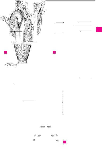
A Pharyngeal opening of auditory tube
15 15
22
27 |
|
29 |
|
32
20
11
13
B
9
10
14
Base of tongue and laryngeal opening
15
23
24
21
25
31 30 26
32
33
34
C |
Pharyngeal muscles |
D |
Pharyngeal muscles |
|
from behind |
|
from right |
E Attachment of the
pharyngobasilar fascia
1
2
3
4
5
6
7
8
9
10
11
12
13
14
15
16
17
18
19
20
21
22
23
24
25