Материал: Атлас Ханц фениш
|
|
|
|
|
|
|
|
|
|
|
|
Digestive system 125 |
|
|||||||
|
246.21 |
216.28 |
|
|
|
|
|
|
|
|
|
|
|
|
||||||
|
|
|
|
|
|
|
|
|
|
|
|
1 |
||||||||
|
|
|
|
|
|
|
|
|
|
|
|
|
|
|
|
|
|
|
|
|
|
|
|
|
|
|
|
|
|
|
|
|
21a 19a |
|
2 |
||||||
|
|
|
|
|
|
|
|
|
|
|
|
|
||||||||
|
248.1 |
|
|
|
|
|
|
|
|
|
3 |
|||||||||
|
|
|
|
|
|
|
|
|
|
|
|
|
|
|
|
|
||||
|
|
|
|
|
|
|
|
|
|
|
|
|
|
|
|
|
||||
|
|
|
|
|
|
|
|
|
|
|
|
|
|
|
|
|
||||
|
7 |
|
|
|
|
|
|
|
|
|
|
|
|
|
|
|
|
|
4 |
|
|
3 |
|
|
|
|
|
|
|
|
|
|
|
|
|
|
|
||||
|
2 |
|
|
|
|
10 |
|
|
|
|
|
21 |
|
|
||||||
|
|
|
|
|
|
|
|
|||||||||||||
|
|
|
|
|
|
|
||||||||||||||
|
|
|
|
|
|
|
|
|
|
|
|
|
|
|
|
|
|
|
5 |
|
|
|
|
|
|
|
|
|
|
|
9 |
|
|
|
|
|
|
|
|
|
|
|
|
|
|
|
|
|
|
|
|
128.1 |
|
|
|
|
|
|
|
|
|
|
|
|
|
|
|
|
|
|
|
|
|
|
|
|
|
|
|
|
|
|
|
|
13 |
|
|
|
|
|
|
|
|
|
|
|
|
|
|
|
|
|
6 |
|
|
|
|
|
|
|
|
|
|
|
|
|
|
|
|
|
|
|
|||
|
|
|
|
|
|
|
|
|
|
|
|
|
|
|
|
|
|
|
||
4 |
11 |
|
|
|
|
|
6 |
|
|
|
|
|
|
|
|
|
|
|
|
|
|
|
|
|
|
|
|
|
|
|
|
|
|||||||||
|
|
|
|
|
|
|
|
|
|
|
7 |
|||||||||
|
|
|
|
|
|
|
|
|
|
|
|
|
|
|
|
|||||
|
12 |
|
|
|
|
|
|
|
|
|
|
|
|
|
|
|
|
|
|
|
|
|
|
|
|
|
|
|
|
|
|
|
|
|
|
|
|
|
|
|
|
|
|
|
|
|
|
|
|
|
|
|
15 |
|
|
|
|
|
|
|
|
|
|
|
|
|
|
|
|
|
|
|
|
|
|
|
|
|
|
|
|
8 |
|
|
5 |
|
|
|
|
|
|
|
|
|
|
|
|
|
||||||
|
|
|
|
|
|
|
|
|
|
|
|
|
|
|
||||||
|
|
|
|
|
|
|
|
|
|
|
|
|
|
|
||||||
|
8 |
|
|
|
|
|
|
|
|
|
|
|
|
|
22 |
|
|
|
9 |
|
|
|
|
|
|
|
|
|
|
|
|
|
|
|
|
|
|
|
|
|
|
|
|
|
|
|
|
|
|
|
|
|
|
|
|
|
|
|
|
|
|
|
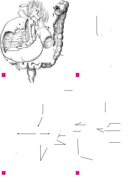
APortal vein, inferior vena cava, aorta and duodenum
27
28
15
26
16
18
22 
31
CSmall and large intestines, anterior view
B Cecum in the living body
29
28
32
30 |
32 |
|
||
|
|
|
33 |
19 |
|
|
|
|
|
|
|
|
|
|
|
|
32 |
21 |
|
|
|
34 |
|
|
|
|
|
|
18 |
22
D Cecum in the cadaver
10
11
12
13
14
15
16
19
17
20
18
2319
20
21
22
23
24
25

|
126 |
Digestive system |
|
|
|
|
|||
|
|
|
Tunica muscularis. Bilayered muscle wall of |
21 |
M. rectococcygeus. Thin plate of smooth |
||||
1 |
|
1 |
|||||||
|
|
|
colon. B |
|
|
|
muscle extending from the 2nd to 3rd coccygeal |
||
2 |
|
2 |
Longitudinal layer. |
Stratum |
longitudinale. |
|
vertebrae to the rectum. C |
||
|
22 |
M. rectourethralis. Smooth muscle fibers ex- |
|||||||
|
|
|
Distinct outer layer of longitudinally oriented |
||||||
|
|
|
|
muscle of variable thickness. B |
|
|
tending from the membranous part of the |
||
3 |
|
3 |
Taeniae coli. About |
1 cm wide, thickened |
|
urethra to the rectum. C |
|||
|
23 |
Transverse rectal folds. Plicae transversae |
|||||||
|
|
|
|
bands of longitudinal muscle fibers. B |
|||||
4 |
|
4 |
Taenia mesocolica. Tenia located at the attach- |
|
recti. Usually three lateral transverse folds. The |
||||
|
|
middle is the largest (Kohlrausch) and projects |
|||||||
|
|
|
ment of the mesocolon in the posteromedial |
|
|||||
|
|
|
|
|
about 6 mm above the anus from the right side, |
||||
|
|
|
|
part of the ascending and descending colon. A |
|
||||
|
|
|
|
|
|||||
5 |
|
|
|
|
the others from the left. C |
||||
5 |
Taenia omentalis. Tenia of transverse colon lo- |
|
|||||||
24 |
ANAL CANAL. Canalis analis. Terminal segment |
||||||||
|
|
|
|
cated at the attachment of the greater omentum |
|||||
|
|
|
|
|
of the digestive tube beginning with the anal |
||||
6 |
|
|
|
in the posteromedial part of the ascending and |
|
||||
|
|
|
|
columns. D |
|||||
|
|
|
descending colon. A |
|
|
|
|||
|
6 |
Taenia libera. Free tenia located between the |
25 |
Anal columns. Columnae anales. Six to ten |
|||||
7 |
|||||||||
|
longitudinal folds provided with abundant |
||||||||
|
|
|
tenia mesocolica and tenia omentalis. A |
|
|||||
|
|
|
|
|
venous plexuses. D |
||||
|
7 |
Circular layer. Stratum circulare. Inner layer of |
|
||||||
8 |
26 |
Anal sinuses. Sinus anales. Recesses between |
|||||||
|
|
|
circularly coursing muscle fibers of the colon. B |
||||||
|
|
|
8 Tela submucosa. Displaceable layer between |
|
the anal columns. D |
||||
9 |
|
|
27 |
Anal valves. Valvulae anales. Small transverse |
|||||
|
|
|
the muscularis mucosa and the muscularis, con- |
||||||
|
|
|
|
folds bordering the anal sinuses distally. D |
|||||
|
|
|
|
sisting mainly of collagenous connective tissue |
|
||||
|
|
|
|
28 |
Anorectal line. Linea anorectalis. Upper border |
||||
10 |
|
|
|
containing nerves and blood vessels. B |
|||||
9 |
Tunica mucosa. Villus-free mucous coat of the |
|
of anal canal formed by the puborectal muscle at |
||||||
|
|
|
|
colon comprised of |
simple, goblet-cell rich, |
|
the level of the levator sling just above the anal |
||
11 |
|
|
|
|
columns. D |
||||
|
|
|
columnar epithelium, connective tissue and |
|
|||||
|
|
|
29 |
Anal pecten. [[Pecten analis]]. A lighter stripe |
|||||
|
|
|
|
lamina muscularis mucosae. B |
|
||||
|
|
|
|
|
|||||
12 |
10 |
Lamina muscularis mucosae. Layer of smooth |
|
between the anal valves and the anocutaneous |
|||||
|
line. It is firmly connected with the underlying |
||||||||
|
|
|
|
muscle cells between the lamina propria and |
|
tissues by fibrous longitudinal muscles. D |
|||
|
|
|
|
|
|||||
13 |
|
|
|
submucosa. Its contraction produces a wrin- |
|
||||
|
|
|
30 |
Anocutaneous line. [[Linea anocutanea]]. Lower |
|||||
|
|
|
kling of the mucosa. B |
|
|
||||
|
11 |
Intestinal glands. |
Glandulae |
intestinales. |
|
border of anal pecten at the level of the lower |
|||
14 |
|
margin of the internal anal sphincter. D |
|||||||
|
|
|
Tubular glands of the colonic mucosa. B |
|
|||||
|
|
|
31 External anal sphincter. M. sphincter ani exter- |
||||||
|
12 |
Solitary lymphatic nodules. Nodi lymphatici |
|||||||
|
|||||||||
|
|
nus. Circular layer of skeletal muscle lying on the |
|||||||
15 |
|
|
|
solitarii. Individual lymphatic nodules dis- |
|
||||
|
|
|
|
internal anal sphincter. C D See p. 174.14−17 |
|||||
|
|
|
|
persed throughout the lamina propria. B |
32 Anus. Lower opening of anal canal surrounded |
||||
|
|
|
|
||||||
16 |
13 |
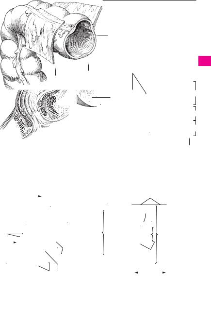
RECTUM. Tenia-free segment, about 15 cm |
|||||||
|
by the subcutaneous and superficial parts of the |
||||||||
|
|
|
|
long, located between the sigmoid colon and the |
|
external anal sphincter. C D |
|||
|
|
|
|
anus. C |
|
|
|
||
|
|
|
|
|
|
|
|
||
1714 Sacral flexure. Flexura sacralis. Anteriorly concave bend in the rectum that conforms with the
18sacrum. C
|
15 |
Perineal flexure. Flexura perinealis. Anteriorly |
|
19 |
|
convex bend in the rectum located just above |
|
|
the anus. C |
||
|
|
||
|
16 |
Ampulla recti. Dilated portion of the rectum |
|
20 |
|||
|
above the anal canal. C |
||
|
|
|
17 Tunica muscularis. Muscular wall of rectum. C
2118 Longitudinal layer. Stratum longitudinale. Layer of longitudinally oriented muscle fibers
22distributed uniformly throughout the entire circumference of the rectum. C
2319 Circular layer. Stratum circulare. Inner layer of circularly oriented muscle fibers of the tunica muscularis; no semilunar folds form in this part
24of the rectum. C
20 Internal anal sphincter. M. sphincter ani inter-
25nus. Thickened muscular ring of the circular layer at the anus, ca. 1−2 cm in height. C
Digestive system 127
1
 178.5
178.5
5
4
11
6
6178.14
|
|
|
|
|
|
|
|
|
|
10 |
|
|
|
|
|
|
|
|
|
|
|
|
|
|
|||
A |
|
Right colic flexure |
|
|
|
|
|
|
|
|
|
|
|
|
|
|
|
|
|||||||||
|
|
|
|
|
|
|
|
|
|
|
|
|
|
|
|
|
|
|
|
|
|||||||
|
|
|
|
|
|
|
|
|
|
|
|
|
|
|
|
|
|
|
|
|
|
|
|
|
|
|
|
|
|
|
|
|
|
|
|
|
|
|
|
|
|
|
|
|
|
|
|
|
|
|
|
|
|||
|
|
|
|
|
|
|
|
|
|
1 |
|
|
|
|
|
|
|
|
|
|
|
|
|||||
|
|
|
|
|
|
|
|
|
|
|
|
|
|
|
|
|
|
|
|
|
|
||||||
|
|
|
|
|
|
|
|
|
|
|
|
|
|
|
|
|
|
|
|
|
|
|
|||||
|
|
|
|
|
|
|
|
|
|
|
|
|
|
|
|
|
|
|
|
|
|
|
|
|
|
|
|
|
|
|
|
|
|
|
|
|
|
|
|
|
|
|
|
|
|
3 |
|
|
|
||||||
|
|
|
|
|
|
|
|
|
|
|
|
|
|
|
|
Wall of colon, histological section |
|||||||||||
|
|
|
|
|
|
|
|
|
|
|
|
|
|
|
B |
||||||||||||
|
|
18 |
|
|
|
|
|
|
|
|
|
|
|
|
|
|
|
|
|
|
|
|
|
||||
|
|
|
|
|
|
|
|
|
|
19 |
|
|
|
|
|
|
|
|
|
|
|
|
|
|
|
|
|
|
|
14 |
|
|
|
|
174.9 |
28 |
|
|
|||||||||||||||||
|
|
|
|
|
|
|
|
|
|
|
|
|
|
|
|
||||||||||||
|
|
23 |
|
|
|
|
|
|
|
|
|
|
|
|
|
|
|||||||||||
|
|
|
|
|
|
25 26 |
|
|
|||||||||||||||||||
|
|
|
|
|
|
|
|
|
|
|
|
|
|
|
|
||||||||||||
|
|
|
|
|
|
|
|
|
|
|
|
|
|
|
|
|
|
|
|
||||||||
|
|
19 |
|
|
|
|
16 |
|
|
21 |
|
|
|
|
|
|
|
|
|
|
|
|
|
|
|||
|
|
|
|
|
|
|
|
|
|
|
|
|
|
|
|
|
|
|
|
|
|
||||||
|
|
|
|
|
|
|
|
|
|
|
|
|
|
|
|
|
|
|
|
|
|
|
|
|
|||
|
|
|
|
|
|
|
|
|
|
|
|
|
|
|
|
|
|
|
|||||||||
|
|
|
|
|
|
|
|
|
|
31 |
|
|
|
|
|
||||||||||||
|
|
|
|
|
|
|
|
|
|
|
|
|
|
|
|
||||||||||||
17 |
|
|
|
|
|
|
|
|
27 29 |
|
24 |
|
|||||||||||||||
15 |
|
|
|
|
|
|
|
|
|
|
|
31 |
|
|
|
|
|
|
|
|
|
|
|
|
|
|
|
|
|
|
|
|
|
|
|
|
|
|
|
|
|
|
|
|
|
|
|
|
|
|
|
|
|||
22 |
|
|
|
|
|
|
|
|
|
|
|
|
|
30 |
|
|
|||||||||||
|
|
|
|
|
|
|
|
|
|
||||||||||||||||||
|
|
|
|
|
|
|
|
|
|
|
|
|
|
|
|
|
|||||||||||
|
|
|
|
|
|
|
|
|
|
|
|
|
|
|
|
|
|
|
|
||||||||
|
|
|
|
|
|
|
|
32 |
20 |
|
|
|
|
|
|
|
|
|
32 |
|
|
||||||
|
|
|
|
|
|
|
|
|
|
|
|
|
|
|
|
|
|
|
|||||||||
|
|
|
|
|
|
|
|
|
|
|
|
|
|
|
|
|
|
||||||||||
|
|
|
|
|
|
|
|
|
|
|
|
|
|
|
|
|
|
|
|
|
|
|
|
||||
|
|
Rectum |
|
|
|
|
|
|
|
|
|
Anus, frontal section |
|
|
|||||||||||||
C |
|
|
|
|
|
|
|
D |
|
|
|||||||||||||||||
2
3
4
5
6
7
9
8
9
8
10
7
11
212
13
14
15
16
17
18
19
20
21
22
23
24
25

|
128 |
Digestive system |
|
|
||||
|
|
|
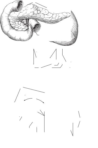
PANCREAS. Organ 13−15 cm in length that lies |
19 |
Accessory pancreatic duct. Ductus pancreati- |
|||
1 |
|
1 |
||||||
|
|
|
partly in the duodenal loop, partly behind the |
|
cus accessorius. Additional excretory duct usu- |
|||
|
|
|
|
omental bursa at the level of L1−2. A B |
|
ally found draining into the minor duodenal |
||
2 |
|
2 |
Head of pancreas. Caput pancreatis. It is nestled |
|
papilla (p. 124.13) above the major duodenal |
|||
|
|
|
|
within the loop of the duodenum. A |
|
papilla. B |
||
3 |
|
3 |
Uncinate process. Processus uncinatus. Hook- |
20 Accessory pancreas. (Pancreas accessorium). |
||||
|
|
Pancreatic tissue dispersed within the wall of |
||||||
|
|
|
|
shaped process passing behind the superior |
|
|||
|
|
|
|
|
the stomach or duodenum. |
|||
4 |
|
|
|
mesenteric vessels. A B |
|
|||
|
|
|
21 Endocrine pancreas. Pars endocrina pancreatis. |
|||||
|
4 |
Pancreatic notch. Incisura pancreatis. Groove |
||||||
|
|
|||||||
|
|
|
Part of the pancreas consisting of about 1 million |
|||||
|
|
|
|
between the uncinate process and the remain- |
|
|||
5 |
|
|
|
|
Langerhans‘ islets, which produce glucagon and |
|||
|
|
|
ing part of the head of the pancreas. A B |
|
||||
|
|
|
|
|
insulin. |
|||
|
|
|
5 Body of pancreas. Corpus pancreatis. Part of the |
|
||||
|
|
|
22 LIVER. Hepar. It is divided into segments on the |
|||||
6 |
|
|
||||||
|
|
|
pancreas lying mainly in front of the vertebral |
|||||
|
|
|
|
basis of the branchings of its blood vessels and |
||||
|
|
|
|
column. It arises from the dorsal anlage of the |
|
biliary ducts. The individual segments are not |
||
7 |
|
|
|
pancreas. A B |
|
|||
|
|
|
|
represented uniformly in the literature. The In- |
||||
|
6 |
Anterior surface. Facies anterior. Anterosupe- |
|
ternational Nomenclature Committee has |
||||
|
|
|||||||
8 |
|
|
|
riorly directed anterior (front) surface of the |
|
adopted Hjortsjö’s classification. C |
||
|
|
|
pancreas. A |
23 Diaphragmatic surface. Facies diaphragmatica. |
||||
|
|
|
|
|||||
|
|
|
|
|
|
|||
9 |
7 |
Posterior surface. Facies posterior. Posteriorly |
|
Surface of liver facing the diaphragm. C |
||||
|
|
|
directed dorsal (back) surface of the pancreas. B |
24 |
Superior part. Pars superior. The cranially |
|||
|
|
|
|
|||||
|
|
|
|
|
|
|||
10 |
8 |
Inferior |
surface. Facies inferior. Anteroinferi- |
|
directed superior portion of the diaphragmatic |
|||
|
|
|
orly directed inferior surface of the pancreas. It |
|
liver surface. C |
|||
|
|
|
|
|
||||
|
|
|
|
is bounded above by the root of the transverse |
25 |
Cardiac impression. Impressio cardiaca. Flat im- |
||
11 |
|
|
|
|||||
|
|
|
mesocolon. A |
|||||
|
|
|
|
pression made by the heart on the left side of the |
||||
|
9 |
Superior |
margin. Margo superior. Superior |
|
liver in front of the inferior vena cava. C |
|||
12 |
|
|
|
margin of the pancreas located between the |
26 |
Anterior part. Pars anterior. The anteriorly |
||
|
|
|
|
anterior and posterior surfaces. A B |
|
directed portion of the diaphragmatic liver sur- |
||
|
|
|
|
|
||||
13 |
10 |
Anterior |
margin. Margo anterior. Anterior |
|
face. C |
|||
|
|
|||||||
|
|
|
|
margin of the pancreas, which corresponds to |
27 |
Right part. Pars dextra. Portion of the diaphrag- |
||
|
|
|
|
|||||
14 |
|
|
|
the line of attachment of the transverse meso- |
|
matic surface directed toward the right side of |
||
|
|
|
colon (p. 178.5). It also forms the lower boun- |
|
the body. C |
|||
|
|
|
|
dary of |
the omental bursa at the posterior |
28 |
Posterior part. Pars posterior. The posteriorly |
|
15 |
|
|
|
abdominal wall. A |
||||
|
|
|
|
directed portion of the diaphragmatic surface. C |
||||
11 |
Inferior margin. Margo inferioris. Inferior mar- |
|
||||||
|
29 |
Bare area. Area nuda. [[Pars affixa]]. Bare por- |
||||||
16 |
|
|
|
gin of the pancreas situated between the lower |
||||
|
|
|
|
tion of the diaphragmatic surface not covered by |
||||
|
|
|
anterior and posterior surfaces. A |
|
||||
|
12 |
Tuber omentale. Prominence on the body of |
|
peritoneum. C |
||||
17 |
30 |
Groove for vena cava. Sulcus venae cavae. Deep |
||||||
|
|
|
pancreas |
near the head. It projects into the |
||||
|
|
|
|
|
groove for reception of the inferior vena cava. C |
|||
|
|
|
|
omental bursa and is caused by the vertebral |
|
|||
18 |
|
|
|
column. A B |
31 |
Fissure for lig. venosum. Fissura ligamenti venosi. |
||
|
13 |
Tail of pancreas. Cauda pancreatis. Upper left |
|
Groove for the venous ligament extending from |
||||
|
|
|||||||
19 |
|
|
|
tail of the pancreas that lies in contact with the |
|
the liver hilum to the inferior vena cava between |
||
|
|
|
|
the caudate lobe and left lobe. C |
||||
|
|
|
|
spleen. A B |
|
|||
|
|
|
|
|
|
|||
2014 Capsule of pancreas. Capsula pancreatis.
15 Exocrine pancreas. Pars exocrina pancreatis.
21The bulk of the pancreas where pancreatic juice is produced.
22 |
16 |
Pancreatic lobule. Lobulus pancreaticus. The |
|
|
macroscopically visible lobule of the pancreas. A |
|
17 |
Pancreatic duct. Ductus pancreaticus. Main ex- |
23 |
||
|
|
cretory duct of the pancreas opening on the |
|
|
greater duodenal papilla together with the bile |
24 |
|
duct. B |
18 Sphincter muscle of pancreatic duct. M.
25sphincter ductus pancreatici. Circular muscle before the duct opening. See p. 135 A
|
|
|
|
|
|
|
|
|
Digestive system |
129 |
|
|
|
|
|
|
|
|
|
|
|
|
|
|
|
|
|
|
|
|
5 |
|
|
|
|
|
|
|
|
1 |
|
|
|
|
12 |
|
9 |
16 |
|
|
|
2 |
|||
|
|
|
|
|
|
|
|
|
|
|
|
|
|
|
|
|
6 |
|
|
|
|
13 |
|
|
|
3 |
|
|
|
|
|
|
|
|
|
|
|
|
|||
4 |
|
|
|
|
|
|
|
|
|
|
|
4 |
|
2 |
|
|
|
|
|
|
|
|
|
|
|
|
|
|
|
|
|
|
|
|
|
|
|
|
|||
3 |
|
|
|
|
|
|
|
|
|
5 |
|||
11 |
8 |
10 |
|
|
|
||||||||
|
|
|
|
||||||||||
6
248.20220.1
|
|
|
|
|
|
|
|
|
|
|
|
|
7 |
|
|
|
|
|
|
|
|
|
|
|
|
|
|
|
|
|
|
|
|
|
|
|
|
|
|
|
8 |
A |
Duodenum and pancreas, anterior view |
|
|
|
|
|
|
|
|||||
|
|
|
|
|
|
|
|
||||||
|
7 |
9 |
12 |
17 |
19 |
|
|
|
|
|
|||
|
|
|
|
9 |
|||||||||
|
|
|
|
|
|
|
|
|
|
|
|
||
|
|
|
|
|
|
|
|
|
|
|
|
||
|
|
|
|
|
|
|
|
|
|
|
|
|
|
|
|
|
|
|
|
|
|
|
|
|
|
|
10 |
13 |
|
|
|
|
|
|
|
|
|
|
|
|
|
|
|
|
|
|
|
|
|
|
|
||||
|
|
|
4 |
|
|
|
|
124.13 |
11 |
||||
|
|
|
|
|
|
|
|
|
|
|
|||
|
|
|
|
|
|
|
|
|
|||||
|
|
|
|
|
|
|
|
|
|
|
|
|
|
|
|
|
|
5 |
17 |
|
|
|
|
124.12 |
12 |
||
|
Pancreas dissected, |
3 |
|
|
|
||||||||
|
|
|
|||||||||||
B |
|
|
|
|
|
|
|
|
|
||||
|
|
|
|
|
|
|
|
|
|||||
|
duodenum fenestrated, |
|
|
|
|
|
|
|
|
|
13 |
||
|
from behind |
|
|
|
|
|
|
|
|
|
|
||
|
|
|
|
|
|
|
|
|
|
||||
|
|
|
|
|
|
|
|
|
|
|
|
|
14 |
|
|
|
|
|
|
|
|
|
|
|
|
|
|
|
|
|
|
|
|
|
|
|
|
|
|
|
|
23 |
|
|
|
|
|
|
|
|
|
|
15 |
||
|
|
|
|
|
|
|
|
|
|
|
|||
178.22 |
|
|
|
|
26 |
|
|
|
|
|
|||
|
|
|
|
|
|
|
|
16 |
|||||
|
|
|
|
|
|
|
|
|
|
|
|
|
|
|
|
|
|
|
|
|
|
|
|
|
|
|
|
25 |
|
|
|
|
|
|
|
|
|
17 |
|||
|
|
|
|
|
|
|
|
|
|
||||
|
|
|
|
|
|
|
|
|
|
|
|
|
|
29 |
|
|
|
|
|
24 |
|
|
|
18 |
|||
|
|
|
|
|
|
|
|
|
|
|
|
|
|
|
|
|
|
|
|
|
|
|
|
|
|
|
19 |
|
|
|
|
|
|
|
|
|
|
|
|
|
|
|
|
|
|
|
|
|
|
|
|
|
|
|
|
|
|
|
|
|
|
|
|
|
|
|
|
|
20 |
|
|
28 |
|
|
|
|
29 |
27 |
|
||||
|
|
|
|
|
|
|
|||||||
|
|
|
|
21 |
|||||||||
|
|
|
|
|
|
|
|
|
|
|
|
|
|
|
|
|
|
|
|
30 |
|
|
|
|
|
|
|
|
|
|
|
|
|
|
|
|
|
|
|
22 |
|
|
|
|
|
|
|
|
|
|
|
|
|
|
|
178.2431
130.26 |
246.24 |
|
23 |
|
|
|
|||
|
Liver, superior view |
|
|
|
C |
|
178.23 |
24 |
|
|
|
|
|
|
|
|
|
|
|
|
|
|
|
|
|
|
|
|
25 |
|
|
|
|
|