Материал: Атлас Ханц фениш
|
110 |
Digestive system |
|
|
|
|
|
|||
|
|
|
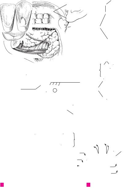
Major salivary glands. Glandulae salivariae |
|
21 Root of tooth. Radix dentis. Portion of the |
|||||
1 |
|
1 |
|
|||||||
|
||||||||||
|
|
|
majores. |
|
|
|
tooth covered by cementum. E |
|
||
2 |
|
2 |
Sublingual gland. Glandula sublingualis. Pre- |
|
22 |
Apex of root of tooth. Apex radicis dentis. E |
||||
|
|
|
dominantly mucous gland situated on the my- |
|
23 Clinical root. Radix clinica. Portion of the tooth |
|||||
|
|
|
|
lohyoid muscle diaphragma oris. It contains |
|
|
situated below the gum. C |
|
||
3 |
|
|
|
several drainage ducts. D |
|
|
|
|||
|
|
|
|
24 |
Occlusal (masticatory) surface. Facies oc- |
|||||
|
3 |
Major sublingual duct. Ductus sublingulis |
|
|||||||
|
|
|
|
clusalis (masticatoria) dentis. B E |
|
|||||
4 |
|
|
|
major. Main drainage duct of the sublingual |
|
25 Vestibular (facial) surface. Facies vestibularis |
||||
|
|
|
gland. It opens at the sublingual caruncle next |
|
||||||
|
|
|
|
|
(facilis) dentis. Tooth surface facing the vesti- |
|||||
|
|
|
|
to the submandibular duct. D |
|
|
||||
|
|
|
|
|
|
bule. D G |
|
|
||
|
|
|
|
|
|
|
|
|||
5 |
4 |
Minor sublingual ducts. Ductus sublinguales |
|
|
|
|
||||
|
25 a Buccal surface. Facies buccalis dentis. Tooth |
|||||||||
|
|
|
|
minores. About 40 small ducts that drain the |
|
|
surface facing the cheek. |
|
|
|
|
|
|
|
|
|
|
|
|||
6 |
|
|
|
sublingual gland and open along the sublingual |
|
|
|
|
||
|
|
|
|
25 b Labial surface. Facies |
labialis |
dentis. Tooth |
||||
|
|
|
fold and the sublingual caruncle. D |
|
||||||
|
|
5 |
Submandibular gland. Glandula submandibu- |
|
|
surface facing the lips. |
|
|
||
7 |
|
|
26 Lingual and palatine surfaces. Facies lingualis/ |
|||||||
|
|
|
laris. Predominantly serous salivary gland lo- |
|
||||||
|
|
|
|
cated almost entirely below the mylohyoid. D F |
|
|
palatalis dentis. Tooth |
surfaces facing the |
||
|
|
|
|
|
|
tongue and palate, respectively. A G |
||||
8 |
|
6 |
Submandibular |
duct. Ductus submandibu- |
|
|
||||
|
|
27 |
Approximal surface. |
Facies |
approximalis. |
|||||
|
|
|
laris. Duct that |
drains the submandibular |
|
|||||
|
|
|
|
|
|
Tooth surface facing the adjacent tooth. G |
||||
9 |
|
|
|
gland. It loops around the posterior margin of |
|
|
||||
|
|
|
|
28 |
Mesial surface. Facies mesialis. Vertical con- |
|||||
|
|
|
the mylohyoid accompanied by glandular |
|
||||||
|
|
|
|
tissue and opens at the sublingual caruncle. D |
|
|
tact surface of a tooth turned away from the last |
|||
10 |
|
7 |
Parotid gland. Glandula parotidea. Salivary |
|
|
molar. G |
|
|
||
|
|
29 |
Distal surface. Facies distalis. Vertical contact |
|||||||
|
|
|
|
gland located behind and on the mandibular |
|
|||||
11 |
|
|
|
ramus. F |
|
|
|
surface of a tooth facing away from the first in- |
||
|
8 |
Superficial part. Pars superficialis. The parts |
|
|
cisor. G |
|
|
|||
|
|
|
29 a Contingent area. Area contingens. Direct contact |
|||||||
|
|
|
|
of the parotid gland located superficial to the |
|
|||||
12 |
|
|
|
facial nerve. F |
|
|
|
surface of adjacent teeth. |
|
|
|
|
9 |
Deep part. Pars produnda. Parts of the parotid |
|
30 Cingulum. Ridge near the neck of a tooth con- |
|||||
13 |
|
|
|
gland located deep with respect to the facial |
|
|
necting both marginal crests at the lingual sur- |
|||
|
|
|
nerve. F |
|
|
|
face of incisor and canine teeth. A |
|||
|
|
|
|
|
|
|
||||
14 |
|
10 |
Accessory parotid gland. Glandula parotidea |
|
31 |
Marginal crest. Crista marginalis. Lateral |
||||
|
|
|
accessoria. Portion of the parotid gland located |
|
|
marginal ridge on the lingual surface of the in- |
||||
|
|
|
|
on the masseter muscle near the parotid ex- |
|
|
cisor and canine teeth which goes over into the |
|||
15 |
|
|
|
cretory duct. F |
|
|
|
cingulum at the neck region. A |
|
|
|
11 |
Parotid duct. Ductus parotideus. Excretory |
|
32 Incisal margin. Margo incisalis. Occlusal edge |
||||||
|
|
|
||||||||
16 |
|
|
|
duct of the parotid gland. It passes around the |
|
|
of incisor and canine teeth. A |
|
||
|
|
|
anterior margin of the masseter and opens near |
|
|
|
|
|
||
17 |
|
|
|
the second upper molar tooth. F |
|
|
|
|
|
|
|
12 |
TEETH. Dentes. A B C D E G |
|
|
|
|
|
|||
|
|
13 Crown of tooth. Corona dentis. Portion of the |
|
|
|
|
|
|||
18 |
|
|
|
tooth covered by enamel. E |
|
|
|
|
|
|
|
|
14 Cusp of tooth. Cuspis dentis [[tuberculum]]. 1− |
|
|
|
|
|
|||
19 |
|
|
|
5 protuberances |
on the occlusal surface of |
|
|
|
|
|
|
|
|
tooth (with the exception of the incisors). E |
|
|
|
|
|
||
|
|
|
|
|
|
|
|
|
||
20 |
|
15 Apex of cusp. Apex cuspidis. E |
|
|
|
|
|
|||
|
16 |
Dental tubercle. Tuberculum dentis. Distinct |
|
|
|
|
|
|||
21 |
|
|
|
eminence on the side of the crown, especially |
|
|
|
|
|
|
|
|
|
in canine and incisor teeth. A |
|
|
|
|
|
||
|
|
17 Transverse ridge. Crista transversalis. Trans- |
|
|
|
|
|
|||
22 |
|
|
|
verse connecting ridge between adjacent |
|
|
|
|
|
|
|
|
|
|
cusps. B |
|
|
|
|
|
|
23 |
|
18 Triangular ridge. Crista triangularis. Triangu- |
|
|
|
|
|
|||
|
|
|
lar connecting ridge between the cusps of the |
|
|
|
|
|
||
24 |
|
|
|
molars. B |
|
|
|
|
|
|
|
19 |
Clinical crown. Corona clinica. Portion of the |
|
|
|
|
|
|||
|
|
|
|
tooth projecting above the gum. C |
|
|
|
|
|
|
25 |
|
20 |
Neck of tooth. Cervix [[collum]] dentis. Portion |
|
|
|
|
|
||
|
|
|
|
of the tooth at the enamel-cementum border. E |
|
|
|
|
|
|
|
|
|
|
|
|
|
|
|
|
|
|
|
|
|
|
|
|
|
|
|
|
|
|
|
|
Digestive system |
111 |
|
|
|||||||||
|
|
|
|
|
|
|
|
|
|
|
|
|
|
|
|
|
|
|
|
|
|
|
|
|
|
|
|
32 |
|
|
|
|
|
|
|
|
|
|
|
|
19 |
|
|
|
|
|
|
1 |
|||||||
|
|
|
|
24 |
|
|
|
17 |
|
|
|
|
|
|
|
|
|
|
|
|
|
||||||
|
|
|
|
|
|
|
|
|
|
|
|
|
|
|
|
|
|
|
|
|
|
2 |
|||||
|
|
|
|
|
|
|
|
|
|
|
|
|
|
|
|
|
|
|
|
|
|
|
|||||
26 |
|
26 |
|
31 |
|
|
|
|
|
|
|
|
|
|
|
|
|
|
|
||||||||
|
|
|
|
|
|
|
|
|
|
|
|
|
|
|
|
|
|
|
|
||||||||
|
|
|
|
|
|
|
|
|
|
|
|
|
|
|
|
|
|
|
|
3 |
|||||||
|
|
|
|
|
16 |
|
|
|
|
18 |
|
|
|
23 |
|
|
|
|
|
|
|
|
|
|
|||
|
|
|
|
|
|
|
|
|
|
|
|
|
|
|
|
|
|
|
|
|
4 |
||||||
|
|
|
|
|
|
|
|
|
|
|
|
|
|
|
|
|
|
|
|
|
|
|
|||||
|
|
|
|
|
|
|
|
|
|
|
|
|
|
|
|
|
|
|
|
|
|
|
|
|
|
|
|
|
|
|
|
|
|
|
|
|
|
|
|
|
|
|
|
|
|
|
|
|
|
|
|
|
|||
30 |
|
|
|
|
|
|
|
|
|
|
|
|
|
|
|
|
|
|
|
|
|
|
|
|
5 |
||
|
|
|
|
|
|
|
|
|
|
|
|
|
|
|
|
|
|
|
|
|
|
|
|
|
|||
|
|
|
|
|
|
|
|
|
|
|
|
|
|
|
|
|
|
|
|
|
|
|
|
|
|
|
|
|
|
|
|
|
|
|
|
|
|
|
|
|
|
|
|
|
|
|
|
|
|
|
|
|
|
||
|
|
|
|
|
|
|
|
|
|
|
|
|
|
|
|
|
|
|
|
|
|
|
|
|
|
6 |
|
|
Incisor tooth and canine tooth, |
|
|
First and second molars, |
|
Incisor, sagittal |
|
|
|
|
|
||||||||||||||||
A |
|
B |
C |
|
|
|
|
|
|||||||||||||||||||
|
|
|
|
|
|||||||||||||||||||||||
|
lingual surface |
|
|
|
|
|
|
occlusal surface |
|
section |
|
|
|
|
|
|
7 |
||||||||||
|
|
|
|
|
|
|
|
|
|
|
|
|
|
|
|
|
|
|
|
|
|
|
|
|
|
|
|
|
|
|
|
|
|
|
|
|
|
|
|
|
|
|
|
|
|
|
|
|
|
|
|
|
|
||
|
|
|
|
|
|
|
|
|
|
|
|
|
|
|
|
14 |
15 |
|
|
|
8 |
||||||
|
|
|
|
|
|
|
|
|
|
|
|
|
|
|
|
|
|
|
|
|
|||||||
|
|
|
|
|
|
|
|
|
|
|
|
|
|
|
|
|
|
|
|
|
|
|
|
|
|
||
|
|
|
|
|
|
|
|
|
|
|
|
|
|
|
|
13 |
|
|
|
|
|
|
|
24 |
|
9 |
|
|
|
|
|
|
|
|
|
|
|
|
|
|
|
|
|
|
|
|
|
|
|
|
|
||||
|
|
|
|
|
|
|
|
|
|
|
|
|
|
|
|
|
|
|
|
|
|
|
|
|
|||
|
|
|
|
|
|
|
|
|
|
|
|
|
|
|
|
|
|
|
|
|
|
|
|||||
|
|
|
|
|
|
|
|
|
|
|
|
|
|
|
|
|
|
|
|
|
|
|
|
|
|||
|
|
|
|
|
|
|
|
|
|
|
|
|
|
|
|
|
|
|
|
|
|
10 |
|||||
|
|
|
|
|
|
|
|
|
|
|
|
|
|
|
|
|
|
|
|
|
|
|
|
|
|
||
25 |
|
|
|
|
|
|
|
|
|
|
|
|
|
|
|
|
|
|
|
|
|
|
|||||
|
|
|
|
|
|
|
|
|
|
|
20 |
|
|
|
|
|
|
|
|
|
|
|
|||||
|
|
|
|
|
|
|
|
|
|
|
|
|
|
|
|
|
|
|
|
|
|
||||||
|
|
|
|
|
|
|
|
|
|
|
|
|
|
|
|
|
|
|
|
|
|
|
|
|
|
||
|
|
|
|
|
|
|
|
|
|
|
4 |
|
|
|
|
|
|
|
|
|
|
11 |
|||||
|
|
|
|
|
|
|
|
|
|
|
|
|
|
|
|
|
|
|
|
|
|||||||
|
|
|
|
|
|
|
|
|
|
|
|
|
|
|
|
|
|
|
|
|
|
||||||
|
|
|
|
|
|
|
|
|
|
|
|
|
|
|
|
|
|
|
|
|
|
|
|
||||
|
|
|
|
|
|
|
|
|
|
|
|
|
|
|
|
|
|
|
|
|
|
|
|
|
|
||
3 |
|
|
|
|
|
|
|
2 |
|
|
|
|
21 |
|
|
|
|
|
|
|
|
|
12 |
||||
|
|
|
|
|
|
|
|
|
|
|
|
|
|
|
|
|
|
|
|
|
|
||||||
|
|
|
|
|
|
|
|
|
|
|
|
|
|
|
|
|
|
|
|
|
|
|
|
|
|
|
|
|
|
|
|
|
|
|
|
|
|
|
|
|
|
|
|
|
|
|
|
|
|
|
|
|
|
13 |
|
|
|
|
|
|
|
|
|
|
|
|
|
|
|
|
22 |
|
|
|
|
|
|
|
|
|
|
|
|
|
|
|
|
|
|
|
|
|
|
|
|
|
|
|
|
|
|
|
|
|
|
||||||
|
|
|
|
|
|
|
|
|
|
|
|
|
|
|
|
|
|
|
|
|
|
|
|
|
|||
|
|
|
|
|
|
|
|
|
|
|
|
|
|
|
|
|
|
|
|
|
14 |
||||||
|
Oral cavity, |
|
|
|
|
|
|
|
|
|
|
|
First lower molar |
|
|
|
|||||||||||
D |
|
|
|
|
|
|
|
|
|
|
E |
|
|
|
|
|
|||||||||||
|
|
|
|
|
|
|
|
|
|
|
|
|
|
|
|||||||||||||
|
medial view |
|
|
|
|
|
|
|
|
|
|
|
|
|
|
|
|
|
|
|
|
|
|
15 |
|||
|
|
|
|
|
|
|
|
|
|
|
|
|
|
|
|
|
|
|
|
|
|
|
|
|
|
|
|
65
|
|
|
|
|
|
|
|
|
|
|
|
|
|
16 |
|
|
|
|
|
|
|
|
|
|
|
|
|
|
|
|
|
|
|
|
|
|
|
|
|
|
|
|
|
17 |
|
|
|
10 |
|
|
|
|
|
|
|
|
|
|
|
|
|
|
|
|
|
|
|
|
|
|
|
|
|
|
11 |
|
|
8 |
|
|
|
18 |
|||||||
|
|
|
|
|
|
|
|
|
|
|
|
|||
|
|
|
|
|
|
|
|
|
|
|||||
|
|
|
|
|
|
|
|
|
||||||
|
|
|
7 |
|
|
|
|
|
|
|
||||
|
|
|
|
|
|
|
|
|
|
|||||
|
|
|
|
|
|
19 |
||||||||
|
|
|
|
|
|
|
|
|
|
|
|
|
|
|
|
|
|
|
|
9 |
|
|
|
|
|
|
|
|
|
|
|
|
|
|
|
|
|
|
|
|
|
|||
|
|
|
|
|
27 |
20 |
||||||||
|
|
|
|
|
|
|
|
|
|
|
|
|
||
|
|
|
28 |
29 |
|
|
||||||||
|
|
|
|
|
|
|||||||||
|
|
|
|
|
21 |
|||||||||
|
|
|
|
|
|
|
|
|
|
26 |
|
|
|
|
|
|
|
|
|
|
|
|
|
|
|
|
|
|
|
|
|
|
|
|
|
|
|
|||||||
|
|
|
5 |
|
|
|
|
|
|
|
29 |
|
|
22 |
|
|
|
|
|
|
|
|
|
|
|
|
|
|
|
|
|
|
|
|
|
|
|
|
|
|
|
|
|
|
|
|
|
|
|
|
|
|
|
|
|
29 |
|
|
23 |
|
|
|
|
|
|
|
|
|
|
|
|
|
|
|
|
|
|
|
|
|
|
|
|
|
|
|
|
|
24 |
F Salivary glands, lateral view |
|
|
|
|
|
|
G Teeth of lower jaw |
|
||||||
|
||||||||||||||
25

|
112 |
Digestive system |
|
|
|
||||
|
|
|
Pulp cavity of tooth. Cavitas dentis (pulparis). |
20 |
Incisor teeth. Dentes incisivi. Cutting teeth lo- |
||||
1 |
|
1 |
|||||||
|
|
|
Cavity in the dentin. Towards the root, it be- |
|
cated on both sides of the midline at the 1st and |
||||
2 |
|
|
|
comes continuous with the root canal. A |
|
|
2nd positions of the dental arch. D |
||
|
2 |
Pulp chamber in the crown. Cavitas coronae. |
21 |
Canine teeth. Dentes canini. Teeth located at |
|||||
3 |
|
|
|
Crown portion of the pulp cavity. A |
|
|
the 3rd position of the dental arch. D |
||
|
3 |
Root canal of tooth. Canalis radicis dentis. |
22 |
Premolar teeth. Dentes premolares. Teeth oc- |
|||||
|
|
|
|
Canal between the pulp cavity and the apical |
|
cupying the 4th and 5th positions of the dental |
|||
4 |
|
|
|
foramen. A |
|
|
|
arch. D |
|
|
4 |
Apical foramen of root of tooth. Foramen apicis |
23 |
Molar teeth. Dentes molares. Teeth located at |
|||||
|
|
||||||||
|
|
|
|
radicis dentalis. The opening of the root canal at |
|
the 6th, 7th and 8th positions of the dental arch. D |
|||
5 |
|
|
|
|
|||||
|
|
|
the apex of the root. A |
|
24 |
Wisdom tooth. Dens serotinus [molaris ter- |
|||
|
|
|
5 Pulp of tooth. Pulpa dentis. Contents of the |
|
tius]. Tooth located at the 8th position of the |
||||
6 |
|
|
|
||||||
|
|
|
pulp cavity consisting of loose, finely fibered |
|
dental arch. D |
||||
|
|
|
|
connective tissue, blood vessels and nerves. |
25 Deciduous (milk) teeth. Dentes decidui. |
||||
|
|
|
|
||||||
7 |
6 |
Crown pulp. Pulpa coronalis. Pulp within the |
|||||||
26 Permanent teeth. Dentes permanentes. Teeth |
|||||||||
|
|
|
|
crown portion of the pulp cavity. |
|
||||
|
|
|
|
|
|
that develop after the deciduous teeth. |
|||
|
7 |
Root pulp. Pulpa radicularis. Pulp within the |
|
||||||
8 |
|
||||||||
27 |
Diastema. Space between adjacent teeth. |
||||||||
|
|
|
root canal portion of the pulp cavity. |
|
|||||
|
8 |
Dental papilla. |
Papilla dentis. A mass |
of |
28 TONGUE. Lingua. D E |
||||
9 |
|||||||||
|
|
||||||||
|
|
|
mesenchyme present in the bell stage of tooth |
29 |
Body of tongue. Corpus linguae. Portion of the |
||||
|
|
|
|
||||||
|
|
|
|
development. B |
|
|
|
tongue situated between the apex and root. E |
|
10 |
|
|
|
|
|
|
|||
9 |
Dentine. Dentinum [[substantia eburnea]]. Pre- |
30 |
Root of tongue. Radix linguae. Anchoring re- |
||||||
|
|
|
|
dominant mass of a tooth consisting of inor- |
|
gion of the tongue at the mandible and hyoid |
|||
11 |
|
|
|
ganic and organic material (especially col- |
|
bone. Also the posterior, vertical segment of the |
|||
|
|
|
|
lagenous fibers). A C |
|
|
tongue. E |
||
|
|
|
|
|
|
||||
12 |
10 |
Enamel. Enamelum [[substantia adamantina]]. |
31 |
Dorsum of tongue. Dorsum linguae. E |
|||||
|
|
|
|
The extremely hard substance surrounding the |
32 |
Anterior, oral portion of tongue. Pars pre- |
|||
|
|
|
|
crown of the tooth like a mantle. A C |
|
||||
13 |
|
|
|
|
|
sulcalis (anterior). The part of the dorsum of |
|||
|
11 GOMPHOSIS. Type of fibrous joint in which a |
|
|||||||
|
|
the tongue situated anterior to the sulcus ter- |
|||||||
|
|
|
|||||||
|
|
|
|
conical process, e. g., a tooth, is inserted into a |
|
minalis. 115 B |
|||
14 |
|
|
|
|
|||||
|
|
|
socket, e. g., alveolus of the jaw (dentoalveolar |
33 |
Posterior, pharyngeal part of tongue. Pars |
||||
|
|
|
|
articulation). |
|
|
|||
|
|
|
|
|
|
|
postsulcalis (posterior). The vertical portion of |
||
|
12 |
Periodontium. Tissues that invest and support |
|
||||||
15 |
|
the dorsum of the tongue between the sulcus |
|||||||
|
|
|
|
the tooth within the alveolus. It consists of the |
|
terminalis and the epiglottis. 115 B |
|||
|
|
|
|
following parts: A |
|
34 Inferior surface of tongue. Facies inferior lin- |
|||
16 |
|
|
|
|
|||||
13 |
Periodontium |
protectoris [[gingiva]]. |
The |
||||||
|
guae. D E |
||||||||
|
|
|
|
outer part of the periodontium with the exter- |
35 |
Fimbriated fold. Plica fimbriata. Serrated fold |
|||
17 |
|
|
|
||||||
|
|
|
nal border epithelium. A |
|
|||||
|
|
|
|
|
lateral to the frenulum. It is a remnant of the in- |
||||
|
14 |
Periodontium insertionis. Portion of the peri- |
|
||||||
|
|
ferior tongue. D |
|||||||
18 |
|
||||||||
|
|
|
odonium touching the tooth. It consists of the |
36 Lateral margin of tongue bordering the teeth. |
|||||
|
|
|
|
inner border epithelium and the periodontal |
|||||
|
|
|
|
ligament. A |
|
|
|
Margo linguae. D |
|
19 |
|
|
|
|
|
|
|||
|
|
|
|
|
|
|
|||
15 |
Periodontal ligament. Desmodontium. All con- |
37 |
Apex of tongue. Apex linguae. D E |
||||||
|
|
|
|
nective tissue fibers which are anchored in the |
|
|
|||
20cementum and, with their vessels and nerves, extend partly into the gum and partly into the
alveolar wall. A
21
16Cementum. Substance similar to bone. It surrounds the tooth from the enamel border to the
22apex of the root and receives the fibers of the periodontal ligament. A
2317 Alveolar bone. Os alveolare. Bony wall of the alveolus. A
2418 Superior dental arch. Arcus dentalis superior. Curved row of teeth of the maxilla.
2519 Inferior dental arch. Arcus dentalis inferior. Curved row of teeth of the mandible.
Digestive system 113
|
|
|
|
|
|
|
|
|
|
|
|
|
|
|
|
|
|
|
|
|
|
|
|
|
|
|
|
|
|
|
1 |
|
|
|
|
|
|
|
|
|
|
|
|
|
|
|
|
|
|
|
|
|
|
|
|
|
|
|
|
|
|
|
2 |
|
|
|
|
|
10 |
108.25 |
|
|
|
|
|
|
|
|
|
|
|
|
|
|
3 |
||||||||||
12 |
|
2 |
|
|
|
|
|
|
|
|
14 |
|
|
|
|
|
|
|
|
|
|
|
|
4 |
|||||||
|
|
|
|
|
9 |
|
|
|
|
|
|
|
|
|
|
|
|
|
|
|
|
|
|||||||||
|
|
|
|
|
|
|
|
13 |
|
|
|
|
|
|
|
|
|
|
|
|
|
||||||||||
|
|
|
|
|
|
|
|
|
|
|
|
|
|
|
|
|
|
|
|
|
|
|
|
|
|
|
|
|
|
||
|
|
|
|
|
|
|
|
|
|
|
|
|
|
|
|
|
|
|
|
|
|
|
|
|
5 |
||||||
|
|
|
|
|
|
|
|
|
|
|
|
|
|
|
|
|
|
|
|
|
|
|
|
|
|
|
|
|
|
|
|
|
|
|
|
|
|
|
|
|
|
|
|
|
|
|
|
|
|
|
|
|
|
|
|
|
|
|
|
|
|
|
|
|
|
|
|
|
|
|
|
|
|
|
|
|
|
|
|
|
|
|
|
|
|
|
|
|
|
|
|
|
|
|
|
|
|
|
|
|
1 |
|
|
|
|
|
|
|
|
|
|
|
|
|
|
|
|
|
|
|
|
|
6 |
||||
15 |
|
|
|
|
|
|
|
|
|
|
|
|
|
|
|
|
17 |
|
|
|
8 |
|
|
|
7 |
||||||
|
|
|
|
|
|
|
|
|
|
|
|
|
|
|
|
|
|
|
|
|
|
||||||||||
|
|
|
|
|
|
|
|
|
|
|
|
|
|
|
|
|
|
|
|
|
|
|
|
|
|
||||||
|
|
|
|
|
|
|
|
|
|
|
|
|
|
|
|
|
|
|
|
|
|
|
|
|
|
|
8 |
||||
|
|
|
|
|
|
|
|
|
|
|
|
|
|
|
|
|
|
|
|
|
|
|
|
|
|
|
|||||
|
|
|
|
|
|
|
|
|
|
|
|
|
|
|
|
|
|
|
|
|
|
|
|
|
|
|
|
|
|
|
|
|
|
|
|
|
|
|
|
|
|
|
|
|
|
|
|
|
|
16 |
|
|
|
|
|
|
|
|
9 |
||||
|
|
|
|
|
|
|
|
|
|
|
|
|
|
|
|
|
|
|
|
|
|
|
|
|
|
||||||
|
|
|
|
|
|
|
|
|
|
|
|
|
|
|
|
|
|
|
|
|
|
|
|
|
|
|
|
|
|
|
|
|
|
|
|
|
3 |
|
|
|
|
|
|
|
|
|
|
|
|
|
B |
Tooth development |
|
|
|
||||||||
|
|
|
|
|
|
|
|
|
|
|
|
|
|
|
|
|
|
|
|
|
|
||||||||||
|
|
|
|
|
|
|
|
|
|
|
|
|
|
|
|
|
|
|
|
|
|
|
|
|
|
|
|
|
|
|
10 |
|
|
|
|
|
|
|
|
|
|
|
|
|
|
|
|
|
|
|
|
|
|
|
|
|
|
|
|
|
|
|
11 |
|
|
|
|
|
|
|
|
|
|
|
|
|
|
|
|
|
|
|
|
|
|
|
|
|
|
|
|
|
|
|
|
|
|
|
|
|
4 |
|
|
|
|
|
|
|
|
|
|
|
|
|
|
|
|
|
|
|
|
12 |
|||||
|
|
|
|
|
|
|
|
|
|
|
|
|
|
|
|
|
|
|
|
|
|
|
|
|
|
|
|
|
|
|
|
|
|
|
|
|
|
|
|
|
|
|
|
|
|
|
|
|
|
|
|
|
|
|
|
|
|
|
9 |
10 |
|
|
|
|
A |
Longitudinal section of tooth |
|
|
|
|
|
|
|
|
|
|
|
|
13 |
||||||||||||||||
|
|
|
|
|
|
|
|
|
|
|
|
|
|
|
|||||||||||||||||
|
|
|
|
|
|
|
|
|
|
|
|
|
|
||||||||||||||||||
|
|
|
|
|
|
|
|
|
|
|
|
|
|
|
|
|
|
|
|
|
|
|
|
|
|
|
|
|
|
|
|
|
|
|
|
|
20 |
|
|
|
|
|
|
|
|
|
|
|
|
|
|
|
|
|
|
|
14 |
||||||
|
|
|
|
|
|
|
|
|
|
|
|
|
|
|
|
|
|
|
|
|
|
|
|
|
|
|
|
|
|
|
|
|
|
|
|
|
|
|
|
|
|
|
|
|
|
|
|
|
|
|
|
|
|
|
|
|
Enamel-dentin border |
|
|
|
|
||
|
|
|
|
|
37 |
|
|
|
|
|
|
|
|
|
|
21 |
|
C |
|
|
|
15 |
|||||||||
|
|
|
|
|
|
|
|
|
|
|
|
|
|
|
|
|
|
|
|
|
|
|
|||||||||
|
|
|
|
|
|
|
|
|
|
|
|
|
|
|
|
|
|
|
|
|
|
|
|||||||||
35 |
|
|
|
|
|
|
|
|
|
|
|
|
|
|
|
|
|
31 |
|
|
|
16 |
|||||||||
|
|
|
|
|
|
|
|
|
|
|
|
|
|
|
|
|
|
|
|
|
|
|
|
|
|
|
|||||
|
|
|
34 |
|
|
|
|
|
|
|
|
|
|
|
|
|
|
|
|||||||||||||
36 |
|
|
|
|
|
|
|
|
|
|
|
|
|
|
|
|
|
|
|
|
17 |
||||||||||
|
|
|
|
|
|
|
|
|
|
|
|
|
|
|
|
|
|
|
|
|
|
|
|
|
|
|
|
|
|||
|
|
|
|
|
|
|
|
|
|
|
|
|
|
|
|
|
|
|
|
|
|
|
|
|
|
|
|
|
|
|
|
23 |
|
|
|
|
|
|
|
114.2 |
|
|
|
|
|
|
|
24 |
37 |
|
29 |
30 |
|
18 |
|||||||||
|
|
|
|
|
|
|
|
|
|
|
|
|
|||||||||||||||||||
|
|
|
|
|
|
|
|
|
|
|
|
|
|
|
|
|
|
|
|
|
|
||||||||||
|
|
|
|
|
|
|
|
|
|
|
|
|
|
|
|
|
|
|
|
|
|
||||||||||
|
|
|
|
|
|
|
|
|
|
|
|
|
|
|
|
|
|
|
|
|
|
|
|
|
|
||||||
|
|
|
|
|
|
|
|
|
|
|
|
|
|
|
|
|
|
|
|
|
34 |
|
|
|
|
19 |
|||||
|
|
|
|
|
|
|
|
|
|
|
|
|
|
|
|
|
|
|
|
|
|
|
|
|
|||||||
|
|
|
|
|
|
|
|
|
|
|
|
|
|
|
|
|
|
|
|
|
|
|
|
|
|
|
|
|
|||
22 |
|
|
|
|
|
|
|
|
|
|
|
|
|
|
|
|
|
|
|
21 |
|
|
|
|
|
|
20 |
||||
|
|
|
|
|
|
|
|
|
|
|
|
|
|
|
|
|
|
|
|
|
|
|
|
|
|
|
|||||
|
|
|
|
|
|
|
|
|
|
|
|
|
|
|
|
|
|
|
|
|
|
|
|
|
|
|
|||||
21 |
|
|
|
20 |
|
|
|
|
|
|
|
|
|
|
|
|
|
|
|
|
|
21 |
|||||||||
|
|
|
|
|
|
|
|
|
|
|
|
|
|
|
|
|
|
|
|
||||||||||||
|
|
|
|
|
|
|
|
|
|
|
|
|
|
|
|
|
|
|
|
|
|
||||||||||
|
|
|
|
|
|
|
|
|
|
|
|
|
|
|
|
|
|
|
|
|
|
|
|
||||||||
|
|
|
|
|
|
|
|
|
|
|
|
|
|
|
|
|
|
|
|
|
|
|
|
|
|
|
|
|
|
|
22 |
|
|
Mouth with tongue elevated |
|
|
|
|
|
|
|
|
Tongue, sagittal section |
|
|
|
|
||||||||||||||||
|
D |
|
|
|
|
|
|
|
E |
|
|
|
23 |
||||||||||||||||||
|
|
|
|
|
|
|
|
|
|
|
|
|
|
|
|
|
|
|
|
|
|
|
|
|
|
|
|
|
|
|
24 |
|
|
|
|
|
|
|
|
|
|
|
|
|
|
|
|
|
|
|
|
|
|
|
|
|
|
|
|
|
|
|
25 |
|
|
|
|
|
|
|
|
|
|
|
|
|
|
|
|
|
|
|
|
|
|
|
|
|
|
|
|
|
|
|
|

|
114 |
Digestive system |
|
|
|
|
|
|||
|
|
|
|
Mucous membrane of tongue. Tunica mucosa |
18 |
TONGUE MUSCULATURE. Musculi linguae (lin- |
||||
1 |
|
1 |
|
|||||||
|
|
|
|
linguae. C |
|
|
guales). The following eight tongue muscles are |
|||
2 |
|
2 |
|
Frenulum of tongue. Frenulum linguae. Mu- |
|
innervated by the hypoglossal nerve (XII). |
||||
|
|
19 |
M. genioglossus. o: Mental spine of mandible. |
|||||||
|
|
|
|
cosal fold extending from the floor of the |
||||||
|
|
|
|
|
mouth to the inferior side of the tongue. D; see |
|
i: Fan-shaped distribution within the tongue |
|||
3 |
|
|
|
|
also p. 113. D |
|
|
from the apex to the base. A: It pulls the tongue |
||
|
3 |
|
Lingual papillae. Papillae linguales. Collective |
|
forward or towards the |
chin. I: Hypoglossal |
||||
|
|
|
|
nerve. C D |
|
|
||||
4 |
|
|
|
|
term for the following five different types of |
|
|
|
||
|
|
|
|
20 |
M. hyoglossus. o: Body and greater horn of |
|||||
|
|
|
|
mucosal formations: A B |
|
|||||
|
|
4 |
Filiform papillae. Papillae filiformes. Fine, al- |
|
hyoid bone. i: Coming from below, it radiates |
|||||
|
|
|
into the lateral parts of the tongue and pene- |
|||||||
5 |
|
|
|
|
most threadlike epithelial elevations on a con- |
|
||||
|
|
|
|
|
trates up to the mucosa. A: It draws the base of |
|||||
|
|
|
|
|
nective tissue core. Their tips that are often |
|
||||
|
|
|
|
|
cleft. A |
|
|
the tongue backward and downward. I: Hypo- |
||
6 |
|
|
|
|
|
|
glossal nerve. D |
|
|
|
5 |
|
Conical papillae. Papillae conicae. Special |
|
|
|
|||||
|
|
21 M. chondroglossus. o: Lesser horn of hyoid. i: |
||||||||
|
|
|
|
|
form of filiform papilla. They are somewhat |
|||||
|
|
|
|
|
|
Same as hyoglossus. I: Hypoglossal nerve. D |
||||
7 |
|
|
|
|
larger, longer and exhibit conical apices which |
|
||||
|
|
|
|
22 |
M. styloglossus. o: Styloid process. i: Coming |
|||||
|
|
|
|
|
bend backwards. A |
|
||||
|
|
|
|
|
|
|||||
|
6 |
|
Fungiform papillae. Papillae |
fungiformes. |
|
from behind and above, it radiates to the lateral |
||||
8 |
|
|
||||||||
|
|
parts of the tongue and interweaves with the |
||||||||
|
|
|
|
Mushroom-like papillae which are not pointed |
|
|||||
|
|
|
|
|
at the tip, but terminate with a small plateau. A |
|
hyoglossus. A: It draws the tongue backward |
|||
9 |
|
|
|
|
|
and upward. I: Hypoglossal nerve. D |
|
|||
|
|
|
|
B |
|
|
|
|||
|
|
|
|
|
23 Superior longitudinal muscle of tongue. M. |
|||||
|
7 |
Vallate (circumvallate) papillae. Papillae val- |
||||||||
|
||||||||||
10 |
|
|
|
|
latae. 7−12 large papillae located in front of the |
|
longitudinalis superior. Longitudinal bundles of |
|||
|
|
|
|
|
muscle just below the mucosa that extend from |
|||||
|
|
|
|
sulcus terminalis. They are circular in cross- |
|
|||||
|
|
|
|
|
section and are surrounded by a moat, the wall |
|
the apex of tongue to the region of the hyoid |
|||
11 |
|
|
|
|
|
bone. I: Hypoglossal nerve. C |
|
|||
|
|
|
|
of which contains taste buds. A B |
|
|
|
|||
|
8 |
Lenticular papillae. Papilla lentiformes. Short |
24 |
Inferior longitudinal muscle of tongue. M. |
||||||
|
||||||||||
12 |
|
|
|
|
fungiform papillae. A |
|
|
longitudinalis inferior. |
Longitudinal |
fibrous |
|
|
|
|
|
|
system situated close to the inferior surface of |
||||
|
9 |
|
Foliate papilla. Papillae foliatae. Several paral- |
|
||||||
|
|
|
the tongue. It passes from the base to the apex |
|||||||
13 |
|
|
|
|
lel folds containing taste buds |
at the post- |
|
of the tongue. I: Hypoglossal nerve. C |
|
|
|
|
|
|
erolateral margin of the tongue. B D |
|
|
||||
|
|
|
|
|
25 Transverse muscle of tongue. M. transversus |
|||||
|
|
10 Median groove of tongue. Sulcus medianus |
||||||||
14 |
|
|
linguae. Transversely oriented muscle |
fibers |
||||||
|
|
|
|
linguae. Shallow, median longitudinal groove |
|
|||||
|
|
|
|
|
extending between the longitudinal system of |
|||||
|
|
|
|
|
situated above the lingual septum. B C |
|
fibers. o: Lingual septum. i: Mucous membrane |
|||
15 |
|
|
|
|
|
|||||
11 |
|
Sulcus terminalis [[”V” linguae]]. Bilateral |
|
along lateral margins of tongue. A: Extension of |
||||||
|
|
|
|
|
groove passing obliquely forward from the |
|
tongue together with the vertical muscle of the |
|||
|
|
|
|
|
|
|||||
16 |
|
|
|
|
foramen caecum. It lies behind the row of val- |
|
tongue. I: Hypoglossal nerve. C |
|
||
|
|
|
|
late papillae which run parallel to it. B |
26 |
Vertical muscle of tongue. M. verticalis lin- |
||||
|
|
|
|
|
||||||
|
12 |
|
Foramen caecum linguae. Pit situated at the |
|
guae. Vertical muscle fibers coursing from the |
|||||
17 |
|
|
||||||||
|
|
|
|
apex of the sulcus terminalis. It is the embryo- |
|
back of the tongue to the inferior surface. I: Hy- |
||||
|
|
|
|
|
logical remains of the thyroglossal duct. B |
|
poglossal nerve. C |
|
|
|
|
|
|
|
|
|
|
|
|||
18 13 Thyroglossal duct. Ductus thyroglossalis. Embryological connection between thyroid gland and tongue. At the site of the future fora-
19men caecum, it extends downward from the base of the tongue as an epithelial cone.
2014 Lingual tonsil. Tonsilla lingualis. Accumulation
|
of lymphatic tissue (lingual follicles) which is |
|
|
irregularly distributed over the root of the |
|
21 |
||
tongue. B D |
||
|
|
15 Lingual follicles. Folliculi linguales. Dome-
22shaped protrusions of the mucosa, 1−5 mm in diameter, caused by masses of lymphatic tissue beneath them. Each has a central crypt. A
2316 Lingual septum. Septum lingualis. Connective tissue plate with a special fibrous architecture
24located in the midsagittal plane. C
17 Lingual aponeurosis. Aponeurosis lingualis.
25Stout connective tissue framework of the tongue between the muscles and the mucosa. C