Материал: Атлас Ханц фениш
Synovial bursae and sheaths 105
4
|
4 |
|
|
|||
|
|
|
|
|
3 |
|
|
|
|||||
|
|
|
|
|
|
|
|
2 |
|
|
|
|
|
2 |
|
|
1 |
|
|
|
|
|
|
||||
|
|
|
|
|
|
|
|
3 |
|
|
|||
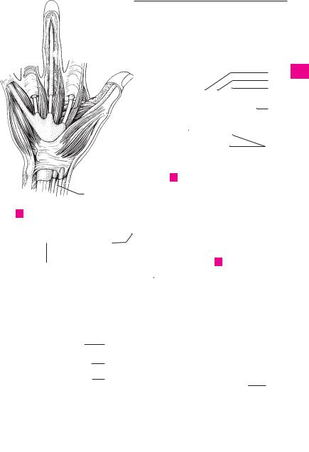
A Palmar view of hand
20
7 
8
 18
18
 17
17  19
19
22
21
23
9
8
7
10 
 11
11
5
|
6 |
|||
13 |
|
|
|
|
|
|
|
||
16 |
|
|
|
|
12 |
||||
|
||||
BDeep hip region, dorsal view
14
C Hip joint, anterior view
 15
15
24
D |
Knee, sagittal section sawed open |
E |
Knee, anterior view |
1
2
3
4
5
6
7
8
9
10
11
12
13
14
15
16
17
18
19
20
21
22
23
24
25

106
1 |
|
1 Anserine bursa. B. anserina. Synovial bursa on |
15 Common tendon sheath for peroneal muscles. |
|
|
|
the tibial collateral ligament below the tendons |
Vag. tendinum mm. peroneorum (fibularium) |
|
|
|
|
of the semitendinosus, gracilis and sartorius |
communis. It lies below the peroneal reti- |
2 |
|
|
muscles. It occasionally communicates with |
naculum and extends to the cuboid bone. C |
|
|
|
the subtendinous bursa of the sartorius. A |
16 Subtendinous bursa of tibialis anterior |
3 |
|
2 Inferior subtendinous bursa of biceps femoris |
muscle. B. subtendinea m. tibialis anterioris. |
|
|
|
muscle. B. subtendinea m. bicipitis femoris in- |
Synovial bursa between the tibialis anterior |
|
|
|
|
ferior. Synovial bursa located partially on the |
tendon and the medial cuneiform bone. D |
|
|
|
||
4 |
|
|
fibular collateral ligament below the tendon of |
17 Subcutaneous calcaneal bursa. B. sucutanea |
|
|
|
insertion of the biceps femoris. B |
calcanea. Synovial bursa between the skin and |
|
|
|
||
5 |
3 |
Subpopliteal recess. Recessus subpopliteus |
the posterior surface of the calcaneus. D |
|
|
|
[bursa m. poplitei]. Synovial bursa on the |
18 Bursa of calcaneal [[Achilles]] tendon. B. ten- |
|
|
|
|
||
|
|
|
lateral femoral condyle below the tendon of |
dinis calcanei [Achilles]. Synovial bursa be- |
6 |
|
|
||
|
|
origin of the popliteal muscle. It always com- |
tween the calcaneus and the Achilles tendon. D |
|
|
|
|
municates with the knee joint cavity, more |
19 Tendon sheath of peroneus longus muscle at |
|
|
|
||
7 |
|
|
rarely with the tibiofibular joint. B |
the sole of the foot. Vag. tendinis m. peronei |
|
4 Lateral subtendinous bursa of gastrocnemius |
|||
|
|
(fibularis) longi plantaris. D |
||
|
|
|
muscle. B. subtendinea m. gastrocnemii later- |
|
|
|
|
20 Tendon sheaths for the flexors of the toes. |
|
8 |
|
|
||
|
|
alis. Synovial bursa between the lateral condyle |
||
|
|
Vagg. tendinum digitorum pedis. D |
||
|
|
|
of the femur and the lateral gastrocnemius ten- |
|
|
|
|
|
|
9 |
|
|
don. B |
|
|
5 Medial subtendinous bursa of gastrocnemius |
|
||
|
|
|
||
|
|
|
muscle. B. subtendinea m. gastrocnemii medi- |
|
10alis. Synovial bursa between the medial condyle of the femur and the medial gastrocne-
11mius tendon. A B
6Bursa of semimembranosus muscle. B. m. semimembranosi. Synovial bursa between the
12semimembranosus tendon and the upper margin of the tibia. A
137 Subcutaneous bursa of lateral malleolus. B. subcutanea malleoli lateralis. Synovial bursa between the skin and the lateral malleolus. C
148 Subcutaneous bursa of medial malleolus. B. subcutanea malleoli medialis. Synovial bursa
15between the skin and the medial malleolus. D
9Tendon sheath of tibialis anterior muscle. Vag.
16tendinis m. tibialis anterioris. It begins just below the extensor retinaculum. D
17 |
10 Tendon sheath of extensor hallucis longus |
||
|
muscle. Vag. tendinis m. extensoris hallucis |
||
|
|
longi. Sheath extending below the extensor ret- |
|
18 |
|
inaculum and further distal. C D |
|
|
11 Tendon sheath of extensor digitorum longus |
||
|
|||
19 |
|
muscle. Vag. tendinum m. extensoris digitorum |
|
|
pedis longi. Sheath extending below the exten- |
||
|
|
||
|
|
sor retinaculum and further distal. C |
|
20 |
12 |
||
Tendon sheath of flexor digitorum longus |
|||
|
|
muscle. Vag. tendinum m. flexoris digitorum |
|
|
|
||
21 |
|
pedis longi. It lies behind and below the medial |
|
|
malleolus covered by the flexor retinaculum. D |
||
|
|
||
|
13 Tendon sheath of tibialis posterior muscle. |
||
22 |
|||
|
Vag. tendinis m. tibialis posterioris. It resides |
||
|
|
below the flexor retinaculum and begins at the |
|
23 |
|
point where it is crossed over by the flexor digi- |
|
|
|
torum longus. D |
|
|
14 |
Tendon sheath of flexor hallucis longus |
|
24 |
|||
|
muscle. Vag. tendinis m. flexoris hallucis longi. |
||
|
|
It extends up to the proximal end of the sole, |
|
25 |
|
where it crosses under the tendon of the flexor |
|
|
|
digitorum longus. D |
|
Synovial bursae and sheaths 107
|
|
|
|
|
|
|
|
|
|
|
|
|
|
|
|
|
|
|
|
|
|
|
|
|
|
|
|
|
|
|
|
|
|
|
1 |
|
|
|
|
|
|
|
|
|
|
|
|
|
|
|
|
|
|
|
|
|
|
|
|
|
|
|
|
|
|
|
|
|
|
|
2 |
|
|
|
|
|
|
|
|
|
|
|
|
|
|
5 |
|
|
|
|
|
|
|
|
|
|
|
|
|
|
|
|
|
|
|
|
3 |
|
|
|
|
|
|
|
|
|
|
|
|
|
|
|
|
|
|
|
|
|
|
|
|
|
|
|
|
|
|||||||
|
|
|
|
|
|
|
|
|
|
|
|
|
|
|
|
|
|
|
|
|
|
|
|
|
|
|
|
|
|
|
|
|
|
|
|
|
|
|
|
|
|
|
6 |
|
|
|
|
|
|
|
|
|
|
|
|
|
|
|
|
|
|
|
|
4 |
|||||||
|
|
|
|
|
|
|
|
|
|
|
|
|
|
|
|
|
|
|
|
|
|
||||||||||||||
|
|
|
|
|
|
|
|
|
|
|
|
|
|
|
|
|
|
|
|
|
|
|
|
|
|
|
|
|
|
|
|
|
|
|
|
|
|
|
|
|
|
|
|
|
|
|
|
|
|
|
|
|
|
|
|
|
|
|
|
|
|
|
|
|
|
|
|
|
|
|
|
|
|
|
|
|
|
|
|
|
|
|
|
|
|
|
|
|
|
|
|
|
|
|
|
|
|
|
|
|
|
|
|
|
|
|
5 |
|
1 |
|
|
|
|
|
|
|
|
|
|
|
|
|
|
|
|
|
|
|
|
|
|
|
|
|
|
|
|
|
|
|
|||
|
|
|
|
|
|
|
|
|
|
|
|
|
|
|
|
|
|
|
|
|
|
|
|
|
|
|
|
|
|
|
|
||||
|
|
|
|
|
|
|
|
|
|
|
|
|
|
|
|
|
6 |
||||||||||||||||||
|
|
|
|
|
|
|
|
|
|
|
|
|
|
|
|
|
|
|
|
|
|
|
|
|
|
|
|
|
|
|
|||||
|
|
|
|
|
|
|
|
|
|
|
|
|
|
|
|
|
|
|
|
|
|
|
|
|
|
|
|
|
|
|
|
|
|
|
|
|
|
|
|
|
|
|
|
|
|
|
|
|
|
|
|
|
|
|
|
|
|
|
|
|
|
|
|
|
|
|
|
|
|
|
|
|
|
|
|
|
|
|
|
|
|
|
|
|
|
|
|
|
|
|
|
|
|
|
|
|
|
|
|
|
|
|
|
|
|
|
|
|
|
|
|
|
|
|
|
|
|
|
|
|
|
|
|
|
|
|
|
|
|
|
|
|
|
|
|
|
|
|
|
|
|
|
7 |
|
|
|
|
|
|
|
|
|
|
|
|
|
|
|
|
|
|
|
|
|
|
|
|
|
|
|
|
|
|
|
|
|
|
|
|
|
|
|
|
|
|
|
|
|
|
|
|
|
|
|
|
|
|
|
|
|
|
|
|
|
|
|
|
|
|
|
|
|
|
|
|
|
|
|
|
|
|
|
|
|
|
|
|
|
|
|
|
|
|
|
|
|
|
|
|
|
|
|
|
|
|
|
|
|
|
|
8 |
|
5 |
|
|
|
|
|
|
|
|
|
|
|
|
|
|
|
4 |
|
|
|
|
||||||||||||||
|
|
|
|
|
|
|
|
|
|
|
|
|
|
|
|
|
|
|
|
||||||||||||||||
|
|
|
|
|
|
|
|
|
|
|
|
|
|
|
9 |
||||||||||||||||||||
|
|
|
|
|
|
|
|
|
|
|
|
|
|
|
|
|
|
|
|||||||||||||||||
|
|
|
|
|
|
|
|
|
|
|
|
|
|
|
|
|
|
|
|
|
|
|
|
|
|
|
|
||||||||
|
|
|
|
|
|
|
|
|
|
|
|
|
|
|
|
|
|
|
|
|
|
|
|
|
|
|
|
|
|
|
|
|
|
|
|
|
|
|
|
|
|
|
|
|
|
|
|
|
|
|
|
|
|
|
|
|
|
|
|
|
|
|
|
|
|
3 |
|
|
|
|
|
|
|
|
|
|
|
|
|
|
|
|
|
|
|
|
|
|
|
|
|
|
|
|
|
|
|
|
|
||||||||
|
|
|
|
|
A |
Right knee joint, posterior view |
|
|
|
|
|
|
|
|
|
|
|
|
|
10 |
|||||||||||||||
|
|
|
|
|
|
|
|
|
|
|
|
||||||||||||||||||||||||
|
|
|
|
|
|
|
|
|
|
|
|
|
|
|
|
|
|
|
|
|
|||||||||||||||
|
|
|
|
|
|
|
|
|
|
|
|
|
|
|
|
|
|
|
|
|
|
|
|
|
|
|
|
|
|
2 |
|
|
|
||
|
|
|
|
|
|
|
|
|
|
|
|
|
|
|
|
|
|
|
|
|
|
|
|
|
|
|
|||||||||
|
|
|
|
|
|
|
|
|
|
|
|
|
|
|
|
|
|
|
|
|
|
||||||||||||||
|
|
|
|
|
|
|
|
|
|
|
|
|
|
|
|
|
|
|
|
|
|
|
|
|
11 |
||||||||||
|
|
|
|
|
|
|
|
|
|
|
|
|
|
|
|
|
|
|
|
|
|
|
|
|
|
|
|
|
|
|
|
|
|
|
|
|
|
|
|
|
|
|
|
|
|
|
|
|
|
|
|
|
|
|
|
|
|
|
|
|
|
|
|
|
|
|
|
|
|
|
|
|
|
|
|
|
|
|
|
|
|
|
|
|
|
|
|
|
|
|
|
|
|
|
|
|
|
|
|
|
|
|
|
|
|
|
|
|
|
|
|
|
|
|
|
|
|
|
|
|
|
|
|
|
|
|
|
|
|
|
|
|
|
|
|
|
|
|
|
|
|
|
12 |
|
|
|
|
|
|
|
|
|
10 |
|
|
|
|
|
|
|
|
|
|
|
|
|
|
|
|
|
|
|
|
|
|
|
|
|
|
|
|
|
|
|
|
|
|
|
|
|
|
|
|
|
|
|
|
|
|
|
|
|
|
|
|
|
|
|
|
|
|
|
|
||
|
|
|
|
|
|
|
|
|
|
|
|
|
|
|
|
|
|
|
|
|
|
|
|
|
|
|
|
|
|
|
|
13 |
|||
|
|
|
|
|
|
|
|
|
|
|
|
|
|
|
|
|
|
|
|
||||||||||||||||
|
|
|
|
|
|
|
|
|
|
|
|
|
|
|
|
|
|
|
|
|
|
|
|
|
|
|
|
|
|
|
|
|
|
|
|
|
|
|
|
|
|
|
|
|
|
|
|
|
|
|
|
|
|
|
|
|
|
|
|
|
|
|
|
|
|
|
|
|
|
|
|
|
|
|
|
|
|
|
|
|
|
|
|
|
|
|
|
|
|
|
|
|
|
|
|
|
|
|
|
|
|
|
|
|
|
|
|
|
|
|
|
|
|
|
|
|
|
|
|
|
|
|
|
|
|
|
|
|
|
|
|
|
|
|
|
|
|
|
|
|
|
|
14 |
|
|
|
|
|
|
|
|
|
|
|
|
|
|
|
|
B |
Right knee joint, posterior view |
||||||||||||||||||
|
|
|
|
|
|
|
|
|
|
|
|
|
|
|
|
|
|||||||||||||||||||
7 |
10 |
|
|
|
|
|
|
|
|
|
|
|
|
|
|
|
|
|
|
|
|
|
|
|
|||||||||||
|
|
|
|
|
|
|
|
|
|
|
|
|
|
|
|
15 |
|||||||||||||||||||
|
|
|
|
|
|
|
|
|
|
|
|
|
|
|
|
|
|
|
|
|
|
|
|
|
|
|
|
|
|
|
|
|
|
||
15 |
|
|
|
|
|
|
|
|
|
|
|
|
11 |
|
|
|
|
|
|
|
|
|
|
|
|
|
|
|
|
|
|
|
|
|
|
|
|
|
|
|
|
|
|
|
|
|
|
|
|
|
|
|
|
|
|
|
|
|
|
|
|
|
|
|
|
|
|
|
|
||
|
|
|
|
|
|
|
|
|
|
|
|
|
|
|
|
|
|
|
|
|
|
|
|
|
|
|
|
|
|
|
16 |
||||
|
|
|
|
|
|
|
|
|
|
|
|
|
|
|
|
|
|
|
|
|
|
|
|
|
|
|
|
|
|
|
|
|
|
|
|
|
|
|
|
|
|
|
|
|
|
|
|
|
|
|
|
|
|
|
|
|
|
|
|
|
|
|
|
|
|
|
|
|
|
|
|
|
|
|
|
|
|
|
|
|
|
|
|
|
|
|
|
|
|
|
|
|
|
|
|
|
|
|
|
|
|
|
|
|
|
|
|
|
|
|
|
|
|
|
|
|
|
|
|
|
|
|
|
|
|
|
|
|
|
|
|
|
|
|
|
|
|
|
|
|
|
|
17 |
|
|
|
|
|
|
|
|
|
|
|
|
|
|
|
|
|
|
|
|
|
|
|
|
|
|
|
|
|
|
|
|
|
|
|
|
|
|
|
|
|
|
|
|
|
|
|
|
|
|
|
|
|
|
|
|
|
|
|
|
|
|
|
|
|
|
|
|
|
|
|
|
|
|
|
|
|
|
|
|
|
|
|
|
|
|
|
|
|
|
|
9 |
|
8 |
|
|
|
|
|
|
|
|
|
18 |
||||
|
|
|
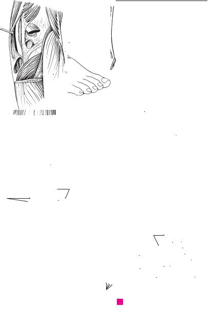
Foot, lateral view |
|
|
|
|
|
|
|
|
|
|
|
|||||||||||||||||||||
|
|
C |
|
|
|
|
|
|
|
|
|
|
|
|
|
|
|
|
|
||||||||||||||||
|
|
|
10 |
|
|
|
|
|
|
13 |
|
19 |
|||||||||||||||||||||||
|
|
|
|
|
|
|
|
|
|
|
|
|
|
|
|
|
|
|
|
|
|
|
|
|
|
|
|
|
|
|
|
12 |
|
|
|
|
|
|
|
|
|
|
|
|
|
|
|
|
|
|
|
|
|
|
|
|
|
|
|
|
|
|
|
|
|
|
14 |
|
20 |
||
|
|
|
|
|
|
|
|
|
|
|
|
|
|
|
|
|
|
|
|
|
|
|
|
|
|
||||||||||
|
|
|
|
|
|
|
|
|
|
|
|
|
|
|
|
|
|
|
|
|
|
|
|
|
|
|
|
|
|
|
|
|
18 |
|
|
|
|
|
|
|
|
|
|
|
|
|
|
|
|
|
|
|
|
|
|
|
|
|
|
|
|
|
|
|
|
|
|
||||
|
|
|
|
|
|
|
|
|
|
|
|
|
|
|
|
|
|
|
|
|
|
|
|
|
|
|
|
|
|
|
|
|
|
|
|
|
|
|
|
|
|
|
|
|
|
|
|
|
|
|
|
|
|
|
|
|
|
|
|
|
|
|
|
|
|
|
|
|
17 |
|
21 |
|
|
|
|
|
|
|
|
|
|
|
|
|
|
|
|
|
|
|
|
|
|
|
|
|
|
|
|
|
|
|
|
|
|
||
|
|
|
|
|
|
|
|
|
|
|
|
|
|
|
|
|
|
|
|
|
|
|
|
|
|
|
|
|
|
|
|
|
|
||
|
|
|
|
|
|
|
|
|
|
|
|
|
|
|
|
|
|
|
|
|
|
|
|
|
|
|
|
|
|
|
|||||
|
|
|
|
|
|
|
|
|
|
|
|
|
|
|
|
|
|
|
|
|
|
||||||||||||||
|
|
|
|
|
|
|
|
|
|
|
|
|
|
|
|
|
|
|
|
|
22 |
||||||||||||||
|
|
|
|
|
|
|
|
|
|
|
|
|
|
|
|
|
|
|
|
|
|
|
|||||||||||||
|
|
|
|
|
|
|
|
|
|
|
|
|
|
|
|
|
|
|
|
|
|
|
|
|
|
|
|
|
|
|
|
|
|
|
|
|
16 |
19 |
12 14 |
|
|
|
|
|
|
|
|
|
|
|
|||||||||||||||||||||
|
20 |
|
|
|
|
|
|
|
|
|
|
|
|
|
|
|
|
|
|
|
|
23 |
|||||||||||||
|
|
|
|
|
|
|
|
|
|
|
|
|
|
|
|
|
|
|
|
|
|
|
|
|
|
|
|
|
|
|
|
|
|
|
|
D Foot, medial view
24
25

|
108 |
|
Digestive system |
|
|
|
|
|
|
|
|
|
|
|
|
|||||
|
|
|
|
|
|
Apparatus |
digestorius 26 |
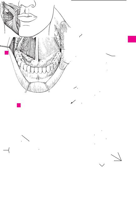
Sublingual papilla. Caruncula sublingualis. A |
||||||||||||
1 |
|
1 |
|
DIGESTIVE SYSTEM. |
|
|||||||||||||||
|
|
|
|
(systema alimentarium) |
. |
|
|
|
|
small mucosal eminence on either side of the |
||||||||||
|
|
2 |
ORAL CAVITY. Cavitas oris. |
|
|
|
|
frenulum. It receives the opening of the sub- |
||||||||||||
2 |
|
|
|
|
|
mandibular |
duct and |
the major sublingual |
||||||||||||
|
3 |
Vestibule of mouth. Vestibulum oris. Space be- |
|
|||||||||||||||||
|
|
duct. B |
|
|
|
|
|
|
||||||||||||
|
|
|
|
tween the rows of teeth and the lips or cheeks. |
|
|
|
|
|
|
|
|||||||||
|
|
|
|
27 Sublingual fold. Plica sublingualis. Mucosal |
||||||||||||||||
3 |
|
|
|
B C |
|
|
|
|
|
|
||||||||||
|
|
|
|
|
|
|
|
|
|
fold overlying the sublingual gland and extend- |
||||||||||
|
|
4 |
Oral fissure. Rima oris. Mouth opening be- |
|
||||||||||||||||
|
|
|
ing posterolaterally from the sublingual papilla. |
|||||||||||||||||
|
|
|
|
tween the lips. A |
|
|
|
|
|
|||||||||||
4 |
|
|
|
|
|
|
|
|
B |
|
|
|
|
|
|
|||||
|
5 |
Lips. Labia oris. |
|
|
|
|
|
|
|
|
|
|
|
|
|
|||||
|
|
|
|
|
|
|
|
28 Parotid papilla. Papilla ductus parotidei. Small |
||||||||||||
|
|
|
|
Upper lip. Labium superius. A B C |
|
|
||||||||||||||
|
6 |
|
|
|||||||||||||||||
5 |
|
|
|
mucosal elevation at the opening of the parotid |
||||||||||||||||
|
|
7 Philtrum. Groove extending from nasal septum |
|
duct lateral to the second upper molar tooth. B |
||||||||||||||||
|
|
|
|
|||||||||||||||||
|
|
|
|
|
|
|
|
|
|
|
||||||||||
|
|
|
|
to upper lip. A |
|
|
|
|
|
|
29 |
Transverse palatine folds. Plicae palatinae |
||||||||
6 |
|
|
|
|
|
|
|
|
|
|||||||||||
8 |
Tuberculum. Small eminence on upper lip mark- |
|
transversae. |
Mucosal |
folds |
running |
trans- |
|||||||||||||
|
|
|
|
ing end of philtrum. A |
|
|
|
|
|
versely on the anterior part of the hard palate. |
||||||||||
7 |
|
|
|
|
|
|
|
|
D |
|
|
|
|
|
|
|||||
9 |
Lower lip. Labium inferius. A B C |
|
|
|
|
|
|
|
|
|
|
|||||||||
|
|
|
30 Incisive papilla. Papilla incisiva. Small mucosal |
|||||||||||||||||
|
10 |
Commissure of lips. Commissura |
labiorum. |
|||||||||||||||||
8 |
|
elevation over the incisive foramen at the ante- |
||||||||||||||||||
|
|
|
Transition of upper lip into lower lip at the |
|
||||||||||||||||
|
|
|
|
rior end of the palatine raphe. D |
|
|||||||||||||||
|
|
|
|
angle of the mouth. A B |
|
|
|
|
|
|||||||||||
|
|
|
|
|
|
|
30 a GLANDULAE ORIS. The glands of the mouth. |
|||||||||||||
9 |
11 |
Angle of mouth. Angulus oris. A |
|
|
|
|||||||||||||||
|
|
|
31 Small glands of the oral cavity. Glandulae |
|||||||||||||||||
|
12 |
Cheek. Bucca. |
Lateral |
wall of |
vestibule |
of |
||||||||||||||
|
|
salivariae minores. |
|
|
|
|
||||||||||||||
10 |
|
|
|
mouth. A |
|
|
|
|
|
|
|
|
|
|
|
|||||
|
|
|
|
|
|
|
|
|
32 |
Labial glands. |
Gll. |
labiales. |
Small |
salivary |
||||||
|
13 Buccal fat pad. Corpus adiposum buccae. [[Bi- |
|||||||||||||||||||
|
|
|
glands at the inner aspect of the lips. B |
|||||||||||||||||
|
|
|
|
chat]]. Encapsulated body of fat between the |
|
|||||||||||||||
11 |
|
|
|
33 |
Buccal glands. Gll. buccales. Small mucous |
|||||||||||||||
|
|
|
buccinator and masseter muscles. A |
|
|
|||||||||||||||
|
14 |
Oral cavity proper. Cavitas oris propria. True |
|
salivary glands |
at |
the inner aspect |
of the |
|||||||||||||
|
|
|||||||||||||||||||
12 |
|
cheeks. B |
|
|
|
|
|
|
||||||||||||
|
|
|
oral cavity enclosed anteriorly and laterally by |
|
|
|
|
|
|
|
||||||||||
|
|
|
34 Molar glands. Gll. molares. Salivary glands cor- |
|||||||||||||||||
|
|
|
|
the teeth and extending as far as the isthmus of |
||||||||||||||||
|
|
|
|
|
responding |
to |
the |
buccal |
glands situated |
|||||||||||
13 |
|
|
|
fauces (oropharyngeal isthmus). C |
|
|
|
|||||||||||||
|
|
|
|
|
|
beneath the mucosal at the level of the molar |
||||||||||||||
15 |
Palate. Palatum. Partition between |
oral |
and |
|
||||||||||||||||
|
|
teeth. B |
|
|
|
|
|
|
||||||||||||
|
|
|
|
nasal cavities. |
|
|
|
|
|
|
|
|
|
|
|
|
|
|||
14 |
|
|
|
|
|
|
|
|
|
35 Palatine glands. Gll. palatinae. Salivary glands |
||||||||||
16 |
Hard palate. Palatum durum. Hard, bony part |
|||||||||||||||||||
|
|
situated beneath the |
mucosa of the |
palate. |
||||||||||||||||
|
|
|
|
of the palate. C D |
|
|
|
|
|
|||||||||||
|
|
|
|
|
|
|
|
|
(Two large groups right and left of the midline.) |
|||||||||||
15 |
|
|
|
|
|
|
|
|
||||||||||||
17 |
Soft palate. Palatum molle (velum palatinum). |
|
||||||||||||||||||
|
D |
|
|
|
|
|
|
|||||||||||||
|
|
|
|
Soft, posterior part of the palate. C D |
|
36 |
Lingual glands. Gll. linguales. Numerous |
|||||||||||||
|
|
|
|
|
||||||||||||||||
16 |
18 |
Palatine raphe. Raphe palati. Median mucosal |
||||||||||||||||||
|
mucous, serous and mixed glands primarily in |
|||||||||||||||||||
|
|
|
|
ridge at the junction of the right and left bony |
|
the lateral and posterior areas of the tongue. B |
||||||||||||||
|
|
|
|
|
||||||||||||||||
|
|
|
|
palatal processes. D |
|
|
|
|
37 |
Anterior lingual glands. Gl. lingualis anterior |
||||||||||
17 |
|
|
|
|
|
|
|
|||||||||||||
19 |
Oral mucosa. Tunica mucosa oris. Mucous |
|||||||||||||||||||
|
|
[[gl. apicis linguae, Nuhn’s glands]]. Mixed |
||||||||||||||||||
|
|
|
|
membrane of oral cavity consisting of |
|
glands near the apex of the tongue providing |
||||||||||||||
18 |
|
|
|
|
||||||||||||||||
|
|
|
stratified, |
nonkeratinized |
|
squamous |
|
several drainage ducts on the undersurface of |
||||||||||||
|
|
|
|
epithelium throughout and underlying mixed |
|
the tongue. B |
|
|
|
|
|
|||||||||
|
|
|
|
|
|
|
|
|
|
|||||||||||
19 |
|
|
|
glands. |
|
|
|
|
|
|
|
|
|
|
|
|
|
|
||
|
20 Frenulum of upper lip. Frenulum labii super- |
|
|
|
|
|
|
|
|
|||||||||||
|
|
|
|
|
|
|
|
|
|
|||||||||||
|
|
|
|
ioris. Median mucosal fold between the gums |
|
|
|
|
|
|
|
|
||||||||
20 and upper lip. B
|
21 Frenulum of lower lip. Frenulum labii inferi- |
||
21 |
|
oris. Median mucosal fold between the gums |
|
|
|
and lower lip. B |
|
|
|
||
22 |
22 |
Gums. Gingivae. Mucous membrane united |
|
|
|
firmly with the teeth and jaw bones. B D |
|
|
23 |
Gingival (gum) margin. Margo gingivalis. B D |
|
23 |
|||
24 |
Gingival (interdental) papilla. Papilla ging- |
||
|
|
ivalis (interdentalis). B D |
|
24 |
|
||
25 |
Gingival sulcus. Sulcus gingivalis. Shallow fur- |
||
|
|
row between the gum margin and the tooth. Its |
|
25 |
|
deepening leads to cavity formation. See p. 113. |
|
|
|
A |
|
Digestive system 109
13 10
|
7 |
12 |
|
6 |
8 |
|
|
11 |
|
||
|
4 |
|
|
|
9 |
|
|
|
|
20 |
22 |
6
A Face, anterior view
37 |
|
|
|
32 |
||||||
|
|
|
|
|
|
|
|
|
|
|
36 |
|
|
|
|
|
|
|
|
|
33 |
|
|
|
|
|
|
|
|
|
||
28 |
|
|
|
|
|
|
||||
|
|
|
|
|
|
|
|
|
34 |
|
|
|
|
|
|
|
|
|
|
||
10 |
|
|
|
|
|
|||||
|
|
|
|
|||||||
|
|
|
|
|
|
|
|
|
|
|
|
|
|
|
27 |
|
|
|
|
|
|
26 |
|
|
|
|
|
|
|
|
|
23 |
|
|
|
|
|
|
|
|
|
||
|
|
|
|
|
|
|
|
|||
|
|
|
|
|
|
|
||||
|
9 |
|
|
|
|
|
|
24 |
||
|
|
|
|
|
|
|||||
|
|
|
|
|
|
|
|
|
||
3  21
21 22
22
B Mouth with tongue elevated
|
16 |
|
|
|
|
|
24 |
|
|
|
|
|
|
|
|
|
|||
|
|
|
|
|
|
30 |
|
||
|
|
|
|
|
29 |
|
|
23 |
|
|
|
|
|
|
|
|
|
||
|
|
|
|
|
|
|
|
|
|
6 |
14 |
|
|
|
|
|
|
18 |
|
16 |
16 |
|
|
|
|||||
|
|
|
|
|
|
||||
3 |
|
|
|
17 |
|
|
|
|
|
|
|
|
|
|
|
|
|
||
9 |
|
|
|
|
|
|
|
|
|
|
|
|
|
|
|
|
22 |
||
|
|
|
|
|
35 |
|
|
|
|
|
|
|
17 |
17 |
|
|
|
||
C |
Sagittal section of oral cavity |
D |
Palate, inferior view |
1
2
3
4
5
6
7
8
9
10
11
12
13
14
15
16
17
18
19
20
21
22
23
24
25