Материал: Атлас Ханц фениш
Muscles 95
7
22
8
10
12
11
13
14
96.3
96.5
26 |
|
|
25 |
|
|
|
|
|
|||
|
96.6
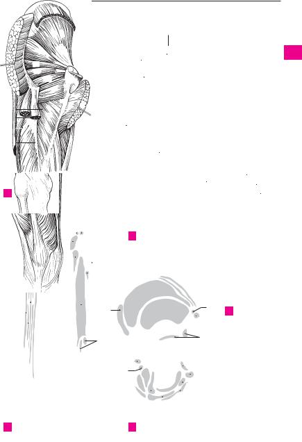
ADeep muscles of hip, posterior view
2 |
|
|
|
|
|
|
|
|
|
96.1 |
4 |
5 |
||
|
|
|
|
|
|
|
|
|
||||||
|
|
|
|
|
|
|
|
|
|
|||||
|
|
|
|
|
24 |
|
|
|
4 |
|||||
|
|
|
|
|
|
|
|
|
|
|
|
|||
22 |
|
|
|
|
|
|
|
|
|
|
|
|||
|
|
|
|
|
|
|
|
|
|
|
|
|
||
|
|
|
|
|
|
|
|
|
|
|
|
|||
|
7 |
|
|
|
|
|
23 |
|
|
3 |
|
|
||
|
|
|
|
|
|
|
|
|
|
|||||
|
|
|
|
|
|
|
|
|
|
|
||||
|
|
|
|
|
|
|
|
|
|
|
|
|
|
|
6 |
|
|
|
|
|
|
25 |
|
|
9 |
|
|
||
|
|
|
|
|
|
|
|
|
|
|
||||
18 |
|
|
|
|
|
|
|
2 |
|
|
||||
|
|
|
|
|
|
|
|
|
|
|
|
|||
19 |
|
|
|
|
|
|
22 |
|
||||||
|
|
|
|
|
|
|
|
|
|
98.18 |
23 |
|
||
|
|
|
|
|
|
|
|
|
|
|
|
|
||
 98.14
98.14
17 |
98.14 |
|
|
|
|
|
|
|
15 |
|
|
|
|
|
|
|
|||
|
|
17 |
|
|
|
|
25 |
||
|
|
|
|
|
|
||||
|
|
|
|
|
|||||
|
|
|
|
|
|
|
|
26 |
|
|
|
|
|
|
|
|
|
||
|
|
|
|
|
|
|
|||
11;12;13 11;12;13
|
|
|
|
|
10 |
|
|
|
|
|
|
|
|
|
|
|
||||
|
|
|
|
|
|
|
|
|
7 |
10 |
|
|
|
|
|
|
|
|
|
|
|
|
|
|
|
|
|
|
|
|
|
|
|
|
|
|
|
|
|
||
|
|
|
|
|
|
|
14 |
8 |
|
|
|
|
|
|
|
|
|
|
||
|
|
|
|
|
|
|
18 |
|
|
|
|
|
|
|
|
|
|
|||
|
|
|
|
|
|
|
|
|
|
|
|
|
|
|
|
|
||||
2 |
|
|
|
|
|
|
6 |
|
|
|
|
|
|
|
|
2 |
||||
|
|
|
|
|
|
|
|
|
|
|
|
|||||||||
|
|
|
|
|||||||||||||||||
|
|
|
|
|
|
|
|
|
|
|||||||||||
22 |
|
|
|
|
|
|
|
|
|
|
|
|
|
|
|
|
20 |
|||
|
|
|
|
|
|
|
|
|
|
|
||||||||||
|
|
|
|
|
|
|
|
|
|
|
|
|
||||||||
24
18
25 |
|
|
|
|
|
|
|
19 |
|
|
|||||
20 |
|
|
|||||
|
|
|
|
|
|
||
|
|
|
|
|
|
||
23 |
|
|
|
|
|
|
|
21 |
|||||||
|
|||||||
25 
D Femur, posterior and anterior views
a
B Thigh, anterior view
7
|
|
|
8 |
|
|
9 |
||||||
|
|
|
|
|
|
|
|
|
|
15 |
||
|
|
|
|
|
|
|
|
|
||||
|
|
|
|
|
|
|
|
|||||
|
|
|
|
|
|
|
17 |
|||||
12 |
|
|
|
|
|
|
|
|
|
|
22 |
|
|
|
|
|
|
|
|
|
|
|
|||
13 |
|
|
|
|
|
|
|
|
||||
|
|
|
|
|
|
|
|
|||||
|
|
|
|
|
|
|
|
|
23 |
|||
|
|
|
|
|
|
|
|
|
|
|||
|
|
|
|
|
|
|
|
|
|
24 |
||
|
|
|
|
|
|
|
|
|
|
|||
|
|
|
|
|
|
|
|
|
|
26 |
||
|
|
|
|
|
|
|
|
|
|
|
|
|
|
|
|
|
|
|
|
|
|
|
|
|
|
14 25
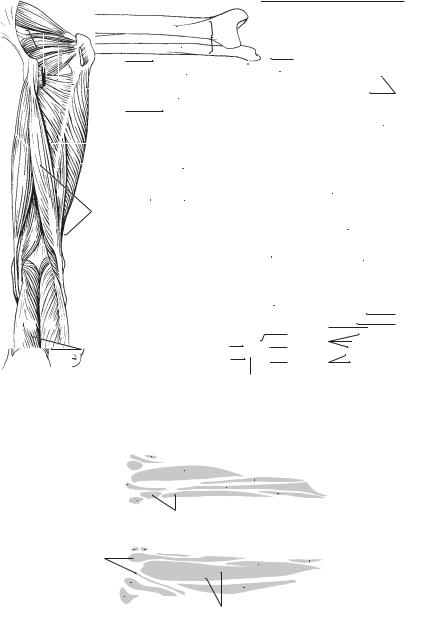
E Hip bone, lateral view
18 20
CThigh, anterior view
3
4
5
6
7
8
9
10
11
12
13
14
15
16
17
18
19
20
21
22
23
24
25

|
96 |
Muscles |
|
|
|||
|
|
|
M. obturator externus. o: External surface of |
14 |
M. gastrocnemius. The superficial calf muscle |
||
1 |
|
1 |
|||||
|
|
|
obturator membrane and environment. i: Tro- |
|
with two heads (lateral, medial). A: Flexes the |
||
|
|
|
|
chanteric fossa. A: Lateral rotation and adduc- |
|
knee joint, plantar flexes and supinates the |
|
2 |
|
|
|
tion of thigh at the hip joint. I: Obturator nerve. A |
|
ankle joint. A B C D |
|
|
|
|
2 M. biceps femoris. o: Arises from the pelvis |
15 Lateral head of gastrocnemius. Caput laterale. o: |
|||
|
|
|
|
and femur via two heads. i: Head of fibula. I: |
|
Proximal to the lateral femoral condyle. i: |
|
3 |
|
|
|
|
|||
|
|
|
Sciatic nerve, tibial part. A B E F |
|
Achilles tendon. A B C |
||
|
|
|
|
|
|||
|
|
|
3 Long head of biceps femoris. Caput longum. |
16 Medial head of gastrocnemius. Caput laterale. o: |
|||
4 |
|
|
|||||
|
|
|
o: Ischial tuberosity. i: Head of fibula. A: Exten- |
|
Proximal to the medial femoral condyle. i: |
||
|
|
|
|
sion, adduction and lateral rotation of thigh at |
|
Achilles tendon. A B C D |
|
5 |
|
|
|
the hip joint; flexion and lateral rotation of the |
17 |
M. soleus. o: Proximal ends of fibula and tibia. |
|
|
|
|
|
knee joint. I: Tibial nerve. A B |
|
i: Achilles tendon. A: Plantar flexes and supi- |
|
|
4 |
Short head of biceps femoris. Caput breve. o: |
|
||||
6 |
|
nates the foot. B F |
|||||
|
|
|
Lateral lip of linea aspera. i: Head of fibula. A: |
|
|||
|
|
|
18 Tendinous arch of soleus muscle. Arcus tendineus |
||||
|
|
|
|
Flexion and lateral rotation of the knee joint. I: |
|||
7 |
|
|
|
Common peroneal nerve. A B |
|
musculi solei. Tendinous arch above the inter- |
|
|
|
|
|
osseous membrane. Passageway for the tibial |
|||
|
|
5 M. semitendinosus. o: Ischial tuberosity. i: Me- |
|
||||
|
|
|
|
nerve and posterior tibial artery and vein. B |
|||
8 |
|
|
|
dial to tibial tuberosity [pes anserinus]. A: Ex- |
|
||
|
|
|
19 |
Tendo calcaneus [[Achilles tendon]]. The ten- |
|||
|
|
|
tension, medial rotation and adduction at the |
||||
|
|
|
|
hip joint; flexion and medial rotation at the |
|
don of the triceps surae at the tuber of the cal- |
|
9 |
|
|
|
knee joint. I: Tibial nerve. A D E |
|
caneus. B C |
|
|
|
|
20 M. plantaris. o: Above the lateral femoral con- |
||||
|
|
|
6 M. semimembranosus. o: Ischial tuberosity. i: |
||||
|
|
|
|||||
10 |
|
|
|
Medial condyle of tibia and oblique popliteal |
|
dyle. i: Achilles tendon or tuberosity of the cal- |
|
|
|
|
ligament. It is partially covered by the semiten- |
|
caneus. I: Tibial nerve. B C |
||
|
|
|
|
|
|||
|
|
|
|
dinosus muscle. A: Extension, adduction and |
21 M. popliteus. o: Lateral femoral condyle. i: |
||
11 |
|
|
|
||||
|
|
|
medial rotation of thigh at the hip joint; flexion |
|
Posterior surface of tibia. A: Flexion of knee |
||
|
|
|
|
and medial rotation of the knee joint. Tenses |
|
joint and medial rotation of leg. B C F |
|
|
|
|
|
|
|||
12 |
|
|
|
knee joint capsule. I: Tibial nerve. A B F |
22 M. tibialis posterior. o: Tibia, fibula, interos- |
||
|
|
7 M. tibialis anterior. o: Lateral surface of tibia, |
|||||
|
|
|
|
seous membrane. i: Navicular, cuneiforms, |
|||
|
|
|
|
interosseous membrane, fascia of leg (crural |
|
||
13 |
|
|
|
|
cuboid and metatarsals 2−4. One bundle of |
||
|
|
|
fascia). i: Medial aspect of medial cuneiform |
|
|||
|
|
|
|
fibers extends backward to the sustentaculum |
|||
|
|
|
|
bone and 1st metatarsal. A: Dorsiflexion and |
|
tali of the calcaneus. A: Plantar flexor and supi- |
|
14 |
|
|
|
supination of foot. I: Deep fibular nerve. D E |
|
nator. I: Tibial nerve. C F |
|
|
|
8 M. extensor digitorum longus. o: Lateral tibial |
|
||||
|
|
|
23 M. flexor digitorum longus. o: Tibia. i: Distal |
||||
|
|
|
|
condyle, interosseous membrane, fibula. i: Dor- |
|||
15 |
|
|
|
|
phalanges of toes 2−5. A: Plantar flexion and |
||
|
|
|
sal aponeurosis of toes 2−5. A: Dorsiflexion and |
|
|||
|
|
|
|
supination of foot, flexion of toes. I: Tibial |
|||
|
|
|
|
pronation of foot, extension of toes. I. Deep |
|
||
|
|
|
|
|
nerve. C F |
||
16 |
|
|
|
fibular nerve. D E |
|
||
|
|
|
24 |
M. flexor hallucis longus. o: Fibula. i: Distal |
|||
9 |
M. peroneus tertius (m. fibularis tertius). |
||||||
|
|||||||
|
|
phalanx of big toe. A: Plantar flexion and supi- |
|||||
|
|
|
|
Muscle split off from the extensor digitorum |
|
||
17 |
|
|
|
|
|||
|
|
|
|
nation of foot, flexion of big toe. I: Tibial nerve. |
|||
|
|
|
longus and inserting into the base of the 5th |
|
|||
|
|
|
|
C F |
|||
|
|
|
|
metacarpal. A: Dorsiflexion and pronation of |
|
||
|
|
|
|
25 M. extensor hallucis brevis. o: Dorsal surface |
|||
18 |
|
|
|
foot. I: Deep fibular nerve. D |
|||
|
10 M. extensor hallicus longus. o: Interosseous |
|
of calcaneus. i: Proximal phalanx of big toe. A: |
||||
|
|
|
|||||
|
|
|
Extends the big toe. I: see p. 26. D |
||||
19 |
|
|
|
membrane and fibula. i: Distal phalanx of big |
|
||
|
|
|
|
|
|||
|
|
|
toe. A: Dorsiflexion of foot, extension of big toe. |
26 M. extensor digitorum brevis. o: Dorsal sur- |
|||
|
|
|
|
||||
|
|
|
|
I: Deep fibular nerve. D E |
|
face of calcaneus. i: Dorsal aponeuroses of toes |
|
20 |
11 |
M. peroneus longus [[m. fibularis longus]]. o: |
|
2−4. A: Extends toes. I: Deep fibular nerve. D |
|||
|
|
|
|
Fibula and crural fascia. i: Medial cuneiform |
|
|
|
|
|
|
|
|
|
||
|
|
|
|
bone and 1st metatarsal after an oblique course |
|
|
|
21below the dorsum of the foot. A: Plantar flexion anf pronation of foot. I: Superficial fibular
22nerve. C D E F
12 M. peroneus brevis [[m. fibularis brevis]]. o:
23Distal 2/3 of fibula. i: Tuberosity of 5th metatarsal. A: Plantar flexion and pronation. I: Superficial fibular nerve. C D E F
2413 M. triceps surae. Muscle group consisting of the gastrocnemius and soleus; it forms the
25Achilles tendon (tendo calcaneus). I: Tibial nerve.
Muscles 97
94.10 |
94.12 |
|
|
|
|
|
|
6 |
|
|
|
|
|
|
|
3 |
|
|
|
|
|
|
|
|
|
|
|
|
|
|
|
|
|
|
|
|
|
|
|||
|
|
|
|
|
|
|
|
|
|
|
|
|
|
|
|
|
|
|
|
|
|
|
|
|
|
|
|
|
|
|
|
||||||||||
|
94.11 |
|
|
|
|
|
|
|
|
|
|
2 |
16 |
|
|
|
|
|
|
|
|
|
|
|
|
|
|
|
|
|
|
||||||||||
|
|
|
|
|
|
|
|
|
|
|
|
|
|
|
|
|
|
|
|
|
|
|
|
|
|
|
|
|
|
|
|
||||||||||
|
|
|
|
94.13 |
|
|
|
|
|
|
|
|
|
|
|
|
4 |
|
|
|
|
|
|
|
|
|
|
|
|
|
|
|
|
|
|
|
|||||
|
|
|
|
16 |
|
|
|
|
|
|
|
|
|
|
|
|
|
20 |
|
|
|
|
|
|
|
|
|
|
|||||||||||||
|
|
|
|
|
|
|
|
|
|
|
|
|
|
|
|
|
|
|
|
|
|
|
|
|
|
|
|
|
|||||||||||||
|
|
|
|
|
|
|
|
|
94.14 |
|
|
|
|
|
|
|
|
|
15 |
|
|
|
|
|
|
|
15 |
|
5 |
3 |
|||||||||||
|
|
|
|
|
|
|
|
|
|
|
|
|
|
|
|
|
|
|
|
|
|
|
|
|
|
|
|
|
|||||||||||||
|
1 |
|
|
|
|
|
|
|
|
|
|
|
|
|
20 |
|
|
|
|
|
|
|
|
|
|
|
|
|
|||||||||||||
|
|
|
|
|
|
|
|
|
|
|
|
|
|
|
|
|
|
|
|
|
|
|
|
|
|
||||||||||||||||
|
|
|
|
|
|
|
|
|
|
|
|
|
|
|
|
|
|
|
|
|
|
|
|
|
|
|
|||||||||||||||
|
|
|
|
|
|
|
|
|
|
|
|
|
|
|
|
|
|
|
|
|
|
|
|
|
4 |
||||||||||||||||
|
|
|
|
|
|
|
|
|
|
|
|
|
|
|
|
|
|
|
|
|
|
|
|
|
|
|
|
||||||||||||||
|
|
|
|
|
|
|
21 |
|
|
|
|
|
|
|
|
|
|
|
|
|
|
|
|
|
|
|
|
|
|
|
|
|
|
|
|
|
|
||||
|
|
|
|
|
|
|
|
|
|
|
|
|
|
|
|
|
|
|
|
|
|
|
|
|
|
|
|
|
|
|
|
|
|
|
|
|
|
|
|||
|
|
|
|
|
|
|
|
|
|
|
|
|
|
|
|
|
|
|
|
|
|
|
|
|
|
|
|
|
|
|
|
|
|
|
|
|
|
|
|||
|
|
|
|
|
|
|
|
|
|
|
|
|
|
|
|
|
|
21 |
|
|
|
|
|
|
|
|
|
|
|
|
|
|
|
||||||||
|
|
|
|
|
|
|
|
|
|
|
|
|
|
|
|
|
|
|
|
18 |
|
|
|
|
|
|
|
|
|
|
|
|
|
16 |
5 |
||||||
|
|
|
|
|
|
|
|
|
|
|
|
|
|
|
|
|
|
|
|
11 |
|
|
|||||||||||||||||||
|
|
|
|
|
|
|
|
|
|
|
|
|
|
|
|
|
|
|
|
|
|||||||||||||||||||||
|
|
|
|
|
|
|
|
94.25 |
|
|
|
|
|
|
|
|
|
|
7 |
|
|
|
|
|
|||||||||||||||||
|
|
|
|
|
|
|
|
|
|
|
|
|
|
|
|
|
|
|
|
|
|
|
|
||||||||||||||||||
|
3 |
|
|
|
|
|
|
|
|
|
|
|
|
|
|
|
|
|
6 |
||||||||||||||||||||||
|
|
|
|
|
|
|
|
|
|
|
|
|
|
|
|
|
|
|
|
|
|
|
|
|
|
|
|
|
|
|
|
|
|
|
|
|
|
||||
|
|
|
|
|
|
|
|
|
|
|
|
|
|
|
|
|
|
|
|
|
|
|
|
|
|
|
|
|
|
|
|
|
|
|
|
|
|
|
|
||
|
|
|
|
|
|
|
94.18 |
|
|
|
|
|
|
|
|
|
|
17 |
|
|
|
|
|
|
|
|
|
|
|
|
|
|
|
|
|
|
|
|
|
||
|
|
|
|
|
|
|
|
|
|
|
|
|
|
|
|
|
|
|
|
|
|
|
|
|
|
|
|
|
|
|
|
|
|||||||||
|
|
|
|
|
|
|
|
|
|
|
|
|
|
|
|
|
|
|
|
|
|
|
|
|
|
|
|
|
|
|
|
|
|
|
|
|
|
|
|||
|
|
|
|
|
|
|
|
|
|
|
|
|
|
|
|
|
|
|
|
|
|
|
|
|
|
|
|
|
|
|
|
|
|
|
|
|
7 |
||||
|
|
|
|
|
|
|
|
|
|
|
|
|
|
|
|
|
|
|
|
|
|
|
|
|
|
|
|
|
|
|
|
|
|
|
|
|
|
|
|
||
|
|
|
|
|
|
|
|
|
|
|
|
|
|
|
|
|
|
|
|
|
|
|
|
|
22 |
|
|
|
|
|
|
|
|
|
|
|
|
|
|||
|
|
|
|
|
|
|
|
|
|
|
|
|
|
|
|
|
|
|
|
|
12 |
|
|
|
|
|
|
|
|
|
|
||||||||||
|
|
|
|
|
|
|
|
|
|
|
|
|
|
|
|
|
|
|
|
|
|
|
|
|
|
|
|
|
|
|
|||||||||||
|
|
|
|
|
|
|
16 |
|
|
|
|
|
|
|
|
|
|
15 |
|
|
|
|
|
|
|
|
8 |
||||||||||||||
|
|
|
|
|
|
|
|
|
|
|
|
|
|
|
|
|
|
|
|
|
|
|
|
|
|
||||||||||||||||
|
|
|
|
|
|
|
|
|
|
|
|
|
|
|
|
|
11 |
|
|
|
|
|
|
|
|
|
|
||||||||||||||
|
|
|
|
|
|
|
|
|
|
|
|
|
|
|
|
|
|
|
|
|
|
|
|
|
|
|
|||||||||||||||
5 |
|
|
|
|
|
|
2 |
|
|
|
|
|
|
|
|
|
|
|
|
23 |
|
|
|
|
|
|
|
|
|
|
|
|
|
|
|
|
|
|
|||
|
|
|
|
|
|
|
|
|
|
|
|
|
|
|
|
|
|
|
|
|
|
|
|
|
|
|
|
|
|
|
|
|
|
|
|||||||
4 |
|
|
|
|
|
|
|
|
|
|
|
|
|
|
|
|
|
|
|
|
|
|
|
|
|
|
|
|
|
|
|
|
|
9 |
|||||||
|
|
|
|
|
|
|
|
|
|
|
|
|
|
|
|
|
|
|
|
|
|
|
|
|
|
|
|
|
|
|
|
|
|
||||||||
|
|
|
|
|
|
|
|
|
|
|
|
|
|
|
|
|
8 |
|
|
|
|
|
|
|
|
||||||||||||||||
|
|
|
|
|
|
|
|
|
|
|
|
|
|
|
|
|
|
|
|
|
|
|
|
|
|
||||||||||||||||
|
|
|
|
|
|
|
|
|
|
|
|
|
|
|
|
|
|
|
|
|
|
|
|
|
|
|
|
|
|
|
|
|
|
|
|
|
|
|
|
|
|
|
2 |
|
|
|
|
|
|
|
|
|
|
|
|
|
|
|
|
|
24 |
|
|
|
|
|
|
|
|
|
|
|
10 |
|
|
|
|
|
10 |
||||
|
6 |
|
|
|
|
|
|
|
|
|
|
|
|
|
|
|
|
|
|
|
|
|
|
|
|
|
|
|
|
|
|
|
|
|
|
|
|
|
|||
|
|
|
|
|
|
|
|
|
|
|
|
|
|
|
|
|
|
|
|
|
|
|
|
|
|
|
|
|
|
|
|
|
|
|
|
|
|
|
|
11 |
|
|
|
|
|
|
|
|
|
|
|
|
|
|
|
|
|
|
|
|
|
|
|
|
|
|
|
|
|
|
|
|
|
|
|
|
|
|
|
|
|
|
|
|
|
|
|
|
|
|
|
|
|
|
|
|
|
|
|
|
|
|
|
|
|
|
|
|
|
|
|
|
|
|
|
|
|
|
|
|
|
|
|
|
|
|
|
|
|
|
|
|
|
|
|
|
|
|
|
|
|
|
|
|
|
|
|
|
|
|
|
|
|
|
|
12 |
|
|
|
|
|
|
|
|
|
12 |
|
|
|
|
|
|
|
|
|
|
|
|
|
|
|
|
|
|
|
|
|
|
|
|
|
|
|
|
|
|
|
|
|
|
|
||||||||
|
|
|
|
|
8 |
13 |
|
19 |
|
19 |
25 |
9 |
|
|
|
|
26 |
|
|
|
|
14 |
22 |
12 |
|
14 |
|
|
|
|
||||
16 |
15 |
23 |
11 |
8 |
|
|
|
|
|
24 |
|
|
15 |
|
|
|
|
|
|
|
|
|
|
|
|
|
|
16 |
|
A |
Thigh, |
B |
Lower leg, |
C |
Deep muscles of |
D |
Lower leg, |
||
|
|||||||||
|
|||||||||
|
posterior view |
|
posterior view |
|
lower leg, posterior view |
|
anterior view |
17 |
|
|
|
|
|
|
|
|
|
||
|
|
|
|
|
|
|
|
|
57
|
|
|
|
|
|
|
|
|
|
|
|
|
|
|
|
|
|
|
|
18 |
|
|
|
|
|
|
|
|
|
|
|
|
|
|
|
|
|
|
|
|
|
|
|
|
|
|
|
|
|
|
|
|
|
|
|
|
|
|
|
|
|
19 |
|
|
|
|
|
|
|
|
|
|
|
|
|
|
|
|
|
|
|
|
|
|
|
|
|
|
|
|
|
|
|
|
|
|
|
|
|
|
|
|
|
|
|
|
|
|
|
|
|
|
|
|
|
|
|
|
|
|
|
|
|
|
20 |
E |
Tibia and fibula, |
|
|
|
|
|
|
|
|
|
|
|
|
|
|
|
|
|
|
|
8 2 |
|
11 |
8 |
10 |
12 |
|
|
|||||||||||||
|
anterior view |
|
|
21 |
||||||||||||||||
|
|
2 11 |
|
|
|
|
|
|
|
|
|
|
||||||||
|
|
|
|
|
|
|
|
|
|
|
|
|
||||||||
|
|
|
|
|
|
|
|
|
|
|
|
|
24 |
12 |
|
|||||
|
|
17 |
|
|
|
|
|
|
|
|
|
|
22 |
|||||||
|
|
|
|
|
|
|
|
|
|
|
|
|
|
|
|
|
|
|
||
|
|
|
|
|
|
|
|
|
|
|
|
|
|
|
|
|
|
|
|
|
|
|
|
|
|
|
|
|
|
|
|
|
|
|
|
|
|
|
|
|
|
|
|
21 |
|
|
|
|
|
|
|
|
|
|
|
|
|
|
|
|
|
23 |
|
|
|
|
|
|
|
|
|
|
|
|
|
|
|
|
|
|
|
||
|
|
|
|
|
|
|
|
|
|
|
|
|
|
|
|
|
|
|
|
|
|
|
|
|
|
|
|
|
|
|
|
|
|
|
|
|
|
|
|
|
|
|
|
6 |
|
|
|
|
|
|
|
|
|
|
|
|
|
|
|
|
|
24 |
F |
Tibia and fibula, |
|
|
|
|
|
|
|
|
|
|
|
|
|
|
|
|
|
||
|
posterior view |
|
|
|
|
|
|
|
|
|
22 |
23 |
|
|
|
|
|
|
||
|
|
|
|
|
|
|
|
|
|
|
|
|
25 |
|||||||
|
|
|
|
|
|
|
|
|
|
|
|
|
|
|
|
|
|
|
|
|
|
|
|
|
|
|
|
|
|
|
|
|
|
|
|
|
|
|
|
|
|
a
|
98 |
Muscles |
|
|
|
|
||
|
|
|
M. abductor hallucis. Abductor muscle of great |
|
11 |
Mm. interossei dorsales pedis. o: Arises by two |
||
1 |
|
1 |
|
|||||
|
||||||||
|
|
|
toe. o: Medial process of tuber calcanei. i: Me- |
|
|
heads from adjacent metatarsal bones. i: Base of |
||
|
|
|
|
dial sesamoid and proximal phalanx of big toe. |
|
|
the proximal phalanx, plantar ligament. A: Ab- |
|
2 |
|
|
|
A: Medial abduction, supports the longitudinal |
|
|
duction and flexion of toes at the metatar- |
|
|
|
|
|
arch. I. See 2. A B |
|
|
sophalangeal joints and extension at the inter- |
|
|
|
|
|
|
|
|
phalangeal joints. I: Lateral plantar nerve. C |
|
|
2 |
M. flexor hallucis brevis. Short flexor muscle of |
|
|
||||
3 |
|
|
||||||
|
12 |
Mm. interossei plantares. o: Single-headed |
||||||
|
|
|
|
the great toe. Origin: Cuneiform I, long plantar |
|
|||
|
|
|
|
ligament, tendon of the posterior tibial m. and |
|
|
from metatarsal bones 3−5. i: Base of proximal |
|
|
|
|
|
|
|
|||
4 |
|
|
|
|
|
phalanges. A: Adduction and flexion of toes at |
||
|
|
|
plantar aponeurosis. Forms a groove for trans- |
|
|
|||
|
|
|
|
mission of the flexor hallucis longus m., stabi- |
|
|
the metatarsophalangeal joints. I: cf. p. 11. C |
|
5 |
|
|
|
lizes the longitudinal arch. A B |
|
13 |
Fascia lata. Fascia of thigh which envelops the |
|
|
|
|
|
|
|
entire thigh musculature. D |
||
|
|
|
2 a Medial head. Caput mediale. o: Tendon of the |
|
|
|||
|
|
|
|
14 |
Iliotibial |
tract. Tractus iliotibialis. Vertical |
||
6 |
|
|
|
abductor m. of the great toe, sesamoid bone |
|
|||
|
|
|
and proximal phalanx. |
|
|
thick band of fascia lata that extends from the |
||
|
|
|
|
|
|
anterior segment of the iliac crest to the lateral |
||
|
|
|
|
|
|
|
||
7 |
|
|
2 b Lateral head. Caput laterale. o: Tendon of the |
|
|
tibial condyle and into which radiate the tensor |
||
|
|
|
adductor m. of the great toe, lateral sesamoid |
|
|
fasciae latae and gluteus maximus. D |
||
|
|
|
|
|
|
|||
8 |
|
|
|
bone and proximal phalanx of the great toe. |
|
15 |
Lateral intermuscular septum of thigh. Sep- |
|
|
|
3 M. adductor hallucis. Important muscle for the |
|
|
tum intermusculare femoris laterale. Firm con- |
|||
|
|
|
|
|
nective tissue layer extending from the fascia |
|||
|
|
|
|
transverse arch of the foot consisting of the fol- |
|
|
||
9 |
|
|
|
|
|
lata to the lateral lip of the linea aspera between |
||
|
|
|
lowing two heads. |
|
|
|||
|
|
|
|
|
|
|
the biceps femoris and vastus lateralis muscles. |
|
10 |
|
4 |
Oblique head. Caput obliquum. o: Metatarsals |
|
16 |
Medial intermuscular septum of thigh. Sep- |
||
|
|
|
2−4, lateral cuneiform and cuboid bones. i: |
|
|
tum intermusculare femoris mediale. Stout con- |
||
|
|
|
|
Lateral sesamoid bone and proximal phalanx of |
|
|
nective tissue layer extending from the fascia |
|
11 |
|
|
|
big toe together with the transverse head. A: |
|
|
lata to the medial lip of the linea aspera between |
|
|
|
|
Important for stabilization of transverse and |
|
|
|||
|
|
|
|
|
|
the vastus medialis, sartorius and adductor |
||
12 |
|
|
|
longitudinal arches. B |
|
|
muscles. |
|
|
|
5 |
Transverse head. Caput transversum. o: Cap- |
|
17 |
Adductor canal. Canalis adductorius. Channel |
||
|
|
|
||||||
|
|
|
|
sules of metatarsophalangeal joints 3−5. i: |
|
|
between adductors, vastus medialis and [vasto- |
|
13 |
|
|
|
Lateral sesamoid bone. A: Primary function is |
|
|
adductor membrane]. It ends with the hiatus |
|
|
|
|
|
to support the transverse arch of the foot. A B |
|
|
tendineus within the adductor magnus. D |
|
14 |
|
6 |
M. abductor digiti minimi. o: Calcaneus and |
|
18 |
Hiatus tendineus (adductorius). Opening near |
||
|
|
|
|
plantar aponeurosis. i: Laterally on proximal |
|
|
the attachment of the adductor magnus at the |
|
|
|
|
|
|
|
level of the inferior margin of the adductor lon- |
||
15 |
|
|
|
phalanx of 5th toe. A: Plantar flexion and abduc- |
|
|
||
|
|
|
|
|
gus. |
|
||
|
|
|
tion of the 5th toe. I: Lateral plantar nerve. A B |
|
|
|
||
|
|
|
7 M. flexor digiti minimi brevis. o: Base of 5th |
|
19 Iliac fascia. Fascia iliaca. Fascia over the iliac and |
|||
16 |
|
|
|
|
inferior portion of the psoas muscles. It attaches |
|||
|
|
|
|
metatarsal, long plantar ligament. i: Proximal |
|
|
to the iliac crest and arcuate line as well as the in- |
|
17 |
|
|
|
phalanx of little toe. A: Flexion and abduction |
|
|
guinal ligament. D |
|
|
|
|
of little toe. I: Lateral plantar nerve. A B |
|
20 |
Muscular lacuna. Lacuna musculorum. Com- |
||
|
|
|
7 a [M. opponens digiti minimi]. Muscle occasion- |
|
|
partment for passage of the iliopsoas muscle and |
||
18 |
|
|
|
ally split off from the flexor digiti minimi |
|
|
the femoral and lateral femoral cutaneous |
|
|
|
|
|
brevis. o: Distal half of 5th metatarsal. |
|
|
nerves between the ilium, inguinal ligament and |
|
19 |
|
|
8 |
M. flexor digitorum brevis. o: Tuber calcanei |
|
|
iliopectineal arch. E |
|
|
|
21 Iliopectineal arch. Arcus iliopectineus. Portion |
||||||
|
|
|
|
and plantar aponeurosis. i: Middle phalanges of |
|
|||
|
|
|
|
|
|
of the iliac fascia between the inguinal ligament |
||
|
|
|
|
toes 2−5 via divided tendons. A: Flexes toes and |
|
|
||
20 |
|
|
|
|
|
and the iliopubic [iliopectineal] eminence. It |
||
|
|
|
supports the longitudinal arch of the foot. I: |
|
|
|||
|
|
|
|
|
|
separates the vascular and muscular lacunae. E |
||
|
|
|
|
Medial plantar nerve. A B |
|
|
||
21 |
|
|
|
|
22 |
Vascular |
lacuna. Lacuna vasorum. Compart- |
|
|
|
9 M. quadratus plantae (m. flexor accessorius). |
|
|||||
|
|
|
|
ment between the pubis, inguinal ligament and |
||||
|
|
|
|
|
||||
22 |
|
|
|
o: Calcaneus. i: Lateral border of tendon of flexor |
|
|
iliopectineal arch for passage of the femoral |
|
|
|
|
digitorum longus. A: Flexes toes and supports |
|
|
artery and the femoral branch of the geni- |
||
|
|
|
|
longitudinal arch of foot. I: Lateral plantar nerve. |
|
|
tofemoral nerve. E |
|
23 |
|
|
|
B |
|
23 |
Femoral triangle. Trigonum femorale. Triangle |
|
|
|
|
|
|
||||
|
|
|
10 |
Mm. lumbricales pedis. Lumbrical muscles of |
|
|
between |
the sartorius and adductor longus |
|
|
|
|
muscles and the inguinal ligament. D |
||||
24 |
|
|
|
the foot. o: Tendons of flexor digitorum longus. i: |
|
|
||
|
|
|
|
24 |
Femoral canal. Canalis femoralis. Passage |
|||
|
|
|
Bases of proximal phalanges 2−5. A: Flexion at |
|
||||
|
|
|
|
the metatarsophalangeal joint. Brings toes |
|
|
within the medial segment of the vascular |
|
25 |
|
|
|
closer to the big toe. I: Medial and lateral plantar |
|
|
lacuna that extends from the inguinal ligament |
|
|
|
|
|
nerves. A B |
|
|
to the saphenous opening. E |
|
|
|
|
|
|
|
|
|
|
Muscles 99
|
|
|
|
|
|
|
|
|
|
|
|
|
|
|
|
|
|
|
|
|
|
|
|
|
|
|
|
|
|
|
|
|
|
|
|
|
|
|
|
|
|
|
|
|
10 |
8 |
|
|
|
3 |
||||
|
|
|
|
|
|
|
|
|
|
|
|
|
|
|
|
|
|
|
|
|
|
|
|
|
|
|
|
|
|
|
|
4 |
|
|
|
|
|
|
|
|
|
2 |
|
|
|
|
|
|
|
|
|
|
|
|
5 |
2 |
5 |
|
|
|
|
|
5 |
||
|
|
|
|
|
|
|||||
|
|
|
|
|
|
|||||
|
|
10 |
|
|
|
|
|
|
|
|
|
|
|
|
|
|
|
|
|
||
|
|
|
|
|
|
|
4 |
|
6 |
|
|
|
|
|
|
|
|
||||
|
|
|
|
|
||||||
|
|
|
|
|
|
|
|
|
|
|
7 |
|
96.24 |
7 |
||
6 |
|
|
|
6 |
|
|
|
|
|
||
8 |
|
|
|||
12
7 |
11 |
|
1 |
|
|
|
|
|
|
|
|
96.23 |
|
|
|
|
8 |
|||
|
|
9 |
1 |
|
|
|
||||||||||||
|
|
|
|
|
|
|
|
|
||||||||||
|
|
|
|
|
|
|
|
|||||||||||
|
|
|
|
|
|
|
9 |
|||||||||||
|
|
|
|
|
|
|
|
|
|
|
|
|||||||
|
|
|
|
|
100.17 |
|
|
|
|
|
|
|
|
|
|
|
|
|
|
|
|
|
|
|
|
|
|
|
|
|
|
|
|
|
|||
|
|
|
|
|
|
|
|
|
|
|
|
|
10 |
|||||
|
|
|
|
|
|
|
|
|
|
|
|
|
|
|||||
|
|
|
|
|
|
|
|
|
|
|
|
|
|
|
|
|
|
|
|
|
|
|
|
|
|
|
|
|
|
|
|
|
|
|
|
|
|
|
|
|
|
|
|
|
|
|
|
|
|
|
|
|
|
|
|
|
|
|
|
|
|
|
|
|
|
|
|
|
8 |
|
|
|
|
|
11 |
|
|
|
|
|
|
|
|
|
|
|
|
|
|
|
|
|
||
|
|
|
|
|
|
|
|
|
|
|
|
|
|
|
|
|
|
|
|
|
|
|
|
|
|
|
|
|
|
|
|
|
|
|
|
|
|
|
|
|
|
|
|
|
|
|
|
|
|
|
|
|
|
|
|
12 |
|
|
|
|
|
|
|
|
|
|
|
|
|
|
|
|
|
|
|
|
|
|
|
|
|
|
|
|
|
|
|
|
|
|
|
|
|
|
|
|
|
|
|
|
|
|
|
|
|
|
|
|
|
|
|
|
13 |
|
|
|
|
|
|
|
|
|
|
|
|
|
|
|
|
|
||
|
|
|
|
|
|
|
|
|
|
|
|
|
|
|
|
|||
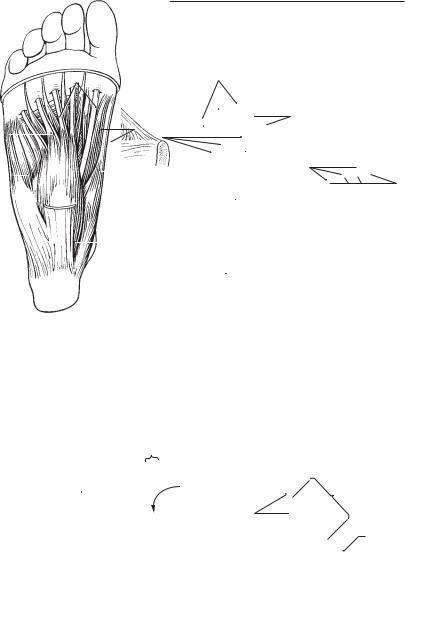
A |
Superficial plantar muscles |
B |
Deep plantar muscles |
|
C |
|
Interosseous muscles |
14 |
||||||||||
|
|
|
|
|
|
|
|
|
|
|
|
|
|
|
|
|
|
|
|
|
|
|
|
|
|
|
|
|
|
|
|
|
|
|
|
|
|
|
|
|
|
|
|
|
|
|
|
|
|
|
|
|
|
|
|
15 |
|
|
|
|
|
|
|
|
|
|
|
|
|
|
|
|
|
|
|
|
|
|
|
|
|
|
|
|
|
|
|
|
|
|
|
|
|
|
|
|
|
|
|
|
|
|
|
|
|
|
|
|
|
|
|
|
16 |
|
|
|
|
|
|
|
|
|
|
|
|
|
|
|
|
|
|
|
|
|
|
|
|
|
|
|
|
|
|
|
|
|
|
|
|
|
|
|
19 |
|
|
|
|
|
|
|
|
|
|
|
|
17 |
||||
|
|
|
|
|
|
|
|
|
|
|
|
|
|
|||||
|
|
|
|
|
|
|
|
|
|
|
|
|
|
|
|
|
|
|
|
23 |
|
|
|
|
|
|
|
|
|
|
|
|
18 |
||||
|
|
|
|
|
|
|
|
|
|
|
|
|
|
|
|
|
|
|
|
|
|
|
|
|
|
|
|
|
|
|
|
|
|
|
|
|
|
|
14 |
|
|
|
|
17 |
|
20 |
|
|
|
|
|
19 |
||||
|
|
|
13 |
|
|
|
|
|
|
|
|
|
|
22 |
|
|||
|
|
|
|
|
|
|
|
|
|
|
|
|
|
|
|
|
||
|
13 |
|
|
|
|
|
|
|
|
|
|
|
20 |
|||||
|
|
|
|
|
|
|
|
|
|
|
|
|
||||||
|
|
|
|
|
|
|
|
|
|
|
|
|
|
|
|
|
||
|
|
|
|
|
|
|
|
21 |
|
|
|
|
|
|
|
|
||
|
|
|
|
|
|
|
|
|
|
|
|
|||||||
|
|
|
|
|
|
|
|
|
|
|
24 |
|
|
21 |
||||
|
|
|
|
|
|
|
|
|
|
|
|
|
|
|
||||
|
|
|
|
|
|
|
|
|
|
|
|
|
|
86.11 |
|
|||
|
|
|
|
|
|
|
|
|
|
|
|
|
|
|
||||
|
|
|
|
|
|
|
|
|
|
|
|
|
|
22 |
||||
|
|
|
|
|
|
|
|
|
|
|
|
|
|
|
|
|
|
|
|
|
|
|
|
|
|
|
|
|
|
|
|
|
|
|
|
|
|
|
|
|
|
|
|
|
|
|
|
|
|
|
|
|
|
|
|
|
|
|
|
|
|
|
|
|
|
|
|
|
|
|
|
|
|
|
23 |
|
|
|
|
|
|
|
|
|
|
|
|
|
|
|
|
|
|
|
|
|
|
|
|
|
|
|
|
|
|
|
|
|
|
|
|
|
|
|
|
|
|
|
|
|
|
|
|
|
|
|
|
|
|
|
|
24 |
D |
Thigh, lateral and anterior medial view |
|
E |
Vascular lacuna |
|
|||||||||||||
|
|
|
||||||||||||||||
|
|
|
|
|
|
|
|
|
|
|
|
|
|
|
|
|
|
|
|
|
|
|
|
|
|
|
|
|
|
|
|
|
|
|
|
|
25 |
|
|
|
|
|
|
|
|
|
|
|
|
|
|
|
|
|
|
|
a