Материал: Атлас Ханц фениш

|
90 |
Muscles |
|
|
|||
|
|
|
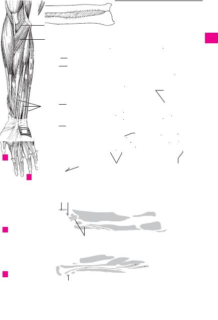
M. flexor carpi radialis. o: Medial epicondyle of |
15 |
M. extensor digitorum. o: Lateral epicondyle of |
||
1 |
|
1 |
|||||
|
|
|
humerus. i: Base of 2nd metacarpal bone. A: Pro- |
|
humerus. i: Distal phalanges 2−5 via their dorsal |
||
|
|
|
|
nation, flexion and radial abduction of the wrist |
|
aponeurosis. A: Extension of fingers and dor- |
|
2 |
|
|
|
joint. I. Median nerve. A |
|
siflexion of the wrist joint. I: Radial nerve. C |
|
|
|
2 |
M. palmaris longus. o: Medial epicondyle of |
16 |
Intertendinous connections. Connexus inter- |
||
|
|
|
|
humerus. i: Palmar aponeurosis. A: Tenses |
|
tendineus [[juncturae tendineum]]. Oblique ten- |
|
3 |
|
|
|
|
|||
|
|
|
aponeurosis and flexes the wrist joint and fin- |
|
dinous connections between the extensor ten- |
||
|
|
|
|
gers 2−5 at the metacarpophalangeal joints. Its |
|
dons of the fingers on the dorsum of the hand. C |
|
4 |
|
|
|
tendon lies above the flexor retinaculum. I: Me- |
17 M. extensor digiti minimi. o: Lateral epicon- |
||
|
|
|
|
dian nerve. A |
|
dyle of humerus. i: Dorsal aponeurosis of 5th fin- |
|
|
|
|
|
|
|||
5 |
3 |
M. flexor carpi ulnaris. o: Medial epicondyle of |
|
ger. A: Extension of little finger, abduction and |
|||
|
|
|
humerus, olecranon and ulna. i: Pisiform bone as |
|
dorsiflexion of the wrist joint. I: Radial nerve. C |
||
|
|
|
|
well as the hamate and 5th metacarpal bones via |
18 |
M. extensor carpi ulnaris. o: Lateral epicondyle |
|
6 |
|
|
|
the pisohamate and pisometacarpal ligaments. |
|
of humerus, radial collateral ligament, posterior |
|
|
|
|
|
A: Ulnar abduction and flexion of the wrist joint. |
|
surface of ulna. i: Base of 5th metacarpal. A: Dor- |
|
|
|
|
|
I: Ulnar nerve. A |
|
siflexion, ulnar abduction. I. Radial nerve. C D |
|
7 |
|
|
|
|
|||
4 |
Humeral head of flexor carpi ulnaris. Caput |
19 |
Humeral head of extensor carpi ulnaris. |
||||
|
|||||||
|
|
|
|
humerale. The part arising from the epicondyle |
|
Caput humerale. The part arising from the |
|
8 |
|
|
|
of the humerus. |
|
humerus. |
|
|
5 |
Ulnar head of flexor carpi ulnaris. Caput ul- |
20 Ulnar head of extensor carpi ulnaris. Caput |
||||
|
|||||||
9 |
|
|
|
nare. The portion originating from the ulna. E |
|
ulnare. The portion arising from the ulna. F |
|
6 |
M. flexor digitorum superficialis. o: Medial |
21 |
M. supinator. o: Lateral epicondyle, radial col- |
||||
|
|||||||
|
|
|
|
epicondyle of humerus, coronoid process of ulna |
|
lateral ligament, supinator crest of ulna. i: Ante- |
|
10 |
|
|
|
|
|||
|
|
|
and anterior surface of radius. i: Middle phalanx |
|
rior surface of radius. A: Supination. I: Radial |
||
|
|
|
|
of fingers 2−5. A: Flexion of all joints it crosses. |
|
nerve. D E F |
|
|
|
|
|
|
|||
11 |
|
|
|
Each tendon is perforated by the corresponding |
22 |
M. abductor pollicis longus. o: Dorsal side of |
|
|
|
|
tendon from the flexor digitorum profundus. I. |
||||
|
|
|
|
|
interosseous membrane and adjacent surfaces |
||
|
|
|
|
Median nerve. A B |
|
||
|
|
|
|
|
of radius and ulna. i: Base of 1st metacarpal. A: |
||
12 |
|
|
|
|
|||
7 |
Humeroulnar head of flexor digitorum su- |
|
Radial abduction and dorsiflexion of the meta- |
||||
|
|
|
|
perficialis. Caput humeroulnare. The portion |
|
carpophalangeal joint of the thumb. Supination. |
|
|
|
|
|
|
|||
13 |
|
|
|
arising from the humerus and ulna. A E F |
|
I: Radial nerve. C D F |
|
8 |
Radial head of flexor digitorum superfi- |
23 |
M. extensor pollicis brevis. o: Radius (extensor |
||||
|
|||||||
|
|
|
|
cialis. Caput radiale. The portion arising from |
|
side) and interosseous membrane. i: Base of pro- |
|
14 |
|
|
|
|
|||
|
|
|
the radius. A E |
|
ximal phalanx of thumb. A: Abduction and ex- |
||
|
9 |
M. flexor digitorum profundus. o: Upper half |
|
tension of thumb at the metacarpophalangeal |
|||
15 |
|
|
|
of ulna. i: Base of distal phalanges of fingers 2−5. |
|
joint. I: Radial nerve. C D F |
|
|
|
|
A: Flexion of all joints crossed. The tendon per- |
24 M. extensor pollicis longus. o: Interosseous |
|||
|
|
|
|
||||
|
|
|
|
forates the corresponding superficial flexor ten- |
|
membrane and dorsal surface of ulna. i: Distal |
|
16 |
|
|
|
|
|||
|
|
|
don. I: Median and ulnar nerves. B E F |
|
phalanx of thumb. A: Adducts and extends the |
||
|
10 |
M. flexor pollicis longus. o: Middle of anterior |
|
thumb. Supinator. I: Radial nerve. C D F |
|||
17 |
|
|
|
surface of radius and usually the medial epicon- |
25 |
M. extensor indicis. o: Interosseous membrane |
|
|
|
|
|
dyle. i: Distal phalanx of thumb. A: Flexion of |
|
and dorsal surface of ulna. i: Dorsal aponeurosis |
|
|
|
|
|
joints crossed. I: Median nerve. B E |
|
of index finger. A: Extends the index finger and |
|
18 |
|
|
|
|
|||
11 |
M. pronator quadratus. o: Lower fourth of |
|
wrist joint. I: Radial nerve. D F |
||||
|
|
|
|
anterior surface of ulna. i: Lower fourth of ante- |
26 M. palmaris brevis. o: Ulnar side of palmar |
||
19 |
|
|
|
rior surface of radius. A: Pronation. I: Median |
|
aponeurosis. i: Skin of ulnar aspect of hand. I. |
|
|
|
|
|
nerve. A B E |
|
Ulnar nerve. A |
|
|
|
|
|
|
|||
2012 and lateral margin of humerus. i: Styloid process of radius. A: Flexion of the elbow joint. It brings
21the arm from the extreme pronation and supination position to the intermediate position. I: Radial nerve. A C E
2213 M. extensor carpi radialis longus. o: Lateral intermuscular septum, lateral epicondyle. i: Ex-M. brachioradialis. o: Intermuscular septum
23 |
tensor side of metacarpal 2. A: Radial abduction |
|
and dorsiflexion of the wrist joint. Flexion of the |
elbow joint. I: Radial nerve. A C
2414 M. extensor carpi radialis brevis. o: Lateral epicondyle of humerus. i: Proximal extensor side of
25third metacarpal. A: Extension and radial abduction of wrist joint. I: Radial nerve. C
Muscles 91
88.18
88.16
88.12 
12 |
|
88.26 |
|
2
1
21
6
13 
9
|
|
|
3 |
|
|
7 |
|
|
|
||
8 |
|
|
|
||
6 |
10 |
||||
|
|||||
11 |
|
|
|
|
11 |
|
|
|
|
||
|
|
|
|
|
|
26 |
|
|
|
||
A Superficial muscles of forearm, anterior view
6
BDeep muscles of forearm, anterior view
5 7
E Radius and ulna with interosseous membrane, anterior view
F Radius and ulna with |
|
interosseous membrane, |
7 |
posterior view |
a
88.19 
|
|
|
|
|
|
12 |
|
|
|
|
|
|
|
|
|
|
|
|
|
|
|
|
|
|
|||||||
88.24 |
|
|
|
|
|
88.24 |
|
|
|
|
|
|
|
|
|
|
|
|
|
||||||||||||
|
|
|
|
|
|
|
|
|
|
|
|
|
|
|
|
|
|||||||||||||||
|
|
|
|
|
|
|
13 |
|
|
|
|
|
|
|
|
|
|
|
|
|
|
|
|
|
|
|
|||||
|
|
|
|
|
|
|
|
|
|
|
|
|
|
|
|
|
|
|
|
21 |
|
|
|
|
|||||||
|
|
|
|
|
|
|
|
|
|
|
|
|
|
|
|
|
|
|
|
|
|
|
|
||||||||
18 |
15 |
88.26 |
|
|
|
|
|
|
|
|
|
|
|
|
|
||||||||||||||||
|
|
|
|
|
|
|
|
|
|
|
|
|
|||||||||||||||||||
|
|
|
|
|
|
|
|
|
|
|
|
|
|
|
|
|
|
|
|
|
|
|
|
|
|
||||||
|
|
|
|
|
|
|
|
|
|
|
|
14 |
24 |
|
|
|
|
|
|
|
|
|
|
|
|
|
|||||
|
|
|
|
|
|
|
|
|
|
|
|
|
|
|
|
|
|
|
|
|
|
||||||||||
|
|
|
|
|
|
|
|
|
|
|
|
|
|
|
|
|
|
|
|
|
|
|
|
|
|
|
|
|
|
||
|
|
|
|
|
|
|
|
|
|
|
|
|
|
|
|
|
|
|
|
|
|
|
|
|
|
|
|
|
|
|
|
|
|
|
|
|
|
|
|
|
|
|
|
|
|
|
|
|
|
|
|
22 |
|||||||||||
17 |
|
|
|
|
|
|
|
22 |
25 |
|
|
|
|
|
|
|
|
|
|
|
|
23 |
|||||||||
|
|
|
|
|
|
|
|
|
|
|
|
|
|
|
|
|
|
||||||||||||||
|
|
|
|
|
|
|
|
|
|
|
|
23 |
|
|
|
|
|
|
|
|
|
|
|||||||||
|
|
|
|
|
|
|
|
|
|
|
|
|
|
|
|
|
|
|
|
|
|
|
|
|
|
||||||
|
|
|
|
|
|
|
|
|
|
|
|
|
|
|
|
|
|
|
|
|
|||||||||||
|
|
|
|
|
|
14 |
18 |
|
|
|
|
|
|
|
|
|
|
|
|
22 |
|||||||||||
|
|
|
|
|
|
|
|
|
|
|
|
|
|
13 |
|
|
|
|
|
|
|||||||||||
|
|
|
|
|
|
|
|
|
|
|
|
|
|
|
|
|
|
|
|
|
|
|
|
|
23 |
||||||
|
|
|
|
|
|
|
|
|
|
|
|
|
|
|
|
24 |
25 |
|
|
|
|
|
|
||||||||
|
|
|
|
|
|
|
|
|
|
|
|
|
|
|
|
|
|
||||||||||||||
|
|
|
|
|
|
|
|
|
|
|
|
|
|
|
|
|
|
|
|
|
|
|
|
|
|
|
|
|
|||
|
|
|
|
|
|
|
|
|
|
|
23 |
|
|
|
|
|
|
|
|
|
|
|
|
|
|
|
|||||
|
|
|
|
|
|
|
|
|
|
|
|
|
|
|
|
|
|
|
|
|
|
||||||||||
|
|
|
|
|
|
|
|
|
|
|
|
|
|
|
|
|
|
|
|
|
|
|
|||||||||
|
|
|
|
16 |
|
|
|
|
|
|
|
|
|
|
|
|
|
|
24 |
|
|||||||||||
|
|
|
|
Superficial extensor |
|
|
Deep extensor muscles |
||||||||||||||||||||||||
|
|
C |
D |
||||||||||||||||||||||||||||
|
|
|
|
muscles of forearm |
|
|
|
|
of forearm |
||||||||||||||||||||||
|
|
|
|
9 |
|
|
|
|
|
|
11 |
|
|
|
|
|
|
|
|
|
|
|
|
|
|
|
|
|
|
|
|
|
|
|
10 |
|
|
|
11 |
|
|
|
|
|
|
|
|
|
|
|
|
|
|
|
|
21 |
8 |
|
|
|
|
|
12 |
||||||
20 |
|
|
|
23 |
|
|
|
|
|||||
|
|
|
|
21 |
|
|
|
|
|
|
|
|
|
|
|
|
|
|
|
|
|
|
|
|
|
|
|
|
|
|
|
|
|
|
|
|
|
|
|||
|
|
|
|
22 |
|
|
|
|
|
|
|||
|
|
|
|
|
|
|
|
|
|
|
|
||
|
|
|
|
|
|
|
|
|
|
|
|
|
|
|
|
|
|
|
|
|
|
|
|
|
|
|
|
|
|
|
|
9 |
24 |
25 |
|
||||||
3
4
5
6
7
8
9
10
11
12
13
14
15
16
17
18
19
20
21
22
23
24
25

92 Muscles
1 M. abductor pollicis brevis. o: Scaphoid bone
1 and flexor retinaculum. i: Lateral sesamoid bone and, radially, the proximal phalanx of the thumb.
2A: Abduction and flexion of the thumb. I: Median nerve. B
3 |
2 M. flexor pollicis brevis. o: Carpal bones, base of |
1st metacarpal and flexor retinaculum. i: Lateral |
sesamoid bone, radially aspect of proximal
4phalanx of thumb. A: Adducts and flexes thumb. B
3Superficial head of flexor pollicis brevis.
Caput superficiale. The portion situated on the 5 tendon of the flexor pollicis longus and innervated
by the median nerve. B
64 Deep head of flexor pollicis brevis. Caput pro-
|
fundum. The portion situated below the tendon of |
7 |
the flexor pollicis longus and innervated by the |
ulnar nerve. B |
5 M. opponens pollicis. o: Trapezium and flexor ret-
8inaculum. i: First metacarpal. A: Adduction and opposition of thumb. I: Median nerve. B
96 M. adductor pollicis. o: Capitate, radiate carpal ligament, metacarpal bone 3. i: Medial sesamoid
bone, ulnar aspect of base of proximal phalanx of 10 thumb. A: Adduction and opposition of thumb. I:
Ulnar nerve. B
11 |
7 Oblique head of adductor pollicis. Caput ob- |
|
liquum. The portion arising from the capitate and |
radiate carpal ligament. B
128 Transverse head of adductor pollicis. Caput transversum. The portion originating from the 3rd
13metcarpal bone. B
9M. abductor digiti minimi. o: Pisoform and flexor
retinaculum. i: Base of proximal phalanx of little
14finger and dorsal aponeurosis. A: Abduction, flexion and extension of the little finger. I. Ulnar nerve.
15B
|
10 M. flexor digiti minimi brevis. o: Hook of hamate |
|
16 |
and flexor retinaculum. i: Base of proximal |
|
phalanx of little finger. A: Flexion of finger at the |
||
|
||
|
metacarpophalangeal joint. I: Ulnar nerve. B |
17 11 M. opponens digiti minimi. o: Hook of hamulus |
||
|
and flexor retinaculum. i: Head and shaft of 5th |
|
18 |
metacarpal. A: Draws the little finger toward the |
|
palm of the hand. I: Ulnar nerve. B |
||
|
||
12 Mm. lumbricales. o: Tendons of flexor digitorum
19profundus. i: Dorsal aponeuroses of fingers 2−5. A: Flexion of finger at the metacarpophalangeal joints, extension at the interphalangeal joints. I:
20Ulnar and median nerves. B C
13 Mm. interossei dorsales. o: Arises by two heads
21from the metacarpals. i: Dorsal aponeurosis of fingers 2−4. A: Spreading of fingers 2−4 away from axis of middle finger, radial and ulnar abduction of
22middle finger, flexion of finger at the metacarpophalangeal joint and extension of the inter-
23phalangeal joints. I. Ulnar nerve. C D E
14 Mm. interossei palmares. o: Metacarpal bones 2, 4 and 5. i: Dorsal aponeuroses of fingers 2, 4 and 5.
24A: Adduction of index, ring and little fingers toward the middle finger, flexion of the metacar-
25pophalangeal joints, extension of the interphalangeal joints. I: Ulnar nerve. B D
15Axillary fascia. Fascia axillaris. Situated on the adipose body of the axilla. It unites the lateral margins of the pectoral and latissimus dorsi muscles. G
16Deltoid fascia. Fascia deltoidea. Strongly fused investing fascia of the deltoid muscle.
17Brachial fascia. Fascie brachii (brachialis). Fascia enclosing the upper arm muscles. F
18Medial intermuscular septum of the arm. Septum intermusculare brachii mediale. Tendinous sheet for muscle origin between the medial margin of the humerus and the brachial fascia. F
19Lateral intermuscular septum of the arm. Septum intermusculare brachii laterale. Tendinous sheet for muscle attachment between the lateral margin of the humerus and the brachial fascia. F
20Antebrachial fascia. Fascia antebrachii. Fascia enveloping the forearm muscles. A
21Fascia of the dorsum of the hand. Fascia dorsalis manus. Fascia situated on the dorsal tendons of the hand. E
22Extensor retinaculum. Retinaculum extensorum [[lig. carpi dors.]]. Transverse fascial fibers over the 6 conduction canals of the 10 extensor tendons. E
23Superficial transverse metacarpal ligament. Lig. metacarpale transversum superficiale. Transverse reinforcement of the palmar fascia of the hand at the level of the heads of the metacarpals. A
24Palmar aponeurosis. Aponeurosis palmaris. Membranous expansion of the tendon of the palmaris longus muscle. A
25Transverse fasciculi. Fasciculi transversi. Transversely oriented fibrous bundles of the palmar aponeurosis. A
26Flexor retinaculum. Retinaculum flexorum [[lig. carpi transversum]]. Stout fibrous band between the scaphoid, trapezoid, pisiform and hamate bones. It binds the flexor tendons. B
27Fibrous sheaths of fingers. Vaginae fibrosae digitorum manus. Fibrous synovial tunnel for the digital flexor tendons. B
28Annular part of fibrous sheath. Pars anularis vaginae fibrosae. Very compact circular fibers of the fibrous sheath located between the joints. B
29Cruciate part of fibrous sheath. Pars cruciformis vaginae fibrosae. Crossed fibers that reinforce the joints. B
30Synovial sheaths of the fingers. Vaginae synoviales digitorum manus. Tendon sheaths of the digital flexor tendons.
31Vincula tendinum. Connective tissue fasciculi (mesotendons) transporting vessels to the tendons. C
32Vinculum longum. Longer fasciculus at the level of the proximal phalanx. C
33Vinculum breve. Shorter fasciculus near the insertions of the tendons. C
34Chiasma tendinum. Crossing of the tendons of the flexor digitorum superficialis and profundus. C
Muscles 93
23
23
|
|
|
|
|
|
|
|
|
|
|
|
|
|
|
|
3 |
|
|
|
|
|
|
|
|
|
|
|
|
|
|
|
|
|
|
|
|
|
|
|
|
|
|
|
|
|
|
|
|
|
4 |
|
|
|
|
|
|
|
|
|
|
|
|
|
|
|
29 |
|
|
|
|
|
|
|
|
|
|
|
|
|
|
|
|
|
|
25 |
|
|
|
|
|
|
|
|
27 |
|
|
|
5 |
|||
|
|
|
|
|
|
|
|
|
|
|
|
|
27 |
|
||
|
|
|
|
|
|
|
|
|
|
|
|
|||||
|
|
|
|
|
|
|
|
|
|
|
|
|
|
|
6 |
|
|
|
|
|
|
|
|
|
|
|
|
|
|
|
|
28 |
|
|
|
|
|
24 |
|
|
|
|
|
|
|
|
|
|
|
|
90.26 |
|
|
|
|
|
|
|
|
|
|
|
12 |
7 |
|||
|
|
|
|
|
|
|
|
|
|
|
|
|||||
|
|
|
|
|
|
|
|
|
|
|
|
|
|
|
|
|
|
|
|
|
|
|
|
|
|
|
|
|
|
|
|
|
8 |
|
|
|
|
|
|
|
14 |
14 |
|
|
|
|
|
|
|
|
|
|
|
|
|
|
|
|
|
|
|
|
|
|
|||
|
|
|
|
20 |
|
8 |
6 |
|
9 |
|||||||
|
|
|
|
|
|
|
|
|
||||||||
|
|
|
|
|
10 |
|
|
|
|
|||||||
|
|
|
|
9 |
|
|
|
|
|
|
|
|
|
|||
|
|
|
|
|
|
7 |
|
|
|
|
10 |
|||||
|
|
Palmar aponeurosis and fasciae |
|
|
|
|
|
|
|
|||||||
|
A |
|
|
|
3 |
|
|
|
|
|
||||||
|
|
|
|
|
|
|
||||||||||
|
|
|
|
11 |
|
|
|
|
|
1 |
5 |
90.10 |
11 |
|||
|
|
|
|
|
|
|
|
|
|
|
|
|
||||
|
|
|
|
|
|
Palm muscles |
|
26 |
|
|
|
|
|
|
|
|
|
|
|
|
|
B |
|
|
|
|
|
|
|
|
|||
|
|
|
|
|
|
|
|
|
|
|
12 |
|||||
|
|
|
|
|
|
|
|
|
|
|
|
|
|
|
|
|
|
|
|
|
|
|
|
90.1 |
|
4 |
|
|
|
|
|||
|
|
|
|
31 |
|
|
|
|
13 |
|||||||
|
|
|
|
|
|
|
|
|
|
|
||||||
|
|
|
|
|
|
|
|
|
|
|
|
|
||||
|
|
|
|
|
|
|
|
|
|
|
|
|
|
|
|
|
|
|
|
90.15 |
|
14 |
|
|
|
|
|
|
|
33 |
32 |
18 |
17 |
15 |
|
|
|
|||
C |
Tendons of finger |
|
|
19 |
16 |
|
|
|
|||
|
|
|
|
||
|
|
34 |
|
|
17 |
|
|
|
|
|
|
|
12 |
13 |
|
|
|
|
|
||||
|
|
F |
Cross section of upper arm |
|
||||||||
|
|
90.6 90.9 |
|
18 |
||||||||
|
|
|
|
|
|
|
|
|
|
|
||
|
|
|
|
|
|
|
|
|
|
|
|
|
|
|
|
|
|
|
|
|
|
|
|
|
|
|
|
14 |
|
|
|
|
|
|
|
19 |
||
|
|
|
13 |
|
|
|
|
|
|
|||
|
|
|
|
|
|
|
|
|
||||
|
|
|
|
|
|
|
|
20 |
||||
13 |
13 |
|
|
|
|
|
|
|
||||
|
|
|
|
|
|
|
|
|||||
|
|
|
|
|
|
|||||||
|
|
|
|
15 |
21 |
|||||||
|
|
|
|
|
|
|
|
|
|
|||
|
|
|
|
|
|
|
|
|||||
|
|
|
|
|
|
|
|
|
|
|
|
|
|
|
21 |
|
|
|
|
|
22 |
||||
|
|
22 |
|
|
|
|
|
|
||||
|
|
|
|
|
|
|
23 |
|||||
|
|
|
|
|
|
|
|
|
|
|
|
|
|
|
|
|
|
|
|
|
|
|
|
|
|
|
|
|
|
|
|
|
|
|
|
|
24 |
|
|
Interosseous muscles, schematic |
|
Dorsum of hand |
|
Cross section of thorax |
|
||||||
D |
E |
|
||||||||||
G |
|
|||||||||||
25 |
||||||||||||
|
|
|
|
|
|
|
|
|
|
|
||
|
|
|
|
|
|
|
|
|
|
|
|
|
a

|
94 |
Muscles |
|
|
|||
|
|
|
1 MUSCLES OF LOWER LIMB. Musculi membri |
14 M. quadratus femoris. o: Ischial tuberosity. i: |
|||
1 |
|
|
|||||
|
|
|
inferioris. |
|
Intertrochanteric crest. A: Lateral rotation and |
||
2 |
|
|
2 M. iliopsoas. Comprised of two muscles, the |
|
adduction of thigh. I: Sacral plexus. A D E |
||
|
|
15 M. sartorius. o: Anterior superior iliac spine. i: |
|||||
|
|
|
psoas major and iliacus. o: Lesser trochanter. A: |
||||
|
|
|
|
Most important flexor and pre-elevator muscle |
|
Medial to tibial tuberosity. A: Flexion, abduc- |
|
|
|
|
|
of the legs; medial and lateral rotation of thigh |
|
tion, lateral rotation of thigh at the hip joint, |
|
3 |
|
|
|
|
|||
|
|
|
at the hip joint. B C D |
|
flexion and medial rotation of leg at the knee |
||
|
|
|
|
|
|||
|
|
|
3 M. iliacus. o: Iliac fossa. i: Lesser trochanter. A: |
|
joint. I: Femoral nerve. C E |
||
4 |
|
|
|
||||
|
|
16 |
M. quadriceps femoris. The muscle group |
||||
|
|
|
Flexion, medial and lateral rotation of thigh at |
||||
|
|
|
|
the hip joint. I: Femoral nerve and lumbar |
|
comprising the three vasti muscles and the rec- |
|
5 |
|
|
|
plexus. C |
|
tus femoris. I: Femoral nerve. |
|
4 |
M. psoas major. o: Bodies and transverse |
17 |
M. rectus femoris. o: Anterior inferior iliac |
||||
|
|||||||
|
|||||||
6 |
|
|
|
processes of L1−4. i: Lesser trochanter. A: Flex- |
|
spine = straight head and upper margin of |
|
|
|
|
ion, medial and lateral rotation of thigh at the |
|
acetabulum. = reflected head. i: Tibial tuberos- |
||
|
|
|
|
hip joint. I: Lumbar plexus. C |
|
ity. A: Flexion of thigh at the hip joint, exten- |
|
7 |
5 |
[M. psoas minor]. o: Bodies of T12 and L1. i: |
|
sion of leg at the knee joint. B C E |
|||
|
|
|
|
Iliac fascia. I: Lumbar plexus. C |
18 |
M. vastus lateralis. o: Greater trochanter, |
|
|
|
|
|
||||
8 |
6 |
M. gluteus maximus. o: Posterior, external sur- |
|
lateral lip of linea aspera. i: Quadriceps tendon. |
|||
|
A: Extension of leg at the knee joint. B C D |
||||||
|
|
|
|
face of ilium, sacrum, coccyx, sacrotuberous |
|
||
|
|
|
|
19 |
M. vastus intermedius. o: Anterior surface of |
||
9 |
|
|
|
ligament. i: Iliotibial tract, gluteal tuberosity, |
|||
|
|
|
lateral intermuscular septum, linea aspera. A: |
|
femur. i: Quadriceps tendon. A: Extension of leg |
||
|
|
|
|
Extension, lateral rotation, abduction and ad- |
|
at the knee joint. B D |
|
10 |
|
|
|
duction of thigh at the hip joint. I: Inferior |
20 |
M. vastus medialis. o: Distal to intertrochan- |
|
|
|
|
|
gluteal nerve. A D E |
|
teric line, medial lip of linea aspera. i: Quadri- |
|
|
|
|
|
|
|||
11 |
7 |
M. gluteus medius. o: External surface of |
|
ceps tendon. A: Extension of leg at the knee |
|||
|
|
|
ilium. i: Greater trochanter. A: Abduction, me- |
|
joint. C D |
||
|
|
|
|
|
|||
|
|
|
|
dial and lateral rotation, flexion and extension |
21 M. articularis genus. o: Anterior surface of |
||
12 |
|
|
|
||||
|
|
|
of thigh at the hip joint. I: Superior gluteal |
|
femur. i: Knee joint capsule. A: Tenses capsule. |
||
|
|
|
|
nerve. A D E |
|
I: Femoral nerve. D |
|
|
|
|
|
|
|||
13 |
8 |
M. gluteus minimus. o: External surface of |
22 M. pectineus. o: Pecten pubis. i: Pectineal line |
||||
|
|
|
|
ilium between anterior and inferior gluteal |
|
below the lesser trochanter. A: Flexion, adduc- |
|
|
|
|
|
|
|||
|
|
|
|
lines. i: Greater trochanter. A: Abduction, me- |
|
tion and lateral rotation of thigh at the hip joint. |
|
14 |
|
|
|
|
|||
|
|
|
dial and lateral rotation, flexion and extension |
|
I: Femoral and obturator nerves. B C D E |
||
|
|
|
|
of the thigh at the hip joint. I: Superior gluteal |
23 M. adductor longus. o: Near the symphysis. i: |
||
15 |
|
|
|
nerve. A D E |
|||
|
|
|
|
Medial lip of linea aspera. A: Adduction and |
|||
|
|
8 a Gluteal aponeurosis. Aponeurosis glutealis. |
|
||||
|
|
|
|
flexion of thigh at the hip joint. I: Obturator |
|||
|
|
|
|
||||
16 |
|
|
|
Deep, sheet-like tendon of origin of the gluteus |
|
nerve. B C D E |
|
|
|
|
maximus lying on the gluteus medius. |
24 M. adductor brevis. o: Inferior ramus of pubis. |
|||
|
|
|
9 M. tensor fasciae latae. o: Near the anterior su- |
||||
|
|
|
|
i: Medial lip of linea aspera. A: Adduction, flex- |
|||
17 |
|
|
|
||||
|
|
|
perior iliac spine. i: Above the iliotibial tract |
|
ion, extension and lateral rotation of thigh at |
||
|
|
|
|
lateral to the tibial tuberosity. A: Flexion, ab- |
|
the hip joint. I: Obturator nerve. B D E |
|
|
|
|
|
|
|||
18 |
|
|
|
duction and medial rotation of thigh at the hip |
25 |
M. adductor magnus. o: Ischial tuberosity, |
|
|
|
|
joint. Flexion, extension and final rotation at |
||||
|
|
|
|
|
ischial ramus. i: Medial lip of linea aspera and |
||
|
|
|
|
the knee joint. I: Superior gluteal nerve. C E |
|
||
19 |
|
|
|
|
with a long tendon to the medial epicondyle. A: |
||
10 |
M. piriformis. o: Anterior surface of sacrum. i: |
|
|||||
|
Adduction and extension of thigh at the hip |
||||||
|
|
|
|
Greater trochanter, inner side of apex. A: Ab- |
|
joint. I: Obturator and sciatic nerves. B C D E |
|
20 |
|
|
|
duction, extension and lateral rotation of thigh |
25 a M. adductor minimus. Uppermost part of the |
||
|
|
|
|
at the hip joint. I. Sacral plexus. A D |
|
adductor magnus muscle. It arises from a more |
|
|
|
11 M. obturator internus. o: Inner surface of ob- |
|
||||
21 |
|
|
anterior part of the pelvis. |
||||
|
|
|
turator membrane and environment. i: Tro- |
26 M. gracilis. o: Inferior ramus of pubis medial to |
|||
|
|
|
|
||||
|
|
|
|
chanteric fossa. A: Lateral rotation, abduction |
|||
|
|
|
|
|
the adductor magnus muscle. i: Medial to tibial |
||
22 |
|
|
|
and adduction of thigh. I: Sacral plexus. A D |
|
||
|
|
|
|
tuberosity. A: Adduction, flexion and extension |
|||
|
|
12 M. gemellus superior. o: Ischial spine. i: Ten- |
|
of thigh at the hip joint. Flexion and medial ro- |
|||
23 |
|
|
|
don of obturator internus and trochanteric |
|
tation of the knee joint. I: Obturator nerve. A C E |
|
|
|
|
fossa. A: Lateral rotation, adduction and abduc- |
|
|
||
|
|
|
|
|
|
||
|
|
|
|
tion of thigh. I: Sacral plexus. A D E |
|
|
|
2413 M. gemellus inferior. o: Ischial tuberosity. i: Tendon of obturator internus, trochanteric
25fossa. A: Lateral rotation, adduction and abduction of thigh. I: Sacral plexus. A D E