Материал: Атлас Ханц фениш

|
80 |
Muscles |
|
|
|||
|
|
|
M. depressor anguli oris. [[Triangularis]]. o: |
16 |
Lateral pterygoid muscle. M. pterygoideus |
||
1 |
|
1 |
|||||
|
|
|
Anterior and lateral part of mandible. i: Angle |
|
lateralis. o: Lateral surface of lateral pterygoid |
||
|
|
|
|
of mouth. I: Facial nerve. A |
|
plate and lower surface of greater wing of sphe- |
|
2 |
|
2 |
M. transversus menti. Transverse muscular |
|
noid. Arises by two heads (variant: three heads), |
||
|
|
|
|
connection between the right and left depres- |
|
one from the discocapsular system, the other |
|
|
|
|
|
|
from the pterygoid fossa. The inferior head pulls |
||
|
|
|
|
sor anguli oris muscles below the chin. I: Facial |
|
||
3 |
|
|
|
|
|||
|
|
|
|
the mandible and discocapsular system forward. |
|||
|
|
|
nerve. A |
|
|||
|
|
|
|
|
The superior head determines the velocity at |
||
|
|
|
3 M. risorius. o: Parotid fascia and buccal skin. i: |
|
|||
4 |
|
|
|
which the discocapsular system is pulled back |
|||
|
|
|
Angle of mouth. I: Facial nerve. A |
|
|||
|
|
|
|
into place. I: Mandibular nerve. B |
|||
|
4 |
M. zygomaticus major. o: Lateral side of zygo- |
|
||||
|
17 |
Medial pterygoid muscle. M. pterygoideus me- |
|||||
5 |
|||||||
|
|
|
matic bone. i: Angle of mouth. I: Facial nerve. A |
|
dialis. o: Pterygoid fossa and tuber of maxilla. i: |
||
|
|
|
5 M. zygomaticus minor. o: Anterior side of zy- |
|
Pterygoid tuberosity and inner aspect of angle of |
||
|
|
|
|
||||
6 |
|
|
|
gomatic bone. i: Upper lip. I: Facial nerve. A |
|
mandible. It courses obliquely downward and |
|
6 |
M. levator labii superioris [[quadratus labii |
|
backward and is a synergist to the temporalis |
||||
|
|
||||||
|
|
and masseter muscles. I: Mandibular nerve. B |
|||||
|
|
|
|
sup., M. levator nasi et labii maxillaris lat.]]. o: |
|
||
7 |
|
|
|
|
|||
|
|
|
18 |
Buccopharyngeal fascia. Fascia buccopharyn- |
|||
|
|
|
Above the infra-orbital foramen. Radiates into |
||||
|
|
|
|
orbicularis oris. I: Facial nerve. A |
|
gea. It lies on the buccinator muscle and extends |
|
8 |
7 |
M. levator labii superioris alaeque nasi [[quad- |
|
from the angle of the mouth deeply as far as the |
|||
|
pharyngeal constrictor muscle. C |
||||||
|
|
|
|
ratus labii sup., M. lev. nasi et labii maxillaris |
|
||
|
|
|
|
19 |
Masseteric fascia. Fascia masseterica. Fascia |
||
9 |
|
|
|
med]]. o: Medial to orbit. i: Nasal ala and upper |
|||
|
|
|
|
covering the masseter muscle, part of which at- |
|||
|
|
|
lip. I: Facial nerve. A |
|
|||
|
|
|
|
|
taches below the parotid at the upper end of the |
||
|
8 |
M. depressor labii inferioris [[quadr. labii inf.]]. |
|
||||
10 |
|
zygomatic arch. D |
|||||
|
|
|
Located below the depressor anguli oris. o: Pla- |
|
|||
|
|
|
20 Parotid fascia. Fascia parotidea. Fascial covering |
||||
|
|
|
|
tysmaandmandible.i:Lowerlip.I:Facialnerve.A |
|||
|
|
|
|
|
of the parotid, partly identical with the masse- |
||
11 |
9 |
M. levator anguli oris [[Caninus]]. o: Canine |
|
||||
|
teric fascia. D |
||||||
|
|
|
|
fossa. i: Angle of mouth. I: Facial nerve. A |
21 |
Temporal fascia. Fascia temporalis. External |
|
|
|
|
|
||||
12 |
|
|
9 a Modiolus. Palpable muscular mass lateral to the |
|
connective tissue investment of the temporalis |
||
|
|
|
angle of the mouth. Point of convergence of adja- |
|
|||
|
|
|
|
|
muscle between the superior temporal line and |
||
|
|
|
|
cent muscles radiating into the orbicularis oris. |
|
the zygomatic arch. It consists of the following |
|
13 |
|
|
|
|
|||
10 |
M. buccinator. Cheek muscle. o: Pterygoman- |
|
two layers. D |
||||
|
|
|
|
dibular raphe and adjoining parts of upper and |
22 |
Superficial layer. Lamina superficialis. Layer of |
|
14 |
|
|
|
lower jaw. i: Angle of mouth and orbicularis oris. |
|
the temporal fascia attached to the outer margin |
|
|
|
|
|
I: Facial nerve. A B |
|
of the zygomatic arch. D |
|
|
|
|
|
|
|||
15 |
11 |
M. mentalis. Arises over the roots of the lower |
23 |
Deep layer. Lamina profunda. Layer of the tem- |
|||
|
|
|
|
incisors. i: Skin of chin (chin dimple). I: Facial |
|
poral fascia attached to the inner margin of the |
|
|
|
|
|
nerve. A |
|
zygomatic arch. D |
|
16 |
|
|
|
|
|||
12 |
M. masseter. Most prominent masticatory |
24 |
NECK MUSCLES. Musculi colli (cervicis). A C |
||||
|
|
|
|
muscle. Closes jaw and, together with the tem- |
25 |
Platysma. Cutaneous muscle occupying an ex- |
|
|
|
|
|
||||
17 |
|
|
|
poral and medial pterygoid muscles, determines |
|
tensive area of the neck. It extends from the |
|
|
|
|
the degree of masticatory power. It is comprised |
|
lower part of the face to the upper thorax. I: Fa- |
||
|
|
|
|
|
|||
|
|
|
|
of a superficial and deep part. I: Mandibular |
|
cial nerve. A D |
|
18 |
|
|
|
|
|||
|
|
|
nerve. A C |
26 |
Sternocleidomastoid muscle. M. sternoclei- |
||
|
13 |
Superficial part. Pars superficialis. o: Anterior |
|
domastoideus. o: Sternum und clavicle. i: Mas- |
|||
19 |
|
|
|
two-thirds of zygomatic arch. i: Angle of |
|
toid process and superior nuchal line. It elevates |
|
|
|
|
|
mandible. It courses obliquely backward and |
|
the chin and rotates it to the opposite side. I: Ac- |
|
20 |
|
|
|
downward. It also draws the mandible some- |
|
cesory nerve, cervical plexus. C |
|
|
|
|
what forward. C |
|
|
||
14 Deep part. Pars profunda. Size varies. Arises
21from the zygomatic arch proximal to the mandibular joint and discocapsular system. i: Mandible. Action: Together with fibers of the
22temporal muscles, it ensures lateral stabilization of the discocapsular system during laterotrusal
23movement. C
15M. temporalis. o: Temporal fossa. i: Coronoid process that extends downward to the occlusal
24plane and to the region of the pterygomandibular raphe. Action: Elevation and retraction of the
25mandible, fixation of the pharynx when swallowing; cf. pp. 12, 14. I: Mandibular nerve. B
Muscles 81
|
|
|
|
|
|
|
|
|
|
|
|
|
|
|
|
7 |
|
3 |
|
6 |
9 |
|
|
4 |
|||
5 |
|||
|
410
3 |
|
|
|
|
12 |
|
|
|
|
|
5 |
||
|
|
|
|
|
|
|
|
|
|
|
|||
|
|
|
|
|
|
|
|
|
|
|
|
|
6 |
1 |
8 |
11 |
|
|
|
|
|
|
|
|
|||
|
|
|
|
|
|
|
|
||||||
|
|
|
|
|
|
|
7 |
||||||
|
|
|
|
|
|
|
|
|
|
|
|
|
|
|
|
|
|
|
|
|
|
|
|
|
|
|
|
|
|
|
|
|
|
|
|
|
|
|
|
|
|
|
|
|
|
|
|
|
|
|
|
|
|
|
8 |
|
|
25 |
2 |
|
|
|
|
15 |
|
|
|
||
|
|
|
|
|
|
|
|
|
|
|
|||
|
|
|
|
|
|
|
|
|
|
|
|
|
|
|
|
|
|
|
|
|
|
|
|
|
|
9 |
|
|
|
|
|
|
|
|
|
|
|
|
|
|
|
|
|
|
|
|
|
|
|
|
|
|
|
|
|
|
|
|
|
|
|
|
|
|
|
|
|
|
|
|
|
|
|
|
|
|
|
|
|
|
|
|
10 |
A |
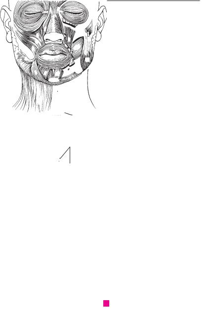
Facial muscles, anterior view |
16 |
|
|
|
||||||||
|
|
|
|||||||||||
|
|
11 |
|||||||||||
|
|
|
|
|
|
|
|
||||||
|
|
|
|
|
|
12 |
|
|
|
|
|
|
|
|
|
|
|
|
|
|
|
|
|
|
|
|
|
|
|
|
|
|
|
|
12 |
||||||
|
|
|
|
|
|
17 |
10 |
||||||
|
|
|
|
|
|
|
|
Masticatory |
|
||||
|
|
|
|
|
|
|
|
|
|||||
14 |
|
|
|
|
|
B |
|
13 |
|||||
|
|
|
|
|
|||||||||
|
|
|
|
|
|
muscles |
|
||||||
|
|
|
|
|
|
|
|
|
|
||||
13 |
|
|
|
|
14 |
|
|
|
|
|
|
||
|
|
18 |
|
|
|
|
26 |
|
|
|
15 |
||
|
|
|
|
|||
|
|
|
|
|
||
|
|
|
|
|
||
|
|
|
|
16 |
||
|
|
|
|
|
|
|
|
|
|
|
|
|
|
|
|
|
|
|
|
|
|
|
|
|
|
|
17 |
|
|
22 |
|
|
|
|
|
|
|
|
|
|
|
|
|
|
|
|
18 |
|
|
|
|
|
|
|
|
|
|
|
|
|
|
|
|
|
|
|
|
|
|
|
|
|
|
|
|
19 |
|
|
|
23 |
|
||
|
|
|
|
|||
|
|
|
20 |
|||
|
|
|
|
|
|
|
|
Muscles of head and neck, |
|
|
|
|
|
|
|
|
|
|
|
|
C |
20 |
|
|
|
21 |
|
|
right inferior view |
|
|
|
||
|
|
|
|
|
|
|
|
|
19 |
|
|
|
|
|
|
|
|
|
22 |
|
|
|
|
|
|
|
|
|
|
|
|
|
|
|
|
|
|
|
|
|
23 |
|
|
|
|
25 |
|
|
|
|
|
|
|
|
|
24
D Fasciae of the head
25
a

|
82 |
Muscles |
|
|
|
|
|
|||
|
|
|
M. longus colli. Arches to connect the 2nd to 5th |
15 |
M. omohyoideus. o: Upper margin of scapula |
|||||
1 |
|
1 |
||||||||
|
|
|
cervical vertebrae with the lower cervical and |
|
medial to scapular notch. i: Body of hyoid bone. |
|||||
|
|
|
|
upper thoracic vertebrae. Its fibers also extend |
|
An intermediate tendon situated above the |
||||
2 |
|
|
|
from the vertebral bodies to the transverse |
|
jugular vein divides it into two bellies. A: Draws |
||||
|
|
|
|
processes with the C6 transverse process as the |
|
the hyoid downward and tenses the cervical fas- |
||||
|
|
|
|
central point. A: Lateral and forward flexion of |
|
cia. I: see p. 13. A C |
|
|
||
|
|
|
|
|
|
|
||||
3 |
|
|
|
|
|
|
||||
|
|
|
the neck. I: Ventral ramus of spinal nerve. D |
16 |
Superior belly. Venter superior. Upper segment |
|||||
|
|
|
|
|
||||||
|
2 |
M. scalenus anterior. o: Transverse processes of |
|
of omohyoid between the hyoid and interme- |
||||||
4 |
|
|||||||||
|
|
|
C3−6. i: Scalene tubercle on 1st rib. A: Elevation of |
|
diate tendon. A |
|
|
|
||
|
|
|
|
1st rib, lateral flexion and rotation of neck. It sep- |
17 |
Inferior belly. Venter inferior. Lower half of |
||||
|
|
|
|
|||||||
5 |
|
|
|
arates the anterior and posterior or scalenus |
||||||
|
|
|
gaps. I: see p. 1. D |
|
omohyoid from the intermediate tendon to the |
|||||
|
|
|
|
|
scapular notch. A |
|
|
|||
|
|
|
3 M. scalenus medius. o: Transverse processes of |
|
|
|
||||
6 |
|
|
18 |
M. sternothyroideus. o: Posterior surface of |
||||||
|
|
|
C2−7. i: 1st rib behind groove for subclavian |
|||||||
|
|
|
|
artery. A: Elevation of 1st rib and lateral flexion of |
|
manubrium and 1st rib. i: Oblique line of thyroid |
||||
7 |
|
|
|
|
cartilage. A: Draws larynx downward. I: see p. 13. |
|||||
|
|
|
neck. I: see p. 1. D |
|
||||||
|
|
|
|
A |
|
|
|
|||
|
|
|
|
|
|
|
|
|
||
|
4 |
M. scalenus posterior. o: Transverse processes |
19 |
M. thyrohyoideus. o: Oblique line of thyroid |
||||||
8 |
||||||||||
|
|
|
of C4−6. i: Upper margin of 2nd rib. A: Elevates the |
|
cartilage. i: Greater horn of hyoid bone. A: Brings |
|||||
|
|
|
|
rib, laterally flexes the neck. I: see p. 1. D |
|
hoyid and thyroid cartilage closer together. I: C1 |
||||
|
|
|
|
|
||||||
9 |
5 |
[M. scalenus minimus]. Extra muscle occasion- |
|
via hypoglossal nerve. A |
|
|
||||
|
|
|
|
ally present between scalenus anterior and me- |
20 [M. levator glandulae thyroideae]. Part of the |
|||||
|
|
|
|
|||||||
10 |
|
|
|
dius. o: Transverse processes of C6 or 7. i: 1st rib |
|
thyrohyoid muscle that extends to the thyroid |
||||
|
|
|
and pleural cupola. |
|
||||||
|
|
|
|
gland. |
|
|
|
|||
|
6 |
SUPRAHYOID MUSCLES M. suprahyoidei. The |
21 CERVICAL FASCIA. Fascia cervicalis. Collective |
|||||||
11 |
||||||||||
|
|
|
following muscles above the hyoid bone. A |
|||||||
|
|
|
|
term for the connective tissue layers of the neck. |
||||||
|
|
|
7 M. digastricus. o: Notch medial to the mastoid |
22 Superficial (investing) layer. Lamina superfi- |
||||||
12 |
|
|
||||||||
|
|
|
process. i: Inner side of mandible. It has an inter- |
|||||||
|
|
|
|
cialis. Superficial layer of cervical fascia that sur- |
||||||
|
|
|
|
mediate tendon which acts on the lesser horn of |
|
rounds the sternocleidomastoid and trapezius |
||||
13 |
|
|
|
the hyoid bone by means of a connective tissue |
|
|||||
|
|
|
|
muscles. It is attached to the anterior margin of |
||||||
|
|
|
|
sling. A: Elevation of hyoid. A |
|
the manubrium, the clavicle and the mandible. C |
||||
|
8 |
Anterior belly. Venter anterior. It extends from |
|
|||||||
14 |
23 |
Pretracheal layer. Lamina pretrachealis. Layer |
||||||||
|
|
|
the mandible to the intermediate tendon. A: |
|||||||
|
|
|
|
Pulls mandible forward and depresses it. I: Mylo- |
|
that spreads |
between |
the |
two omohyoid |
|
15 |
|
|
|
|
muscles and is attached to the posterior margin |
|||||
|
|
|
hyoid nerve. A E |
|
||||||
|
|
|
|
of the manubrium and clavicle. It surrounds the |
||||||
|
|
|
|
|
|
|||||
|
9 |
Posterior belly. Venter posterior. It passes from |
|
infrahyoid muscles. C |
|
|
||||
16 |
|
|
|
|||||||
|
|
|
the mastoid process to the intermediate tendon. |
24 |
Prevertebral |
layer. Lamina |
prevertebralis. |
|||
|
|
|
|
A: It draws back the hyoid bone. I: Facial nerve. A |
||||||
|
|
|
|
|
Layer that lies between the vertebral column and |
|||||
17 |
|
|
|
E |
|
pharyngeal constrictors as well as the |
||||
|
|
|
|
|
||||||
|
10 |
M. stylohyoideus. o: Styloid process. i: Lesser |
|
esophagus, covers the scaleni muscles and con- |
||||||
|
|
|||||||||
18 |
|
|
|
horn of hyoid bone. It accompanies the posterior |
|
tains the sympathetic trunk and phrenic nerve. C |
||||
|
|
|
|
|
|
|
|
|||
|
|
|
belly of the digastric muscle and can pass |
25 |
Carotid sheath. Vagina |
carotica. Connective |
||||
|
|
|
|
|||||||
|
|
|
|
through it via a fissure. A: It pulls the hyoid back- |
||||||
|
|
|
|
|
tissue investing the neurovascular bundle |
|||||
19 |
|
|
|
|
||||||
|
|
|
ward and upward. I: Facial nerve. A E |
|
||||||
|
|
|
|
(carotid artery, jugular vein and vagus nerve) |
||||||
|
11 |
M. mylohyoideus. Muscle of the floor of the |
|
and continuous with the pretracheal layer. C |
||||||
|
|
|
|
|
|
|||||
20mouth. o: Mylohyoid line of mandible. i: Body of hyoid bone. A: Draws the hyoid forward and upward and forms the diaphragma oris. I: Mylohy-
21oid nerve. A B
22 |
12 |
M. geniohyoideus. o: Mental spine. i: Body of |
|
hyoid bone. A: Draws the hyoid forward and up- |
|
|
|
ward. I: C1 via the hypoglossal nerve. B |
23 |
|
|
13 |
Infrahyoid muscles. Mm. infrahyoidei. The |
|
|
|
muscles below the hyoid bone (infrahyoid m.). I: |
24 |
|
Ansa cervicalis. A |
14 M. sternohyoideus. o: Posterior surface of ma-
25nubrium sterni. i: Body of hyoid bone. A: Draws the hyoid downward. I: see p. 13. A
Muscles 83
|
|
|
|
|
|
|
|
|
|
|
|
|
10 |
|
8 |
|||||
|
|
|
|
|
|
|||||||||||||||
|
|
|
|
|
|
|
|
|
|
|
|
|
|
|
9 |
|
||||
9 |
10 |
|
|
|
|
|
|
|
|
|
|
|
|
|
||||||
|
|
|
|
|
|
|
|
|
|
|
|
|
|
|
|
|||||
|
|
|
|
|
|
|
|
|
|
|
|
|
|
|
|
|||||
|
|
|
8 |
|
|
|
|
|
|
|
|
|
|
|
|
|
||||
|
|
|
|
|
|
|
|
|
|
|
|
|
Segment of A |
|||||||
|
|
|
|
|
|
|
|
|
|
|
|
E |
||||||||
|
|
|
11 |
|
|
|
|
|
|
|
|
|
|
|
|
|
|
|||
|
|
|
19 |
|
|
|
16 |
|
|
|
|
|
|
|
|
|
|
|
|
|
|
|
|
14 |
|
|
|
|
|
|
|
|
|
|
|
|
|
|
|
||
|
|
|
|
|
|
|
|
|
|
|
|
|
|
|
|
|
||||
|
|
|
|
|
|
|
|
|
|
|
|
|
|
|
|
|
|
|
||
|
|
|
|
18 |
|
|
|
|
|
|
|
|
|
|
|
11 |
|
|
||
|
|
|
|
|
|
|
|
|
|
|
|
|
|
|
|
|
||||
|
|
|
11 |
12 |
|
|
|
|||||||||||||
|
|
|
|
|
|
|
|
|
|
|
|
|
|
|
|
|
|
|
|
|
|
|
|
|
|
|
|
|
|
|
|
|
|
|
|
|
|
|
|
|
|
17 |
|
|
|
|
|
|
|
|
|
|
|
|
|
|
|
|
|
|
||
|
Muscles of hyoid bone |
|
|
|
|
|
|
Muscles of floor of mouth |
||||||||||||
A |
|
|
|
|
B |
|||||||||||||||
23 |
22 |
|
|
|
|
|
|
|
from above and behind |
|||||||||||
|
|
|
|
|
|
|
|
|
|
|
|
|
|
|
|
|
||||
80.26 
25
15
23
22
24
3
2
|
|
5 |
C |
Fasciae of the neck |
4 |
|
|
1 1
DDeep neck muscles, anterior view
a
3
4
5
6
7
8
9
10
11
12
13
14
15
16
17
18
19
20
21
22
23
24
25

|
84 |
Muscles |
|
|
|||
|
|
|
MUSCLES OF THORAX. Musculi thoracis. A−F |
18 |
Internal intercostal membrane. Membrana in- |
||
1 |
|
1 |
|||||
|
2 |
[M. sternalis]. Muscle that occasionally (4%) |
|
tercostalis interna. Continuation of the internal |
|||
|
|
|
intercostal muscles from the rib angle to the |
||||
|
|
|
|
crosses the pectoralis major muscle parallel and |
|
||
2 |
|
|
|
|
vertebrae. E |
||
|
|
|
proximal to the sternum. A |
|
|||
|
|
3 |
M. pectoralis major. Consists of the three parts |
19 |
Innermost intercostal muscles. Mm. inter- |
||
|
|
||||||
3 |
|
costales intimi. Internal portion of the internal |
|||||
|
|
|
listed below (nos. 4−6). o: Clavicle, sternum, first |
|
|||
|
|
|
|
intercostal muscles separated by the intercostal |
|||
|
|
|
|
4−6 costal cartilages and rectus sheath. i: Crest of |
|
||
|
|
|
|
|
vessels. F |
||
4 |
|
|
|
greater tubercle and humerus. A: Adduction and |
|
||
|
|
|
20 |
Subcostal muscles. Mm. subcostales. Internal |
|||
|
|
|
medial rotation of arm. I: Medial and lateral pec- |
||||
|
|
|
|
toral nerves. A |
|
intercostal muscles that pass over 1−2 ribs. I: |
|
5 |
4 |
Clavicular part. Pars clavicularis. The portion |
|
see p. 17. E |
|||
21 |
M. transversus thoracis. Situated on the inner |
||||||
|
|
|
|
originating from the clavicle. A |
|||
|
|
|
|
||||
6 |
5 |
Sternocostal part. Pars stenocostalis. The por- |
|
surface of the anterior thoracic wall, it radiates |
|||
|
obliquely upward from the sternum to costal |
||||||
|
|
|
|
tion arising from the sternum and ribs. A |
|
cartilages 2−6. I: see p. 17. C |
|
|
|
|
|
|
|||
7 |
6 |
Abdominal part. Pars abdominalis. The portion |
|
||||
22 |
Thoracic fascia. Fascia thoracica. Epimysium of |
||||||
|
|
|
|
arising from the rectus sheath. A |
|
the inner thoracic musculature. |
|
|
|
|
|
|
|||
87 M. pectoralis minor. It lies beneath the pec- 23 Diaphragm. Diaphragma [thoraco-abdomi- toralis major. o: Ribs 3−5. i: Coracoid process. A: nale]. Dome-shaped, muscular partition be-
|
|
Draws scapula forward and downward and ribs |
|
tween the thoracic and abdominal cavities. I: |
||
9 |
|
|
||||
|
upward. Accessory respiratory muscle. I: see |
|
Phrenic nerve. C D |
|||
|
|
p. 3. A |
24 Lumbar part of diaphragm. Pars lumbalis dia- |
|||
|
|
|||||
10 |
8 |
M. subclavius. o: 1st costal cartilage. i: Lower |
||||
|
phragmatis. Medial part of the diaphragm aris- |
|||||
|
|
surface of clavicle. A: Stabilizes sternoclavicular |
|
ing from the lumbar vertebral bodies, inter- |
||
|
|
|
||||
|
|
joint against tension. I: Subclavian nerve. A |
|
vertebral discs and fibrous arches. D |
||
11 |
|
|
||||
9 |
Pectoral fascia. Fascia pectoralis. It covers the |
25 |
Right crus of lumbar part. Crus dextrum. o: |
|||
|
||||||
|
|
pectoralis major muscle, is attached to the |
|
L1−3(4). D |
|
|
12 |
|
|
|
|||
|
clavicle and sternum and is continuous with the |
26 |
Left crus of |
lumbar part. Crus sinistrum. o: |
||
|
|
axillary fascia. |
||||
|
|
|
L1−2(3). D |
|
||
13 |
10 |
Clavipectoral fascia. Fascia clavipectoralis. Fas- |
|
|
||
27 |
Costal part of diaphragm. Pars costalis dia- |
|||||
|
|
cia attached to the coracoid process and the |
|
phragmatis. The part of the diaphragm originat- |
||
|
|
|
||||
14 |
|
clavicle. It covers the pectoralis minor and sub- |
|
ing from ribs 7−12. C D |
||
|
clavius muscles. A |
28 Sternal part of diaphragm. Pars sternalis dia- |
||||
|
11 |
M. serratus anterior. o: Ribs 1−9. i: Medial mar- |
||||
|
|
phragmatis. The part of the diaphragm arising |
||||
15 |
|
|||||
|
gin of scapula. A: Supports, lowers and rotates |
|
from the sternum. C D |
|||
|
|
the scapula and draws it forward. It assists in |
29 Aortic (opening) hiatus. Hiatus aorticus. Pas- |
|||
16 |
|
raising the arm high above the horizontal plane. |
||||
|
|
sageway for the aorta between the right and left |
||||
|
I: Long thoracic nerve. A |
|
||||
|
|
|
crus of the lumbar part. D |
|||
|
12 Mm. levatores costarum. Muscles behind and |
|
||||
17 |
30 |
Esophageal |
(opening) hiatus. Hiatus oe- |
|||
|
below the long back muscles. o: Thoracic trans- |
|||||
|
|
|
sophageus. Passageway for the esophagus and |
|||
|
|
verse processes. i: Ribs. I: Posterior ramus of spi- |
|
the vagus nerves above the aortic opening. D |
||
18 |
|
nal nerve. B |
|
|||
|
31 Central tendon. Centrum tendineum. Clover- |
|||||
|
13 |
Mm. levatores costarum longi. o: Transverse |
||||
|
|
leaf-shaped, tendinous central area of the dia- |
||||
19 |
|
processes. i: Passes over a rib to insert on the next |
|
|||
|
|
phragm. D |
|
|||
|
|
lower rib. A: Elevates the ribs. B |
32 Foramen for the inferior vena cava. Foramen |
|||
|
14 |
Mm. levatores costarum breves. o: Transverse |
||||
20 |
|
venae cavae. Opening in the central tendon for |
||||
|
|
process. i: Next lower rib. B |
|
the inferior vena cava. D |
||
|
15 |
External intercostal muscles. Mm. inter33 |
Medial arcuate ligament. Lig. arcuatum medi- |
|||
21 |
||||||
|
costales externi. They extend obliquely forward |
|
ale. Tendinous arch between the body and trans- |
|||
|
|
and downward between the ribs. A: Inspiration, |
|
verse process of L1 or L2 forming the passageway |
||
22 |
|
bracing of the ribs. I: Intercostal nerves. A E F |
|
for the psoas muscle. D |
||
|
16 |
External intercostal membrane. Membrana in- |
34 |
Lateral arcuate ligament. Lig. arcuatum |
||
|
||||||
23 |
|
tercostalis externa. Membrane that replaces the |
|
laterale. Tendinous arch over the quadratus lum- |
||
|
external intercostal muscles anteriorly between |
|
borum muscle between the transverse process |
|||
|
|
|
||||
|
|
the costal cartilages. A |
|
of L1 and the 12th rib. D |
||
24 |
|
|
||||
17 |
Internal intercostal muscles. Mm. intercostales |
35 |
Median arcuate ligament. Lig. arcuatum medi- |
|||
|
|
interni. They pass obliquely backward and |
|
anum. Tendinous arch over the aortic hiatus. D |
||
25downward between the ribs. A: Partially expiratory, bracing of the ribs. I: Intercostal nerves. E F