Материал: Атлас Ханц фениш
Muscles 75
3 |
|
|
|
|
|
|
80.26 |
|
|
|
|
|
|
|
|
|
||||
|
|
|
|
|
|
|
|
|
|
|
|
|
|
|
||||||
20 |
|
|
|
|
|
|
|
|
|
|
|
|||||||||
|
|
|
|
|
|
|
|
|||||||||||||
|
|
|
|
|
|
|
|
|
|
|
|
|
||||||||
|
|
|
|
|
|
|
|
9 |
|
|
|
|
|
|
|
|
||||
|
|
|
|
|
|
|
|
2 |
|
|
|
|
|
|
|
|
|
|||
2 |
|
|
|
|
|
|
|
|
|
|||||||||||
|
|
|
|
|
|
|
|
|
|
|||||||||||
|
|
|
|
|
|
|
|
6 |
|
|
|
|
|
|
|
|
|
|
|
|
|
|
|
|
|
|
|
|
5 |
|
|
|
88.10 |
|
|
|
|||||
|
|
|
|
|
|
|
|
|
|
|
|
|
|
|||||||
|
|
|
|
|
|
|
|
4 |
|
|
|
|||||||||
|
|
|
|
|
|
|
|
|
|
|
|
|
|
|
|
|||||
4 |
|
|
|
|
|
|
|
|
|
|
|
|
|
|
|
|
|
|||
|
|
|
|
|
|
|
|
8 |
|
|
|
|
|
|
|
|
|
|
||
4 |
|
|
|
|
|
|
|
|
|
|
|
|
|
|||||||
|
|
|
|
|
|
|
|
20 |
|
|
|
|||||||||
86.9 |
|
|
|
|
|
|
|
|
|
|
|
|
|
|
|
|
|
19 |
76.4 |
|
|
|
|
|
|
|
|
|
|
|
|
|
|
|
|
|
|
|
|
||
|
|
|
|
|
|
|
|
86.18 |
|
|
|
|
|
|
|
|
|
|||
|
|
|
|
|
|
|
|
9 |
|
|
|
|
||||||||
|
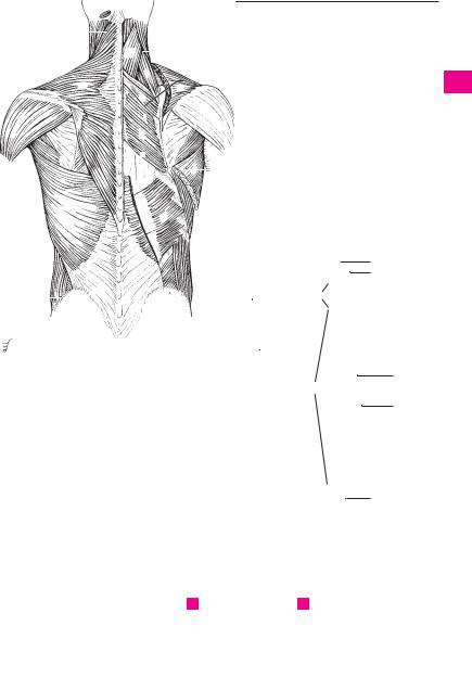
Superficial dorsal muscles |
|
|
|
|
|
|
|
|
19 |
|
|||||||||
|
|
|
||||||||||||||||||
|
|
|
|
|
|
|
|
|
||||||||||||
A |
17 |
|
|
|
|
|
|
|
||||||||||||
|
|
|
|
|
|
|||||||||||||||
76.3 
 76.2
76.2
 11 12
11 12
16
86.9 
14
13
 18
18
12
17
 16
16
12
B Deep dorsal muscles C Autochthonous
dorsal muscles, schematic view
a
3
4
5
6
7
8
9
10
11
12
13
14
15
16
17
18
19
20
21
22
23
24
25

|
76 |
Muscles |
|
|
|
|
||||
|
|
|
[[Medial tract]]. [Tractus mediale]. |
|
20 |
Thoracolumbar fascia. Fascia thoracolumbalis. |
||||
1 |
|
1 |
|
|||||||
|
2 |
M. spinalis. Muscular system attached to the |
|
Encasing fascia of the erector spinae muscle. It |
||||||
|
|
|
is attached to the spinous processes with a su- |
|||||||
|
|
|
|
spinous processes and consisting of the follow- |
|
|||||
2 |
|
|
|
|
perficial layer, to the costal processes with a |
|||||
|
|
|
ing three segments: See p. 75 B |
|
|
|||||
|
|
3 |
M. spinalis thoracis. o: Transverse processes |
|
deep layer, both layers being united laterally. |
|||||
|
|
|
Associated muscles: transversus abdominis, |
|||||||
|
|
|
||||||||
3 |
|
|||||||||
|
|
|
of T11−L2. i: Spinous processes of T2−11. A: |
|
serrati posteriores, latissimus dorsi and, in |
|||||
|
|
|
|
Back flexion. See p. 75 C |
|
|
some cases, the internal oblique muscle of the |
|||
|
|
|
|
|
|
|||||
4 |
4 |
M. spinalis cervicis. o: Transverse processes of |
|
abdomen. F |
||||||
|
|
|||||||||
|
|
|
|
C6−T2. i: Spinous processes of C2−4. A: Back |
21 |
Nuchal fascia. Fascia nuchae (nuchalis). Dorsal |
||||
|
|
|
|
flexion. See p. 75 C |
|
|
|
continuation of the superficial layer of the cer- |
||
5 |
|
|
|
|
|
|
||||
5 |
M. spinalis capitis. Inconstant part of semispi- |
|
vical fascia. (Investing fascia of the true neck |
|||||||
|
|
|
|
nalis capitis with additional origins from the |
|
musculature.) |
||||
6 |
|
|
|
|
||||||
|
|
|
|
|
||||||
|
|
|
upper thoracic and lower cervical spinous |
22 |
HEAD MUSCLES. Musculi capitis. |
|||||
|
|
|
|
processes. |
|
|
23 |
MM. SUBOCCIPITALES. The following seven |
||
|
|
|
|
|
|
|||||
7 |
6 |
MM. INTERSPINALES. Unlike the spinal muscles, |
||||||||
|
muscles: |
|||||||||
|
|
|
|
these muscles extend only between the |
24 |
M. rectus capitis anterior. o: Lateral mass of |
||||
|
|
|
|
|||||||
|
|
|
|
spinous processes of continuous vertebrae. A: |
||||||
8 |
|
|
|
|||||||
|
|
|
Dorsiflexion. C D |
|
|
|
the atlas. i: Basilar part of occipital bone. A: For- |
|||
|
7 |
Mm. interspinales cervicis. These are paired |
|
ward flexion of head. I: Anterior rami of spinal |
||||||
|
|
|||||||||
|
|
nerves. E |
||||||||
9 |
|
|||||||||
|
|
|
because of the bifid cervical spinous processes. |
25 M. rectus capitis posterior major. o: Spinous |
||||||
|
|
|
|
D |
|
|
|
|||
|
|
|
|
|
|
|
|
process of axis. i: Middle of inferior nuchal line. |
||
10 |
8 |
Mm. interspinales thoracis. Usually absent. C |
|
|||||||
|
A: Outward rotation and dorsiflexion of head. I: |
|||||||||
|
9 |
Mm. |
interspinales |
lumborum. |
Especially |
|
Suboccipital nerve. D. See also p. 79 A |
|||
|
|
|||||||||
11 |
|
|
|
strong muscle bands. C |
|
26 |
M. rectus capitis posterior minor. o: Posterior |
|||
|
10 |
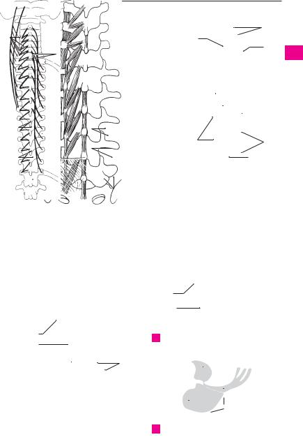
MM. TRANSVERSOSPINALES. Collective term for |
|
tubercle of atlas. i: Inner third of inferior nuchal |
||||||
|
|
|||||||||
12 |
|
|
|
the following nine muscles. A B C |
|
|
line. A: Mainly dorsiflexion of head. I: Sub- |
|||
|
|
|
|
|
occipital nerve. D. See also p. 79 A |
|||||
|
11 M. semispinalis. The longest superficial por- |
|
||||||||
|
|
|
|
|||||||
13 |
|
|
|
tion of the transversospinales. It spans four or |
|
|
||||
|
|
|
more vertebrae and comprises the following |
|
|
|||||
|
|
|
|
|
|
|||||
|
|
|
|
three segments: A B |
|
|
|
|
||
14 |
|
|
|
|
|
|
|
|||
12 |
M. semispinalis thoracis. o: Transverse |
|
|
|||||||
|
|
|
|
processes of T7−I2. i: Spinous processes of C6− |
|
|
||||
15 |
|
|
|
T6. A: Primarily dorsiflexion. A |
|
|
|
|||
|
13 |
M. |
semispinalis |
cervicis. o: |
Transverse |
|
|
|||
|
|
|
||||||||
16processes of T1−6. i: Spinous processes of C2−5. A: Mainly dorsiflexion. A
17 |
14 |
M. semispinalis capitis. o: Transverse |
|
|
processes of C4−T6. i: Occipital bone between |
||
|
|
superior and inferior nuchal lines. A: Dorsiflex- |
|
18 |
|
ion of head and rotation, depending on starting |
|
|
|
position. A |
|
19 |
15 |
Mm. multifidi. Portion of the transversospinal |
|
|
|
system spanning 2−4 vertebrae. A: Dor- |
|
|
|
solateral flexion and slight rotation. B |
|
20 |
|
||
16 |
Mm. rotatores. Deepest layer of the transver- |
||
|
|
sospinal system with short fibers taking an |
|
21 |
|
especially transverse course, thereby providing |
|
|
|
stronger rotation. They attach to an adjacent or |
|
|
|
||
|
|
superior vertebra. A B C |
|
22 |
|
||
17 |
Mm. rotatores cervicis. o: Inferior articular |
||
|
|
process. i: Arch or root of spinous process of |
|
23 |
|
||
|
cervical vertebrae. A |
||
|
18 |
Mm. rotatores thoracis. o: Transverse process |
|
24 |
|||
|
of thoracic vertebrae. i: Spinous process. A C |
19 Mm. rotatores lumborum. o: Mamillary
25process. i: Roots of spinous process of lumbar vertebrae. A C
Muscles 77
18
14 |
|
|
|
74.24 |
|
|
|
|
|
|
||
|
|
|
|
|
|
|
|
|
|
|||
|
|
|
|
|
|
|
|
8 |
||||
|
|
|
|
17 |
|
|
|
|
|
|
|
|
|
|
|
|
|
13 |
|
|
|
|
|
|
|
|
|
|
|
|
|
|
|
|
|
|
|
|
|
|
|
|
|
|
|
|
|
|
|
|
|
|
|
|
|
74.22 |
|
|
|
|
|
|
||
|
|
|
|
|
|
|
|
|
||||
|
|
|
|
74.23 |
|
|
|
|
|
9 |
||
|
|
|
|
|
|
|
|
|
||||
|
|
|
|
20 |
|
|
|
|||||
|
|
|
|
|
|
|||||||
|
|
|
|
|
|
|
|
|
|
|||
18 |
|
|
|
11 |
|
|
|
|
|
|
|
|
|
|
|
|
|
12;11 |
|
|
|
|
|
|
|
|
|
|
|
|
|
|
|
|
|
|
|
|
|
|
|
|
74.21 |
|
6 |
||||||
|
|
|
|
15 |
|
|
||||||
|
|
|
|
|
|
|
|
|
|
|
||
19 |
|
|
|
|
|
|
|
19 |
|
|||
16 |
|
|
|
20 |
|
|
|
|
|
|
||
|
|
|
16 |
|
|
|
|
|
|
|
||
|
|
|
|
|
|
|
|
|
|
|||
|
|
|
|
|
|
|
|
|
|
|
||
A |
Systems of mm. |
B |
Transversospinal |
C |
Short muscles of the |
|
transversospinales |
|
system |
|
vertebral column |
24
|
78.1 |
26 |
74.28 |
25
78.2
E Deep neck muscles, anterior view
78.3
|
|
|
|
94.4 |
|
|
|
|||||
74.25 |
|
|
7;6 |
|
|
|
|
86.32 |
||||
|
|
|
|
|
|
|||||||
|
|
|
|
|
||||||||
|
Short neck muscles |
74.10 |
|
|
|
|
|
|
|
|
||
|
|
|
|
|
|
|
|
|
||||
|
|
|
|
|
|
|
|
|
||||
D |
|
|
20 |
|
||||||||
F Thoracolumbar fascia
a
3
4
5
6
7
8
9
10
11
12
13
14
15
16
17
18
19
20
21
22
23
24
25

|
78 |
Muscles |
|
|
|||
|
|
|
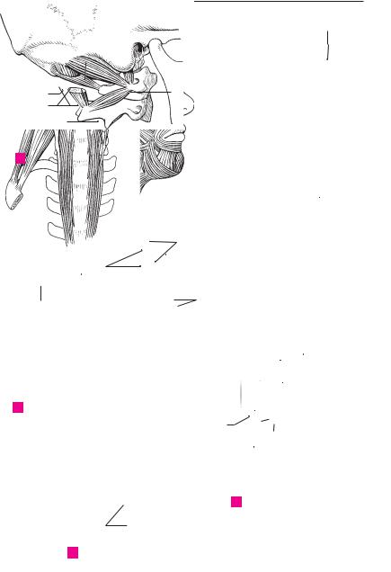
M. rectus capitis lateralis. o: Transverse |
|
of the eye. Comprises three segments. It closes |
||
1 |
|
1 |
|
||||
|
|
|
process of atlas. i: Jugular process of occipital |
|
the eyelids and assists the flow of tears into the |
||
|
|
|
|
bone. A: Lateral flexion of head. I: Anterior rami |
|
lacrimal sac and into the nose. i: Facial nerve. C |
|
2 |
|
|
|
of spinal nerves C1−2. A B, see also p. 77 E |
|
D |
|
|
|
2 |
M. obliquus capitis superior. o: Transverse |
18 |
Palpebral part. Pars palpebralis. Fibers sit- |
||
3 |
|
|
|
process of atlas. i: Field above attachment of |
|
uated in the eyelids passing from the medial |
|
|
|
|
rectus capitis posterior major. A: Backward and |
|
palpebral ligament and the adjacent bones to |
||
|
|
|
|
lateral flexion of head. I: Posterior rami of spi- |
|
the lateral palpebral ligament. C |
|
4 |
|
|
|
|
|||
|
|
|
nal nerves C1−2. A, see also p. 77 D |
19 |
Orbital part. Pars orbitalis. Arises from the |
||
|
3 |
M. obliquus inferior. o: Spinous process of axis. |
|||||
|
|
medial palpebral ligament and adjacent bones, |
|||||
5 |
|
|
|
i: Transverse process of atlas. A: Lateral rotation |
|
thus encircling the eye. C |
|
|
|
|
of atlas and face toward the same side. I: Poste- |
|
|||
|
|
|
20 |
Lacrimal part. Pars lacrimalis. o: Posterior |
|||
|
|
|
|
rior rami of spinal nerves C1−2. A, see also p. 77 |
|||
6 |
|
|
|
D |
|
lacrimal crest. It curves around the lacrimal |
|
|
|
|
|
canaliculus, extends partially behind the lacri- |
|||
|
|
|
4 M. longus capitis. o: Anterior tubercle of trans- |
|
|||
|
|
|
|
mal sac and radiates into the palpebral part of |
|||
7 |
|
|
|
verse processes of C3−6. i: Basal part of occipi- |
|
||
|
|
|
|
the orbicularis oculi muscle below the medial |
|||
|
|
|
tal bone. A: Forward and lateral flexion of head |
|
|||
|
|
|
|
and cervical vertebral column. I: Anterior rami |
|
palpebral ligament. D |
|
|
|
|
|
|
|||
8 |
|
|
|
of spinal nerves C1−2. B |
21 |
M. corrugator supercilii. o: Nasal part of fron- |
|
|
|
|
5 FACIAL AND MASTICATORY MUSCLES. Musculi |
|
tal bone. i: Skin over the middle of the eyebrow. |
||
|
|
|
|
||||
|
|
|
|
faciales et masticatorii. |
|
Located below the orbicularis oculi muscle. I: |
|
9 |
|
|
|
|
|||
|
|
6 M. epicranius. Collective term for the muscles |
|
Facial nerve. D |
|||
|
|
|
|
attaching to the galea aponeurotica. I: Facial |
22 |
M. depressor supercilii. Muscle medial to the |
|
10 |
|
|
|
||||
|
|
|
nerve. C |
|
corrugator supercilii that radiates from the |
||
|
7 |
M. occipitofrontalis. Muscle extending ante- |
|
orbicular oculi into the skin of the medial part |
|||
|
|
||||||
11 |
|
|
|
riorly and posteriorly into the galea |
|
of the eyebrow. I: Facial nerve. D |
|
|
|
|
|
|
|||
|
|
|
|
aponeurotica. C |
23 |
M. auricularis anterior. Muscle in front of the |
|
|
|
|
|
||||
|
|
|
8 Frontal belly. Venter frontalis. The portion of the |
|
ear. o: Temporal fascia. i: Spine of helix. I: Facial |
||
12 |
|
|
|
||||
|
|
|
occipitofrontalis which passes from the galea |
|
nerve. C |
||
|
|
|
|
aponeurotica to the eyebrows. A: Moves scalp |
24 M. auricularis superior. o: Galea aponeurotica. |
||
13 |
|
|
|
forward and raises the eyebrows. C |
|||
|
|
|
|
i: Root of pinna. I: Facial nerve. C |
|||
|
|
|
9 Occipital belly. Venter occipitalis. The portion of |
25 M. auricularis posterior. o: Mastoid process. i: |
|||
|
|
|
|||||
14 |
|
|
|
the occipitofrontalis which passes from the su- |
|||
|
|
|
|
Root of pinna. I: Facial nerve. C |
|||
|
|
|
preme nuchal line to the galea aponeurotica. A: |
|
|||
|
|
|
|
Moves galea aponeurotica backward. C |
26 |
M. orbicularis oris. Encircles the mouth open- |
|
15 |
10 |
M. temporoparietalis. o. Region of superior |
|
ing and consists of two parts (see nos. 27, 28). It |
|||
|
|
|
|
auricular muscle. i: Galea aponeurotica. C |
|
assists in closing the lips and helps to empty |
|
|
|
|
|
|
|||
|
11 |
Galea aponeurotica (aponeurosis epicrania- |
|
the vestibule of the mouth. I: Facial nerve. C D E |
|||
16 |
|
||||||
27 |
Marginal part. Pars marginalis. The peripheral |
||||||
|
|
|
|
lis). Displaceable, helmet-like, superficial ten- |
|||
|
|
|
|
don for the two parts of the epicranius. It lies |
|
margin radiating into the neighboring muscles. |
|
17 |
|
|
|
|
|||
|
|
|
against the periosteum and is attached to the |
|
D |
||
|
|
|
|
supreme nuchal line and to the external occipi- |
28 |
Labial part. Pars labialis. The main part of the |
|
|
|
|
|
||||
18 |
|
|
|
tal protuberance. C |
|
orbicularis oris including the portion which |
|
12 |
M. procerus. o: Dorsum of nose. i: Skin above |
|
|||||
|
takes a hook-like bend externally below the red |
||||||
|
|
|
|
the nose. A: Depression of frontal skin. I: Facial |
|
portion of the lips. C D E |
|
19 |
|
|
|
|
|||
|
|
|
nerve. C |
|
|
||
|
|
13 M. nasalis. Common term for the following two |
|
|
|||
|
|
|
|||||
20 |
|
|
|
nasal muscles: I: Facial nerve. D |
|
|
|
|
|
14 Transverse part of nasalis. Pars transversa |
|
|
|||
|
|
|
|||||
|
|
|
|
[[compressor naris]]. o: Field over root of canine |
|
|
|
21 |
|
|
|
|
|
||
|
|
|
tooth. i: Superficial tendon on dorsum of nose. |
|
|
||
|
|
|
|
D |
|
|
|
22 |
|
|
|
|
|
||
15 |
Alar part of nasalis. Pars alaris [[dilatator |
|
|
||||
|
|
|
|
naris]]. o: Above the lateral incisor tooth. i: |
|
|
|
|
|
|
|
|
|
||
23 |
|
|
|
Margins of the nasal openings and the adjacent |
|
|
|
|
|
|
region. D |
|
|
||
|
|
|
|
|
|
|
|
16 M. depressor septi. o: Above the medial incisor
24tooth. i: Cartilaginous nasal septum. A: Depresses tip of nose. I: Facial nerve. D
25
17 M. orbicularis oculi. Circular sphincter muscle
Muscles 79
2
76.26 |
1 |
|
76.25
3
A Deep suboccipital muscles, lateral view
|
11 |
|
|
|
|
|
|
|
8 |
|
|
|
10 |
19 |
|||
|
|
|
|||
24 |
|
|
|
|
12 |
|
|
|
|||
9 |
18 |
|
|
|
|
|
|
|
19 |
|
|
|
|
|
|
|
|
|
23 |
|
|
|
|
 28 25
28 25 
C Superficial muscles of head
28 

28
ESagittal section through the lips
|
|
|
|
|
|
|
|
|
|
|
76.24 |
|
|
|
|
|
|
|
|
1 |
|
|
|
|
|
|
|
||
|
|
|
|
|
|
|
|
|
|
|
|
|||
|
|
|
|
|
|
|
|
|
|
|
|
|||
|
|
|
|
|
|
|
|
|
|
3 |
||||
|
|
|
|
|
|
|
|
|
|
|
|
|
|
|
|
|
|
|
|
|
|
|
|
|
|
|
|
|
|
|
|
|
|
|
|
|
|
|
|
|
|
|
|
|
|
|
|
|
|
|
|
|
|
|
|
|
|
|
4 |
|
|
|
|
|
4 |
|
|
|
|
|
|
|||
|
|
|
|
|
|
|
|
|
|
|
||||
|
|
|
|
|
|
|
|
|
|
5 |
||||
|
|
|
|
|
|
|
|
|
|
|
|
|
|
|
|
|
|
|
|
|
|
|
|
|
|
|
|
|
|
|
|
|
|
|
|
|
|
|
|
|
|
|
|
|
|
|
|
|
|
|
|
|
|
|
|
|
|
|
6 |
|
|
|
|
|
|
|
|
|
|
|
|
|
|
|
|
|
|
|
|
|
|
|
|
|
|
|
|
|
|
|
|
|
|
|
|
|
|
|
|
|
|
|
|
7 |
|
|
|
|
|
|
|
|
|
|
|
|
|
|
|
|
|
|
|
|
|
|
|
|
|
|
|
|
|
|
|
|
|
|
|
|
|
|
|
|
|
|
|
|
8 |
|
|
|
|
|
|
|
|
|
|
|
|
|
82.1 |
|
|
|
|
|
|
|
|
|
|
|
|
|
|
|
|
|
|
|
|
|
|
|
|
|
|
|
9 |
|||
|
|
|
|
|
|
|
|
|
|
|
|
|||
|
|
|
|
|
|
|
|
|
|
|
|
|
|
|
|
|
|
|
|
|
|
|
|
|
|
|
|
|
|
|
|
|
|
|
|
|
|
|
|
|
|
|
|
|
|
|
|
|
|
|
|
|
|
|
|
|
|
|
10 |
|
|
|
|
|
|
|
|
|
|
|
|
|
|
|
|
|
|
|
|
|
|
|
|
|
|
|
|
|
|
|
|
|
|
|
|
|
|
|
|
|
|
|
|
11 |
|
|
|
|
|
|
|
|
|
|
|
|
|
|
|
|
|
|
|
|
|
|
|
|
|
|
|
|
|
|
|
|
|
|
|
|
|
|
|
|
|
|
|
|
12 |
|
|
|
|
Neck, anterior view |
|
|
|
|
||||||
|
|
B |
|
|
|
|
||||||||
|
|
|
13 |
|||||||||||
|
|
|
|
|
|
|
|
|
|
|
|
|
|
|
|
|
|
|
|
|
|
|
|
|
|
|
|
|
|
|
|
|
|
|
|
|
|
|
|
|
|
|
|
|
|
|
|
|
|
|
|
|
|
|
|
|
|
|
14 |
|
|
|
|
|
|
|
|
|
|
|
|
|
|
|
|
|
|
|
|
|
|
|
|
|
|
|
|
|
|
|
|
|
|
|
22 |
21 |
|
|
|
15 |
||||
|
|
|
|
|
|
|
|
|
||||||
|
|
|
|
|
|
|
|
|
|
|
|
|
|
|
|
|
|
|
|
|
|
|
|
|
|
|
|
|
16 |
|
|
|
|
|
|
|
|
|
|
|
|
|
|
|
|
|
|
|
|
|
|
|
|
|
|
|
|
|
|
|
|
|
|
|
|
|
|
|
|
|
|
|
|
|
|
|
|
|
|
|
|
|
|
|
|
|
|
|
|
|
|
|
|
|
|
|
|
|
|
|
|
|
17 |
|
13 |
|
|
|
|
20 |
|
|
|
|
|||||
|
|
|
|
|
|
|
|
|
|
|
||||
|
|
|
|
|
|
|
|
|
|
|
|
|
|
|
|
|
|
|
|
|
|
|
|
|
|
|
|
|
|
|
|
|
|
|
|
|
|
|
|
|
|
|
|
|
15 |
|
|
|
|
|
|
|
|
|
|
|
|
|
18 |
|
|
|
|
27 |
|
|
|
|
|
|
|
|||
16 |
|
|
|
|
|
|
|
|
|
|
|
|||
|
|
|
|
|
|
|
|
|
19 |
|||||
|
|
|
|
|
|
|
|
|
|
|
|
|
||
28 |
|
|
|
|
|
|
|
|
|
|
|
|
|
|
|
|
|
|
|
|
|
|
|
|
|
|
|
|
|
|
|
|
|
|
|
|
|
|
|
|
|
|
20 |
|
|
|
|
|
|
|
|
|
|
|
|
|
|
||
|
|
|
|
|
|
|
|
|
|
|
|
|
|
|
|
|
|
|
|
|
|
|
|
|
|
|
|
|
|
|
|
|
|
|
|
|
|
|
|
|
|
|
|
21 |
|
|
|
|
|
|
|
|
|
|
|
|
|
|
|
22
D Deep mimic muscles
23
24
25
a