Материал: Атлас Ханц фениш
Sutures, joints and ligaments 65
15 |
15 |
|
15 |
|
14 |
|
|
14 |
|
|
|
|
||
|
|
|
|
|
|
11 |
|
|
|
7 |
7 |
|
7 |
7 |
|
|
|
|
7 |
|
|
|
2 |
|
3
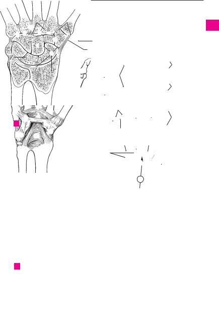
ACarpal joints in horizontal section
15 15
15
12 12
8 8 8 8
10
11 |
|
|
|
|
|
|
|
|
|
|
|
|
|
|
|
|
|
|
|
20 |
|||
|
|
|
|
|
|
|
|
|
|
|
|
21 |
22 |
|
|
|
|
|
|
|
|||
|
|
|
|
|
|
|
20 |
||||
|
|
|
|
|
|
|
|
||||
21 |
|
|
|
|
|
|
|
|
|
|
|
17 |
|
|
|
|
|
|
|
||||
|
|
|
|
|
|
|
|
||||
18 |
|
|
|
|
|
|
|
|
16 |
||
|
|
|
|
|
|
|
|
|
|
||
|
19 |
|
19 |
19 |
|
|
|||||
|
13 |
13 |
13 |
|
|
||||||
|
|
|
|
|
|||||||
|
9 |
|
|
|
|
|
9 |
|
|
||
|
|
|
|
|
|
||||||
|
|
|
|
|
|
|
|
|
|
||
|
5 |
|
4 |
|
|
|
10 |
||||
|
|
|
|
||||||||
|
|
|
|
|
|
|
|||||
|
|
|
|
|
|
|
|
6 |
|
|
|
1
2
3
4
5
6
7
8
9
10
11
12
13
14
15
16
17
CCarpal joints of right hand, dorsal view
|
Carpal joints, palmar view |
18 |
B |
||
|
|
|
|
|
19 |
|
|
|
|
|
|
|
|
20 |
|
|
|
21
22
23
24
25
a |
a |

66 Sutures, joints and ligaments
1 |
|
1 |
JOINTS OF THE PELVIC GIRDLE. Articulationes |
14 |
Dorsal sacroiliac ligaments. Ligg. sacroiliaca |
||||||||
|
|
cinguli pelvici. |
|
|
|
|
|
|
posteriora (dorsalia). Superficial bundle of liga- |
||||
|
|
2 Obturator membrane. Membrana obturatoria. |
|
ments attached dorsally |
to the interosseous |
||||||||
|
|
|
|||||||||||
2 |
|
|
sacroiliac ligaments between the sacrum and |
||||||||||
|
|
Membrane which closes off the obturator fora- |
|
||||||||||
|
|
|
men except for the obturator canal. It is covered |
|
ilium. B |
|
|||||||
|
|
|
15 |
Pubic symphsis. Symphysis pubica. Synchon- |
|||||||||
3 |
|
|
by the obturator externus and internus muscles. |
||||||||||
|
|
A B C D |
|
|
|
|
|
|
|
drosis that articulates with the interpubic disc. A |
|||
|
3 |
Obturator canal. Canalis obturatorius. Opening |
16 |
Superior pubic ligament. Lig. pubicum su- |
|||||||||
4 |
|||||||||||||
|
|
in the supralateral part of the obturator mem- |
|
perius. Fibrous connection between the two |
|||||||||
|
|
|
brane. It corresponds to the obturator groove be- |
|
halves of the symphysis emanating from the pec- |
||||||||
|
|
|
|
||||||||||
5 |
|
|
tween the |
two obturator |
tubercles |
and is |
|
ten ossis pubis on either side. A |
|||||
|
|
traversed by the obturator artery, vein and nerve. |
17 |
Arcuate pubic ligament. Lig. arcuatum pubis. |
|||||||||
|
|
|
|||||||||||
|
|
|
A C D |
|
|
|
|
|
|
|
Strong, curved ligament below the symphysis. A |
||
6 |
|
|
|
|
|
|
|
|
|
||||
4 |
Lumbosacral joint. Articulatio lumbosacralis. |
18 |
Interpubic disc. Discus interpubicus. Fibrocar- |
||||||||||
|
|
|
Articulation between the sacrum and lumbar |
|
tilage plate with a synovia-filled median groove, |
||||||||
7 |
|
|
vertebra 5 (4). A |
|
|
|
|
|
|
located between the articular surfaces made of |
|||
|
5 |
Iliolumbar ligament. Lig. iliolumbale. Strong |
|
hyaline cartilage on the right and left pubic |
|||||||||
|
|
||||||||||||
|
|
|
ligament thatpasses to the ilium mainly from the |
|
bones. A |
|
|||||||
8 |
|
|
|
|
|||||||||
|
|
transverse processes of L4 and 5. A B |
|
19 |
JOINTS OF THE FREE LOWER LIMB. Articula- |
||||||||
|
|
|
|
||||||||||
|
6 |
Sacrotuberous |
ligament. Lig. sacrotuberale. |
|
tiones membri inferioris liberi. |
||||||||
9 |
|
||||||||||||
|
|
Strong ligament that extends from the medial |
20 |
HIP JOINT. Articulatio coxae (iliofemoralis). Joint |
|||||||||
|
|
|
margin of the ischial tuberosity to the sacrum |
|
formed by the acetabulum and the head of the |
||||||||
|
|
|
|
||||||||||
10 |
|
|
and ilium. B D |
|
|
|
|
|
|
femur. A B C |
|
||
7 |
Falciform |
process. |
Processus |
falciformis. |
21 |
Joint capsule. Capsula articularis. It is attached |
|||||||
|
|||||||||||||
|
|||||||||||||
|
|
|
Slender extension of fibers from the sacro- |
|
anteriorly to the intertrochanteric line, posteri- |
||||||||
11 |
|
|
|
||||||||||
|
|
tuberous ligament to the inner aspect of the |
|
orly above to the intertrochanteric crest. A frac- |
|||||||||
|
|
|
ischium. B D |
|
|
|
|
|
|
ture of the neck of the femur can therefore be in- |
|||
12 |
8 |
Sacrospinous |
ligament. |
Lig. |
sacrospinale. |
|
tracapsular when in the anterior region or extra- |
||||||
|
|
|
Fibrous band medial to the sacrotuberous liga- |
|
capsular when in the posterior region. A |
||||||||
|
|
|
|
||||||||||
13 |
|
|
ment. It passes from the ischial spine to the |
22 |
Orbicular zone. Zona orbicularis. Ligamentous |
||||||||
|
|
sacrum and coccyx and separates the greater |
|
fibers encircling the neck of the femur. B |
|||||||||
|
|
|
from the lesser sciatic foramen. B D |
|
23 Iliofemoral ligament. Lig. iliofemorale. Strong |
||||||||
14 |
|
|
|
||||||||||
|
|
|
|
|
|
|
|
|
|||||
9 |
Greater sciatic foramen. Foramen sciaticum |
|
anteriorligamentofthehipjointcapsuleextend- |
||||||||||
|
|
|
(ischiadicum) majus. Foramen between the |
|
ing from the ilium to the intertrochanteric line. A |
||||||||
|
|
|
|
||||||||||
15 |
|
|
greater sciatic notch, sacrum, sacrospinous liga- |
|
B |
|
|||||||
|
|
ment and the upper part of the sacrotuberous |
24 Ischiofemoral ligament. Lig. ischiofemorale. It |
||||||||||
|
|
|
|||||||||||
|
|
|
ligament. It is traversed by the piriformis muscle, |
|
radiates into the orbicular zone from the poste- |
||||||||
|
|
|
|
||||||||||
16 |
|
|
superior and inferior gluteal arteries, veins and |
|
|||||||||
|
|
|
rior margin of the acetabulum and is also at- |
||||||||||
|
|
|
nerves, the internal pudendal vein, pudendal |
|
tached to the anterior margin of the greater tro- |
||||||||
|
|
|
|
||||||||||
17 |
|
|
nerve, sciatic nerve and posterior femoral cu- |
|
chanter and to the intertrochanteric line. B |
||||||||
|
|
taneous nerve. A B D |
|
|
|
|
|
||||||
|
|
|
|
|
|
25 |
Pubofemoral ligament. |
Lig. pubofemorale. |
|||||
|
10 |
Lesser sciatic foramen. Foramen sciaticum |
|||||||||||
|
|
Ligament that arises medially from the joint cap- |
|||||||||||
18 |
|
||||||||||||
|
|
(ischiadicum) minus. Foramen between the |
|
sule of the pubic bone and extends to the orbicu- |
|||||||||
|
|
|
lesser sciatic notch and the sacrospinous and |
|
lar zone and to the part of the femur proximal to |
||||||||
|
|
|
|
||||||||||
19 |
|
|
sacrotuberous ligaments. It transmits the obtu- |
|
the lesser trochanter. A |
|
|||||||
|
|
rator internus muscle as well as the internal pu- |
|
|
|||||||||
|
|
|
26 Acetabular lip. Labrum acetabulare. A ring of fi- |
||||||||||
|
|
|
dendal artery and vein and the pudendal nerve to |
||||||||||
|
|
|
|
brocartilage and connective tissue that |
|||||||||
20 |
|
|
the ischiorectal fossa. B D |
|
|
|
|
||||||
|
|
|
|
|
|
completes and deepens the bony acetabulum. C |
|||||||
|
11 |
Sacroiliac |
joint. Articulatio sacroiliaca. Joint |
27 |
Transverse acetabular |
ligament. Lig. trans- |
|||||||
21 |
|
|
connected by fibers that permits little motion |
||||||||||
|
|
|
versumacetabuli.Itbridgestheacetabularnotch. |
||||||||||
|
|
[[amphiarthrosis]]. It |
is located |
between the |
|
||||||||
|
|
|
|
C |
|
||||||||
|
|
|
sacrum and the ilium and may become syn- |
|
|
||||||||
22 |
|
|
28 |
Ligament of head of femur. Lig. capitis femoris. |
|||||||||
|
|
osteotic. A |
|
|
|
|
|
|
|||||
|
12 |
Ventral sacroiliac ligaments. Ligg. sacroiliaca |
|
A smooth ligament extending from the acetabu- |
|||||||||
|
|
lar notch to the pit on the head of the femur. It |
|||||||||||
23 |
|
|
anteriora (ventralia). Thin but broad fibrous |
|
|||||||||
|
|
|
transmits blood vessels and has no direct me- |
||||||||||
|
|
|
bandsthatextendfromtheanteriorsurfaceofthe |
|
chanical action. C |
|
|||||||
|
|
|
first and second sacral vertebrae to the ilium. A D |
|
|
||||||||
24 |
|
|
|
|
|
||||||||
13 |
Interosseous |
sacroiliac |
ligaments. |
Ligg. |
|
|
|
||||||
|
|
|
sacroiliaca interossea. Dorsal mass of ligaments |
|
|
|
|||||||
25that pass from the tuberosity of the sacrum to the tuberosity of the ilium. B
Sutures, joints and ligaments 67
5 |
|
1 |
|
|
|
|
|
|
|
|
|
|
|
|
|
|
|
|
|
|
|
|
2 |
|
|
|
|
11 |
|
|
|
4 |
||||
|
12 |
|||||||
86.10 |
|
|
|
|
|
|
||
|
|
|
|
|
|
|||
20;21 |
|
|
9 |
|
|
|
||
23 |
|
|
3 |
16 |
|
|||
23 |
|
25 |
|
|
|
|
||
|
|
|
|
|
||||
|
|
|
|
|
|
|
|
|
|
|
|
|
|
|
|
|
18 |
|
|
|
|
|
|
|
||
21 |
|
|
|
|
17 |
|||
|
|
15 |
||||||
|
|
|
|
|
|
|
||
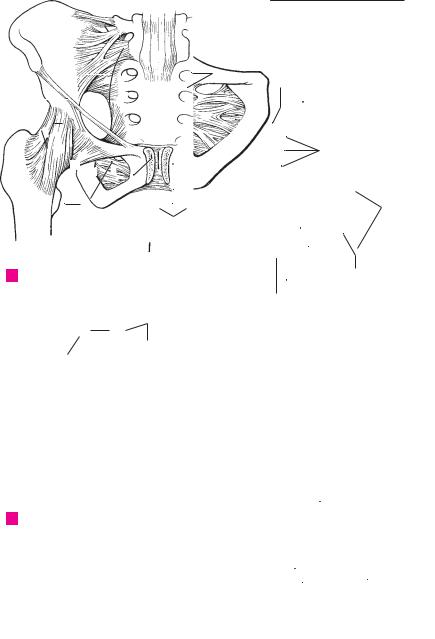
A Pelvic ligaments, anterior view
26
20
28 3
27
2
C Hip joint, opened
13 |
|
5 |
|
|
|
|
3 |
||||
|
|
|
|
|
|
||||||
|
|
|
|
|
|
|
|
|
|
|
|
|
|
|
|
|
|
|
|
|
|
|
4 |
|
|
|
|
|
|
|
|
|
|
|
|
|
|
|
|
|
|
|
|
|
|
|
|
|
|
|
|
|
|
|
|
|
|
|
|
|
|
|
|
|
|
|
|
|
|
|
5 |
|
|
|
|
|
|
|
|
|
|
|
|
|
|
|
|
|
|
|
|
|
|
|
|
|
|
|
|
|
|
|
|
14 |
|
6 |
|
|
|
|
|
|
|
|
|
|
|
|
|
|
|
|
|
|
|
|
|
|
|
|
|
|
|
|
|
|
|
|
|
|
|
|
7 |
|
|
|
|
9 |
|
|
|
|
|||
|
|
|
|
|
|
|
|
||||
|
|
|
|
23 |
20;21 |
8 |
|||||
|
|
|
|
8 |
|
|
|||||
|
|
|
|
|
|
|
|
|
|||
|
|
6 |
|
|
|
|
|
24 |
|
9 |
|
|
|
|
|
|
|
|
|
||||
|
|
|
|
|
|
|
|
||||
|
|
|
|
|
|
|
|
|
|
||
|
|
|
|
|
|
|
|
|
|
||
|
|
|
|
|
|
|
|
|
10 |
||
|
|
|
|
|
|
|
|
|
|
|
|
|
|
|
|
|
|
|
|
|
|
|
|
|
|
|
|
|
|
|
|
|
|
|
|
|
|
|
|
|
|
|
|
22 |
|
11 |
|
|
|
|
|
|
|
|
|
|
|
|
|
|
|
|
|
|
|
|
|
|
|
|
|
|
|
|
|
|
|
|
|
|
12 |
||
2 |
7 |
10 |
|
|
|
||||||
|
|
|
|
|
|
|
|
|
|
|
|
|
|
|
|
|
|
|
|
|
|
|
|
|
|
|
|
|
|
|
|
|
|
|
13 |
B |
Pelvic ligaments, |
|
|
|
|||||||
|
|
|
|
||||||||
|
posterior view |
|
|
|
|
||||||
|
|
|
14 |
||||||||
|
|
|
|
|
|
|
|
|
|
|
|
|
|
|
|
|
|
|
|
|
|
|
|
|
|
|
|
|
|
|
|
|
|
|
|
|
|
|
|
|
|
|
|
|
|
|
15 |
|
|
|
|
|
|
|
|
|
|
|
|
|
|
|
|
|
|
|
|
|
|
|
|
|
|
|
|
|
|
|
|
|
|
|
16 |
|
|
|
|
|
|
|
|
|
|
|
|
|
|
|
|
|
|
|
|
|
|
|
|
|
|
|
|
|
|
|
|
|
|
|
17 |
|
|
|
|
|
|
|
|
|
|
|
|
|
|
|
|
|
|
|
|
|
|
|
|
|
|
|
|
|
|
|
|
|
|
|
18 |
|
|
|
|
|
|
|
|
|
|
|
|
|
|
|
|
|
|
|
|
|
|
|
|
|
|
|
|
|
|
|
|
|
|
|
19 |
|
|
|
|
|
|
|
|
|
|
|
|
|
|
|
|
|
|
|
|
|
|
|
|
|
|
12 |
|
|
|
|
|
|
|
20 |
|
|
|
|
|
|
|
|
|
|
|||
|
|
|
|
|
|
|
|
|
|
|
|
|
|
|
|
|
|
|
|
9 |
|
21 |
|
|
|
|
|
|
|
|
|
|
|
||
|
|
|
|
|
|
|
|
|
|
||
|
|
|
|
|
|
8 |
22 |
|
|
|
3 |
|
|
|
|
||
|
|
|
|
|
|
|||
|
|
|
|
|
|
|
23 |
|
|
|
|
|
|
|
|
||
|
|
10 |
|
|
||||
|
|
2 |
|
|
|
|
|
|
|
|
|
|
|
|
|
|
24 |
|
Pelvic ligaments, |
|
|
|
|
6 |
||
D |
|
|
|
|
|
|
|
|
|
|
|
|
|
|
|
||
|
medial view |
|
|
|
|
|
|
25 |
a |
|
|
|
a |
|
|
|
|
|
|
|
|
|
|
|||

68 Sutures, joints and ligaments
1 |
|
1 |
Knee joint. Articulatio genus [genualis]. A B C D E |
14 |
Oblique popliteal ligament. Lig. popliteum ob- |
||||
|
2 |
Lateral meniscus. Meniscus lateralis. Roughly |
|
liquum. Fibrous band originating in the posterior |
|||||
|
|
|
wall of the capsule, extending upward and out- |
||||||
|
|
|
|
circular ring, the superficial layer of which is |
|
||||
|
|
|
|
|
|||||
2 |
|
|
|
|
ward from the tendon of the semimembranous |
||||
|
|
|
made of fibrocartilage, whereas the deep layer is |
|
|||||
|
|
|
|
more tendinous. It is located below the lateral |
|
muscle, thereby reinforcing the capsule. C |
|||
|
|
|
|
|
|
||||
3 |
|
|
|
femoral condyle and has close-set bases. It is |
15 |
Arcuate popliteal ligament. Lig. popliteum ar- |
|||
|
|
|
relatively mobile because it is not fused with the |
|
cuatum. Curved band of fibers extending from |
||||
|
|
|
|
|
|||||
4 |
|
|
|
lateral collateral ligament. B D E |
|
|
the epicondyle, across the origin of the popliteal |
||
|
|
3 |
Anterior |
meniscofemoral |
ligament. |
Lig. |
|
muscle to the head of the fibula, thus reinforcing |
|
|
|
|
|
meniscofemorale anterius. Fibrous band oc- |
|
the posterior wall of the capsule. C |
|||
|
|
|
|
|
|
||||
5 |
|
|
|
casionallyfoundconnectingtheposteriorpartof |
16 |
Patellar ligament. Ligamentum patellae. Wide |
|||
|
|
|
|
the lateral meniscus with the anterior cruciate |
|
(2−3 cm) and thick (ca. 0.5 cm) fibrous band |
|||
6 |
|
|
|
ligament. It passes in front of the posterior |
|
that forms the continuation of the tendon of the |
|||
|
|
|
cruciate ligament. D E |
|
|
|
quadriceps femoris muscle. It extends from the |
||
|
|
|
4 |
Posterior |
meniscofemoral |
ligament. |
Lig. |
|
apex of the patella to the tibial tuberosity. A |
7 |
|
|
|
meniscofemorale posterius. It passes posterior |
17 |
Medial retinaculum of patella. Retinaculum |
|||
|
|
|
|
to the lateral meniscus to the fibular surface of |
|
patellae mediale. Aponeurosis from a part of the |
|||
8 |
|
|
|
the medial femoral condyle behind the posterior |
|
vastus medialis muscle that extends medially |
|||
|
|
|
cruciate ligament. D E |
|
|
|
from the patella and attaches to the medial mar- |
||
|
|
|
|
|
|
|
|||
9 |
|
|
5 |
Medial meniscus. Meniscus medialis. A cres- |
|
gin of the tibial tuberosity. It maintains the path- |
|||
|
|
|
cent-shaped ring segment located below the |
|
way of movement of the patella via muscular |
||||
|
|
|
|
medial femoral condyle. It is not very mobile be- |
|
contraction and serves as a reserve extension ap- |
|||
10 |
|
|
|
cause it is fused with the medial collateral liga- |
|
paratus. A |
|||
|
|
|
ment. Its histological structure is like that of the |
18 |
Lateral retinaculum of patella. Retinaculum |
||||
|
|
|
|
||||||
|
|
|
|
lateral meniscus. B D E |
|
|
|||
11 |
|
|
|
|
|
|
patellae laterale. Aponeurosis of a part of the |
||
|
|
|
|
|
|
|
|
||
|
|
6 |
Transverse ligament of knee. Lig. transversum |
|
vastus lateralis lateral to the patella with attach- |
||||
|
|
|
|
genus [genuale]. Transverse ligament joining |
|
ment lateral to the tibial tuberosity. Its action is |
|||
12 |
|
|
|
the anterior ends of the lateral and medial |
|
comparable to that of no. 17. A |
|||
|
|
|
|
menisci. B D |
|
|
19 Infrapatellar fat pad. Corpus adiposum infra- |
||
|
|
|
7 |
|
ligaments of knee. Ligg. cruciata |
||||
13 |
|
|
Cruciate |
|
patellare. Large, wedge-shaped mass of fatty |
||||
|
|
|
genus [geniualia]. They prevent displacement of |
|
tissue in front of the knee joint space. It includes |
||||
|
|
|
|
|
|||||
14 |
|
|
|
the tibia and femur in the sagittal plane. B E |
|
|
the infrapatellar alar and synovial folds. A |
||
|
|
8 |
Anterior cruciate ligament. Lig. cruciatum an- |
20 |
Tibiofibular joint. Articulatio tibiofibularis. Ar- |
||||
|
|
|
|
terius. It passes from the inner surface of the |
|
ticulation between the head of the fibula and the |
|||
15 |
|
|
|
lateral femoral condyle obliquely forward and |
|
lateral condyle of the tibia. E |
|||
|
|
|
|
inferomedially to the anterior intercondylar |
21 Anterior ligament of head of fibula. Lig. capitis |
||||
|
|
|
|
area. It prevents inward rotation and forward |
|||||
16 |
|
|
|
|
fibulae anterius. Group of fibers passing anteri- |
||||
|
|
|
displacement of the tibia toward the femur. B D E |
|
|||||
|
|
|
9 |
Posterior cruciate ligament. Lig. cruciatum |
|
orly from the head of the fibula to the tibia, thus |
|||
17 |
|
|
|
holding the two bones together. A |
|||||
|
|
|
posterius. It passes from the inner surface of the |
22 |
Posterior ligament of head of fibula. Lig. |
||||
|
|
|
|
medial femoral condyle to the posterior condy- |
|||||
|
|
|
|
|
capitis fibulae posterius. Weaker group of fibers |
||||
18 |
|
|
|
lar area, stabilizes the joint when flexed, and pre- |
|
||||
|
|
|
|
extending from the posterior part of the head of |
|||||
|
|
|
vents backward displacement of the tibia away |
|
|||||
19 |
|
|
|
from the femur. B D E |
|
|
|
the fibula to the tibia. C D E |
|
|
|
|
|
|
23 |
Tibiofibular syndesmosis [joint]. Syndesmosis |
|||
|
|
10 |
Infrapatellar synovial fold. Plica synovialis in- |
||||||
|
|
|
|
frapatellaris. Connective tissue that often con- |
|
[articulatio] tibiofibularis. Distal union of tibia |
|||
20 |
|
|
|
tains fat. |
It extends from |
the infrapatellar |
|
with fibula. |
|
|
|
|
adipose body to the intercondylar fossa. B |
|
24 |
Interosseous membrane. Membrana interos- |
|||
|
|
|
|
|
|||||
21 |
|
|
11 |
Alar folds. Plicae alares. Deformable, paired |
|
sea cruris. Membrane attached to the interosser- |
|||
|
|
|
bulges of the adipose body that fill empty spaces |
|
ous margins of the tibia and fibula. It is the field |
||||
|
|
|
|
in the anterior part of the joint cavity. B |
|
|
of origin of the lower leg muscles and ensures the |
||
22 |
|
|
12 |
Fibular collateral ligament. Lig. collaterale |
|
stability of the malleolar bifurcation. A C F G |
|||
|
|
25 |
Anterior tibiofibular ligament. Lig. tibiofibu- |
||||||
|
|
|
|
fibulare. Lateral collateral ligament that extends |
|||||
23 |
|
|
|
from the lateral epicondyle to the head of the |
|
lare anterius. Anterior fibrous bands connecting |
|||
|
|
|
fibula independent of the capsule and meniscus. |
|
the fibular incisure to the lateral malleolus, thus |
||||
|
|
|
|
|
|||||
24 |
|
|
|
A B C D E |
|
|
|
|
stabilizing the malleolar bifurcation. F |
|
|
13 |
Tibial collateral ligament. Lig. collaterale ti- |
26 |
Posterior tibiofibular ligament. Lig. tibiofibu- |
||||
|
|
|
|
biale. Medial collateral ligament extending from |
|
lare posterius. Posterior fibrous bands connect- |
|||
25 |
|
|
|
the medial epicondyle to the tibia. It is fused with |
|
ing the fibular incisure to the lateral malleolus, |
|||
|
|
|
|
the joint capsule and the meniscus. A B C D E |
|
thus stabilizing the malleolar bifurcation. G |
|||
|
|
|
|
|
|
|
|
|
|
Sutures, joints and ligaments 69
12 |
|
|
|
17 |
|
|
13 |
7 |
|
9 |
10 |
|
|
|
|
|
|
|
|
|
|
||||||||
18 |
|
|
12 |
|
|
|
|
|
|
|||||
|
|
|
|
|
8 |
|
|
|
||||||
19 |
|
|
16 |
|
|
2 |
|
5 |
|
|
||||
|
|
|
|
|
|
|
|
|
|
|||||
|
|
|
|
|
|
|
|
|
|
|||||
|
|
|
|
|
|
|
6 |
|
|
13 |
||||
21 |
|
|
|
|
|
|||||||||
|
|
|
|
|
11 |
|
|
11 |
|
|||||
|
|
|
|
|
|
|
|
|
||||||
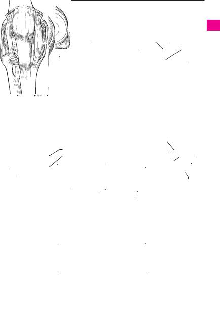
A |
Right knee joint, |
24 |
B |
Right knee joint, |
|
anterior view |
|
|
opened, anterior view |
|
|
|
|
|
|
|
|
|
|
|
|
|
|
|
|
|
|
|
|
|
|
|
|
|
|
|
|
|
|
|
7 |
|
|
|
|
|
|
|
|
|
|
|
|
|
96.21 |
|
|
|
6 |
|
|
|
|
|
|
|
|
|
|
|
|
|
|
|
|||||||||
14 |
|
15 |
|
|
|
|
|
|
|
|
|
|
|
|
|
|
|
|
4 |
8 |
3 |
|||||||||||||||
|
|
|
|
|
|
|
|
|
|
|
|
|
|
|
|
|
|
|||||||||||||||||||
|
|
|
|
|
|
|
|
|
|
12 |
|
|
|
|
|
|
|
|
|
|
|
|
|
|
|
5 |
9 |
2 |
|
|
12 |
|||||
13 |
|
|
|
|
|
|
|
|
|
|
|
13 |
|
|
|
|
|
|
||||||||||||||||||
|
|
|
|
|
|
|
|
|
|
|
|
|
|
|
|
|
|
|
|
|
|
|
22 |
|
|
|
||||||||||
|
|
|
|
|
|
|
|
|
|
|
|
|
|
|
8 |
|
|
|
|
|
|
|
|
|
||||||||||||
|
|
|
|
|
|
|
|
|
|
|
|
|
|
|
|
|
|
|
|
|
|
|
||||||||||||||
|
|
|
|
|
|
|
|
|
|
|
|
|
|
|
|
|
|
|
|
|
|
|
|
|
|
|
|
|
|
|
|
|
||||
96.6 |
|
|
|
|
|
|
|
|
|
|
|
|
|
|
|
|
|
|
|
|
|
|
|
|
|
|
|
|
|
|
|
|||||
|
|
|
|
|
22 |
|
|
|
|
|
|
|
2 |
|
|
|
|
|
|
|
|
|
|
|
20 |
|
|
|||||||||
|
|
|
|
|
|
|
|
|
|
|
|
|
|
|
|
|
|
|
|
|
|
|
|
|
|
|
|
|||||||||
|
|
|
|
|
|
|
|
|
|
|
|
|
|
|
|
|
|
|
|
|
|
|
|
|
|
|
|
|
|
|
|
|
||||
|
|
|
|
|
|
|
|
|
|
|
|
|
|
|
|
|
|
|
|
|
|
|
|
|
|
|
|
|
|
|
|
|
||||
|
|
|
|
|
|
|
|
|
|
|
|
|
|
|
|
5 |
|
|
|
|
|
|
|
12 |
|
|
|
|
|
|
|
|
|
|
||
|
|
|
|
|
|
|
|
|
|
|
|
|
|
9 |
|
|
|
|
|
22 |
|
|
|
|
|
|
|
|
|
|||||||
|
|
|
|
|
|
|
|
13 |
|
|
|
|
|
|
|
|
|
|
|
|
||||||||||||||||
|
|
|
|
|
|
|
|
|
|
|
|
|
|
|
|
|
|
|
|
|
||||||||||||||||
|
|
|
|
|
|
|
|
|
24 |
|
|
|
|
|
|
|
|
|
|
|
|
|
|
|
|
|
|
|
|
|
|
|
|
|
|
|
|
|
|
|
|
|
|
|
|
|
|
|
|
|
|
|
4 3 |
|
|
|
|
|
|
|
|
|
|
|
|
|
|
|
|
|
|||
|
|
|
|
|
|
|
|
|
|
|
|
|
|
|
|
|
|
|
|
|
|
|
|
|
|
|
|
|
|
|
|
|
||||
|
Right knee joint, |
|
|
|
|
|
|
|
|
|
Right knee joint, opened, |
|
Right knee joint, opened, |
|||||||||||||||||||||||
C |
|
|
|
|
|
|
|
|
D |
E |
||||||||||||||||||||||||||
|
|
posterior view |
|
|
|
|
|
|
|
|
|
superior view |
|
|
|
|
|
|
|
|
|
|
posterior view |
|
|
|
||||||||||
24 |
|
|
|
|
|
|
|
|
|
|
|
|
|
|
|
|
|
|
|
|
|
|
|
|
24 |
|
|
|
|
|||||||
|
|
|
|
|
|
|
|
|
|
|
|
|
|
|
|
|
|
|
|
|
|
|
|
|
|
|
|
|||||||||
|
|
|
|
|
|
|
|
|
|
|
|
|
|
|
|
|
|
|
|
|
|
|
|
|
|
|
|
|||||||||
25 |
|
|
|
|
|
|
|
|
|
|
|
|
|
|
|
|
|
|
|
|
|
|
|
|
26 |
|
|
|
|
|||||||
|
|
|
|
|
|
|
|
|
|
|
|
|
|
|
|
|
|
|
|
|
|
|
|
|
|
|
|
|||||||||
|
|
|
|
|
|
|
|
|
|
|
|
|
|
|
|
|
|
|
|
|
|
|
|
|
|
|
|
|||||||||
|
|
|
|
|
|
|
Distal area of right leg, |
|
|
Distal area of right leg, |
|
|
|
|
|
|
||||||||||||||||||||
F |
|
G |
|
|
|
|
|
|
||||||||||||||||||||||||||||
|
|
|
|
|
|
|
anterior view |
|
|
|
|
|
|
posterior view |
|
|
|
|
|
|
|
|
|
|||||||||||||
1
2
3
4
5
6
7
8
9
10
11
12
13
14
15
16
17
18
19
20
21
22
23
24
25
a |
a |