Материал: Атлас Ханц фениш

70 Sutures, joints and ligaments
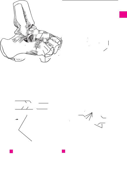
1Ankle (talocrural) joint. Articulatio talocruralis. 1 Upper ankle joint between the talus, tibia and
fibula. D
22 Medial or deltoid ligament. Lig. mediale (del-
|
toideum) articulationis talocruralis. Ligament on |
3 |
the medial side of the ankle which consists of the |
four segments described below. D |
3 Tibionavicular part. Pars tibionavicularis. Group
4of fibers connecting the medial malleolus to the dorsal and medial surfaces of the navicular bone. D
54 Tibiocalcaneal part. Pars tibiocalcanea. Group of fibers connecting the medial malleolus to the sustentaculum tali. B D
65 Anterior tibiotalar part. Pars tibiotalaris ante-
rior. Segment of the deltoid ligament that connects
7the medial malleolus to the medial surface of the talus as far as the neck of the talus. D
6 Posterior tibiotalar part. Pars tibiotalaris poste-
8rior. Fibers extending posteriorly from the medial malleolus almost as far as the posterior process of
9the talus. B D
7Anterior talofibular ligament. Lig. talofibulare
anterius. Ligament extending from the lateral mal- 10 leolustothelateralsurfaceoftheneckofthetalus.A
8Posterior talofibular ligament. Lig. talofibulare
11posterius.Itoriginatesinthelateralmalleolarfossa and inserts at the lateral tubercle of the posterior
12 |
|
process of the talus. A B |
|
9 Calcaneofibular ligament. Lig. calcaneofibulare. |
|||
|
|||
|
|
It passes obliquely and posteriorly from the apex of |
|
13 |
|
||
|
the alteral malleolus to the calcaneus. A B |
||
|
10 |
Intertarsal joints. Articulationes intertarseae. |
|
|
|||
14 |
|
Joints between the tarsal bones. |
|
|
11 |
JOINTS OF THE FOOT. Articulationes pedis. |
|
|
|||
1512 Talocalcaneonavicular joint. Articulatio talocalcaneonavicularis. The anterior portion of the lower ankle joint in which the talus articulates with the
16calcaneus and navicular bones. A C
13 Subtalar joint. Articulatio subtalaris (talocal-
17canea). Joint between the talus and calcaneus that represents the posterior part of the lower ankle joint. A B C D
1814 Lateral talocalcaneal ligament. Lig. talocal-
caneum laterale. Ligament that passes from the
19trochlea of the talus to the lateral surface of the calcaneus. It is partially covered by the calcaneofibular ligament. A
2015 Medial talocalcaneal ligament. Lig. talocal-
caneum mediale. Ligament on the medial side of
21the foot that extends from the medial tubercle of the posterior process of the talus to the sustantaculum tali. B D
2215 a Posterior talocalcaneal ligament. [[Lig. talocal-
caneum posterior.]] Fibrous band extending from
23the posterior process of the talus to the calcaneus, thereby bridging the sulcus of the tendon of the flexor hallucis longus muscle.
2416 Transverse tarsal (midtarsal) joint [[Chopart’s joint]] Articulatio tarsi transversa. Joint situated in
25front of the talus and calcaneus but proximal to the cuboid and navicular bones. C
17Calcaneocuboid joint. Articulatio calcaneocuboidea. Joint between the calcaneus and cuboid bones. A C
17 a Cuneocuboid joint. Articulatio cuneocuboidea. Articulation between the cuboid bone and lateral cuneiform bone. C
18Cuneonavicular joint. Articulatio cuneonavicularis. Joint between the navicular bone and cuneiform bones. C D
18 a Intercuneiform joints. Articulationes intercuneiformes. Joint between the cuneiform bones.
19Interosseous ligaments of the tarsus. Ligg. tarsi interossea. The following three interosseous ligaments are present between the tarsal bones:
20Interrosseous talocalcaneal ligament. Lig. talocalcaneum interosseum. Strong mass of ligaments in the sinus tarsi. A C
21Interosseous cuneocuboid ligament. Lig. cuneocuboideum interosseum. Taut connection betweenthelateralcuneiformboneandthecuboid bone. A C
22Interosseous intercuneiform ligaments. Ligg. intercuneiformia interossea. Taut ligaments between the three cuneiform bones. C
23Dorsal ligaments of the tarsus. Ligg. tarsi dorsalia. The following eight dorsal ligaments are present between the tarsal bones.
24Talonavicular ligament. Lig. talonaviculare. Dorsal ligament between the head of the talus and the navicular bone. A D
25Dorsal intercuneiform ligaments. Ligg. intercuneiformia dorsalia. Dorsal ligaments between the cuneiform bones. A
26Dorsal cuneocuboid ligaments. Lig. cuneocuboideum dorsale. Dorsal ligaments between the lateral cuneiform bone and the cuboid bone. A
27Dorsal cuboideonavicular ligament. Lig. cuboideonaviculare dorsale. Ligament between the cuboid and navicular bones. A
28Bifurcate ligament. Lig. bifurcatum. V-shaped double ligament in front of the sinus tarsi on the dorsum of the foot. It extends forward from the calcaneus and consists of the following two parts. A
29 Calcaneonavicular ligament. Lig. calcaneonaviculare. It extends laterally from the head of the talus to the navicular bone. A
30 Calcaneocuboid ligament. Lig. calcaneocuboideum. It extends from the calcaneus and attaches near the middle of the cuboid bone. A
31Dorsal cuneonavicular ligaments. Ligg. cuneonavicularia dorsalia. Broad group of ligaments on the dorsum of the foot connecting the navicular bone with the three cuneiform bones. A
31 a Dorsal calcaneocuboid ligament. Lig. calcaneocuboideum dorsale. Moderate reinforcement of the joint capsule lateral to the bifurcate ligament. A
Sutures, joints and ligaments 71
8 |
|
|
|
|
12 |
31 |
|
|
|
|
|
|
|
|
|
|
|
||
|
|
|
|
|
|
|
|
|
|
|
|
|
|
|
|
||||
|
|
|
7 |
24 |
|
25 |
|
|
|
|
|
|
|
|
|
|
|||
13 |
|
|
|
|
|
6 |
|
|
|
|
8 |
|
|||||||
|
|
|
|
|
|
|
|
|
|
|
|
||||||||
|
|
|
|
|
|
|
|
|
|
|
|
|
|
||||||
9 |
|
|
27 |
|
|
|
|
4 |
|
|
|
|
|
||||||
|
|
|
|
|
|
|
|
|
|
|
|
|
|||||||
|
|
|
|
|
|
|
|
|
|
|
|
|
|||||||
|
|
|
|
|
|
15 |
|
|
|
|
|
|
9 |
||||||
|
|
|
|
|
|
|
|
|
|
26 |
|
|
|
|
13 |
||||
|
|
|
|
|
|
|
|
|
|
|
|
|
|
|
|
||||
14 |
20 |
30 29 |
|
|
|
|
|
|
|
|
|
|
|
|
|
||||
|
|
|
|
|
17 |
|
|
|
21 |
|
|
|
|
|
|
|
|
|
|
|
|
|
|
|
28 |
|
|
|
|
|
|
|
|
|
|
|
|
||
|
|
|
|
|
|
|
|
|
|
|
|
|
|
|
|
|
|||
|
|
|
|
|
|
|
|
|
|
|
|
|
|
|
|
|
|
|
|
|
|
|
|
|
|
31a |
|
|
|
|
|
|
|
|
|
|
|
|
|
|
Ligaments of right foot, |
|
|
|
|
|
|
Ligaments of right ankle |
|||||||||||
A |
|
|
|
|
|
B |
|||||||||||||
|
lateral view |
|
|
|
|
|
|
|
(talocrural) joint, |
|
|||||||||
|
|
|
|
|
|
|
|
|
|
|
|
posterior view |
|
|
|
||||
1
2
3
4
5
6
7
8
9
10
11
12
13
22 |
21 |
|
|
18 |
17a |
16 |
17 |
|
20
12
13
CTarsometatarsal bones of right foot, horizontal section
|
24 |
1 |
|
|
2 |
|
|
|
|
|
|||
|
5 |
|
|
|||
|
|
|
|
|
||
|
|
|
|
|
|
6 |
18 |
|
|
|
3 |
4 |
|
|
|
|
|
|
||
18
14
15
16
1
15 |
17 |
1318
19
20
D |
Ligaments of right foot, |
21 |
|
medial view |
|
|
|
22
23
24
25
a |
a |

72 Sutures, joints and ligaments
1 |
|
1 |
Plantar ligaments of tarsus. Ligg. tarsi 14 |
Interosseous |
metatarsal ligaments. Ligg. |
|
|
plantaria. Ligaments on the palmar aspect of |
metatarsalia |
interossea. Ligaments between |
|
|
|
|
the foot. They are particularly important for the |
the bases of the metatarsal bones. They form |
|
|
|
|
|||
2 |
|
|
bracing of both plantar arches of the foot. |
the distal limits of the articular spaces between |
|
|
2 |
Long plantar ligament. Lig. plantare longum. |
the metatarsal bones. C |
||
|
|
|
|||
3Stout ligament which passes from the cal15 Dorsal metatarsal ligaments. Ligg. metatarcaneus closely in front of its tuber to the cuboid salia dorsalia. Ligaments between the bases of
|
|
bone and to the bases of metatarsals II−V. It |
|
the metatarsal bones on the dorsum of the foot. |
|||||
4 |
|
|
|||||||
|
supports the longitudinal arch. A |
|
|
B |
|||||
|
3 |
Plantar calcaneocuboid ligament or short |
16 |
Plantar metatarsal ligaments. Ligg. metatar- |
|||||
|
|||||||||
5 |
|||||||||
|
plantar |
ligament. |
Lig. calcaneocuboideum |
|
salia plantaria. Ligaments found between the |
||||
|
|
plantare. Shorter portion of the long plantar |
|
bases of the metatarsal bones on the plantar |
|||||
|
|
|
|||||||
6 |
|
ligament. A |
|
|
|
|
aspect of the foot. A |
||
4 Plantar |
calcaneonavicular |
(spring) |
liga- |
17 |
Metatarsal interosseous spaces. Spatia inter- |
||||
|
|||||||||
|
|||||||||
7 |
|
ment. Lig. calcaneonaviculare plantare. It lies |
|
ossea metatarsi. Spaces between the shafts of |
|||||
|
medial to the above-mentionend ligament and |
|
the metatarsal bones. They are occupied by the |
||||||
|
|
supports, according to more traditional view, |
|
corresponding muscles. A |
|||||
8 |
|
the articular cavity for the head of the talus. |
18 |
Metatarsophalangeal joints. Articulationes |
|||||
|
|
Since the talar side of the ligament is quite |
|
metatarsophalangeales. A |
|||||
|
|
loose and contains no fibrocartilage, this con- |
|
||||||
9 |
|
|
|||||||
|
|
|
|||||||
|
cept is questionable. A |
|
|
19 |
Collateral ligaments. Ligg. collateralia. A |
||||
|
5 |
Plantar cuneonavicular ligaments. Ligg. 20 |
Plantar ligaments. Ligg. plantaria. Connective |
||||||
10 |
|||||||||
|
cuneonavicularia plantaria. Groups of liga- |
|
tissue reinforcement of the capsule of the |
||||||
|
|
ments that connect the navicular bone with the |
|
metatarsophalangeal joint. It is more firmly |
|||||
|
|
|
|||||||
11 |
|
cuneiform bones lodged in front of it. A |
|
|
fused with the proximal phalanx than with the |
||||
6 |
Plantar |
cuboideonavicular |
ligament. |
Lig. |
|
head of the metatarsals and forms a stroma for |
|||
|
|
||||||||
|
|
the flexor tendons. A |
|||||||
12 |
|
cuboideonaviculare |
plantare. A plantar |
liga- |
|
||||
|
21 |
Deep transverse metatarsal ligament. Lig. |
|||||||
|
ment coursing somewhat obliquely to the axis |
||||||||
|
|
||||||||
|
|
of the foot connecting the cuboid and navicular |
|
metatarsale transversum profundum. Trans- |
|||||
13 |
|
|
|||||||
|
bones. It supports the transverse plantar arch of |
|
versely oriented ligament connecting the heads |
||||||
|
|
the foot. A |
|
|
|
|
of the metatarsal bones. A |
||
|
|
|
|
|
|
||||
14 |
7 |
Plantar intercuneiform ligaments. Ligg. in- |
22 |
Interphalangeal joints of the foot. Articula- |
|||||
|
|
tercuniformia plantare. Fibrous bands lying on |
|
tiones interphalangeales pedis. The middle and |
|||||
|
|
|
|||||||
15 |
|
the plantar aspect of the foot between the |
|
terminal joints between the phalanges of the |
|||||
|
cuneiform bones. They support the transverse |
|
foot. A |
||||||
|
|
plantar arch of the foot. A |
|
|
23 Collateral ligaments. Ligg. collateralia. A |
||||
16 |
|
|
|
||||||
|
|
|
|
|
|
||||
8 |
Plantar |
cuneocuboid ligament. Lig. cuneo- |
24 |
Plantar ligaments. Ligg. plantaria. Fibrous |
|||||
|
|
cuboideum plantare. Fibrous brace on the plan- |
|||||||
|
|
|
bands that reinforce the plantar aspect of the |
||||||
17 |
|
tar aspect of the foot between the lateral |
|
||||||
|
|
interphalangeal articular capsules. |
|||||||
|
|
cuneiform and cuboid bones. A |
|
|
|
|
|||
189 Tarsometatarsal joints. Articulationes tarsometatarsales. Joints between the tarsal and metatarsal bones of the foot. A B C
1910 Dorsal tarsometatarsal ligaments. Ligg. tarsometatarsalia dorsalia. Ligaments located on
20the dorsum of the foot between the tarsal and metatarsal bones. B
2111 Plantar tarsometatarsal ligaments. Ligg. tarsometatarsalia plantaria. Ligaments located on the plantar aspect of the foot between the tar-
22sal and metatarsal bones. A
23 |
12 |
Interosseous cuneometatarsal ligaments. |
|
|
Ligg. cuneometatarsalia interossea. Ligaments |
|
|
occupying the joint spaces between the |
24 |
|
cuneiform and metatarsal bones. C |
13 Intermetatarsal joints. Articulationes inter-
25metatarsales. Joints between the bases of the metatarsal bones. B C
Sutures, joints and ligaments 73
|
|
22 |
|
|
|
|
|
|
|
|
|||||
|
23 |
|
|
|
|
|
|
|
22 |
|
|
||||
|
|
|
|
|
|
|
|
|
|
|
|||||
22 |
|
|
|
|
|
|
|
|
|
|
|
|
|
|
|
|
|
21 |
|
|
|
|
|
20 |
|
|
|
|
|
||
|
|
|
|
|
|
|
|
|
|
|
|
|
|
||
|
19 |
|
|
|
|
|
|
|
|
|
|
18 |
|||
|
|
|
|
|
|
|
|
|
|
||||||
18 |
|
|
|
|
|
|
|
|
19 |
|
|
|
|
|
|
|
17 |
17 |
|
|
17 |
|
17 |
|
|
|
|
|
|||
|
|
|
|
|
|
|
|
|
|
|
|
|
|
|
|
|
|
|
|
|
|
16 |
|
|
|
|
|
|
|
||
16 |
|
2 |
2 |
|
|
|
|
|
9 |
||||||
|
|
|
|
||||||||||||
|
|
2 |
|
|
|
|
11 |
|
|
|
|
|
11 |
||
11 |
|
11 |
|
|
|
|
|
|
|||||||
2 |
|
|
|
|
|
|
|
|
|
|
7 |
||||
8 |
|
|
|
|
|
|
|
|
|
|
|
|
|||
|
|
|
|
|
|
|
|
|
|
|
|
||||
|
|
|
|
|
|
|
|
|
|
||||||
|
|
|
|
|
|
|
|
|
|||||||
|
|
|
|
|
|
|
|
|
|
|
|
|
|
|
|
36
 5
5
3 2 4
4
50.27
 50.3
50.3
ALigaments of right foot, plantar view
C Ligaments of sectioned foot, dorsal view
15 |
|
13 |
15 |
9 |
|
10
B Ligaments of foot, dorsal view
12 13
9
14
9
13  14
14
9  13
13  14
14
9
913
14 12
1
2
3
4
5
6
7
8
9
10
11
12
13
14
15
16
17
18
19
20
21
22
23
24
25
a |
a |

|
74 |
|
Muscles |
|
|
|
|
|
|
|
|
|||
|
|
|
|
|
|
|
13 |
M. longissimus cervicis. o: Transverse proces- |
||||||
|
|
|
|
|
|
|
||||||||
1 |
|
|
|
|
MUSCULAR SYSTEM |
|||||||||
|
|
1 DORSAL MUSCLES. Musculi dorsi. The muscles |
|
ses T1−6. i: Transverse processes of vertebrae |
||||||||||
|
|
|
|
of the back. True back muscles are innervated |
|
C2−7. It lies between the iliocostalis cervicis |
||||||||
2 |
|
|
|
by the dorsal rami of spinal nerves, whereas the |
|
and longissimus capitis muscles. C |
|
|
||||||
|
|
|
|
muscles of the shoulder girdle are not. A B C |
14 |
M. longissimus capitis. o: Transverse |
||||||||
|
|
|
2 M. trapezius. o: Spinous processes of vertebrae |
|
processes |
of |
vertebrae |
C3−T3. |
i: |
Mastoid |
||||
3 |
|
|
|
|||||||||||
|
|
|
T1-T12 and C1, the nuchal ligament, occipital |
|
process. It lies between the longissimus cervi- |
|||||||||
|
|
|
|
protuberance, and superior nuchal line. i: Spine |
|
cis and semispinalis capitis muscles. A: Lateral |
||||||||
4 |
|
|
|
of scapula, acrimonion, and clavicle. A: Rotates, |
|
and backward flexion of the head. It rotates the |
||||||||
|
|
|
|
raises, lowers, and adducts the scapula; rotates |
|
face toward the ipsilateral side. C |
|
|
||||||
|
|
|
|
|
|
|
||||||||
5 |
|
|
|
the head. I: Accessory nerve, cervical plexus. A |
15 |
M. iliocostalis. Iliocostal muscle, which con- |
||||||||
|
|
3 M. transversus nuchae. (Rare, 25%). Platysmal |
|
sists of the following three segments. |
|
|||||||||
|
|
|
|
|
||||||||||
|
|
|
|
muscle situated between the insertions of the |
16 |
M. iliocostalis lumborum. o: Iliac crest. i: |
||||||||
6 |
|
|
|
|||||||||||
|
|
|
trapezius and sternocleidomastoid. It passes |
|
Angle of ribs 5−12. A: Extenson and lateral flex- |
|||||||||
|
|
|
|
transversely, either superficial or deep, to the |
|
ion of lower vertebral column. B C |
|
|
||||||
|
|
|
|
|
|
|
||||||||
7 |
|
|
|
trapezius. A |
17 |
M. iliocostalis thoracis. o: Medial sides of 6 |
||||||||
|
|
4 M. latissimus dorsi. o: Thoracolumbar fascia, |
||||||||||||
|
|
|
|
lower rib angles. i: Six uppermost rib angles. A: |
||||||||||
|
|
|
|
spinous processes of vertebrae T7−L5, sacrum, |
|
Flattening of thoracic kyphosis, lateral flexion. |
||||||||
8 |
|
|
|
|
||||||||||
|
|
|
iliac crest, and four lower ribs. i: Crest of lesser |
|
B C |
|
|
|
|
|
|
|||
|
|
|
|
tubercle of humerus. A: Adduction and medial |
18 |
M. iliocostalis cervicis. o: Upper and middle |
||||||||
|
|
|
|
|||||||||||
9 |
|
|
|
rotation of the arm. I: Thoracodorsal nerve. A |
|
ribs. i: Transverse processes of middle cervical |
||||||||
|
|
5 M. rhomboideus major. o: Spinous processes |
|
|||||||||||
|
|
|
|
vertebrae. C |
|
|
|
|
||||||
|
|
|
|
of vertebrae T1−4. i: Medial margin of scapula. |
19 |
M. splenius cervicis. o: Spinous processes of |
||||||||
10 |
|
|
|
|||||||||||
|
|
|
A: Medial and upward movement of scapula. I: |
|
T3−5. |
i: |
Posterior tubercle of |
transverse |
||||||
|
|
|
|
Dorsal scapular nerve. A |
|
processes of C1−2. A: Backward flexion and ro- |
||||||||
|
|
|
|
|
||||||||||
11 |
6 |
M. rhomboideus minor. o: Spinous processes |
|
tation of head. B |
|
|
|
|||||||
|
|
|
|
of cervical vertebrae 6−7. i: Medial margin of |
20 M. splenius capitis. o: Spinous processes of C4− |
|||||||||
|
|
|
|
scapula above the spine. A: Medial and upward |
||||||||||
12 |
|
|
|
|
T3. i: External half of superior nuchal line and |
|||||||||
|
|
|
movement of scapula. I: Dorsal scapular nerve. |
|
||||||||||
|
|
|
|
mastoid process. A: Backward flexion and rota- |
||||||||||
|
|
|
|
A |
|
|||||||||
|
|
|
|
|
tion of head. A B |
|
|
|
||||||
13 |
|
|
7 M. levator scapulae. o: Posterior tubercle of |
|
|
|
|
|||||||
|
|
21 |
MM. INTERTRANSVERSARII. Muscular connec- |
|||||||||||
|
|
|
transverse processes of cervical vertebrae 1−4. |
|||||||||||
|
|
|
|
|
tion of adjacent transverse processes. A: Lateral |
|||||||||
|
|
|
|
i: Superior angle of scapula. A: Elevates super- |
|
|||||||||
14 |
|
|
|
|
flexion. See p. 77 C D E |
|
|
|
||||||
|
|
|
ior angle of scapula and rotates neck. I: Dorsal |
|
|
|
|
|||||||
|
|
|
22 Mm. intertransversarii laterales lumborum. |
|||||||||||
|
|
|
|
scapular nerve and cervical plexus. A |
||||||||||
|
|
|
|
|
Muscles between adjacent costal processes. I: |
|||||||||
15 |
8 |
M. serratus posterior inferior. o: Spinous |
|
|||||||||||
|
Ventral rami of spinal nerves. See p. 77 C |
|||||||||||||
|
|
|
|
processes of vertebrae T11−L2. i: Four lower |
23 Mm. intertransversarii mediales lumborum. |
|||||||||
|
|
|
|
ribs. A: Retroversion of four lower ribs. I: Inter- |
||||||||||
16 |
|
|
|
|
Muscles between the mamillary processes. See |
|||||||||
|
|
|
costal nerves. A |
|
||||||||||
|
9 |
M. serratus posterior superior. o: Spinous |
|
p. 77 C |
|
|
|
|
|
|||||
|
24 Mm. intertransversarii thoracis. Usually ab- |
|||||||||||||
17 |
|
|
|
processes of vertebrae C6−T2. i: Second to fifth |
||||||||||
|
|
|
|
sent. See p. 77 C |
|
|
|
|||||||
|
|
|
ribs. A: Raises ribs in inspiration. I: Intercostal |
|
|
|
|
|||||||
|
|
|
|
nerves. A B |
25 |
Mm. intertransversarii |
posteriores |
cervicis. |
||||||
18 |
|
|
|
|||||||||||
10 |
M. ERECTOR SPINAE. Collective term for the |
|
Muscles between posterior tubercles of trans- |
|||||||||||
|
|
|
|
muscles of the lateral and medial tracts of the |
|
verse processes of cervical vertebrae. See p. 77 |
||||||||
|
|
|
|
|
||||||||||
19 |
|
|
|
back. I: Posterior rami of spinal nerves. o: Iliac |
26 |
Pars medialis. Medial part of 25. |
|
|
||||||
|
|
|
|
crest, spinous processes of L1−S4, mamillary |
27 |
Pars lateralis. Lateral part of 25. I: Ventral |
||||||||
|
|
|
|
processes of L1−2, transverse processes of T7− |
|
ramus of spinal nerves. |
|
|
|
|||||
20 |
|
|
|
|
|
|
|
|||||||
|
|
|
12. i: Costal and accessory processes of lumbar |
28 |
Mm. |
intertransversarii |
anteriores |
cervicis. |
||||||
|
|
|
|
vertebrae, angle of 11 lower ribs, all thoracic |
||||||||||
|
|
|
|
|
Muscles connecting the anterior tubercles of |
|||||||||
21 |
|
|
|
transverse processes. A: Lateral and backward |
|
|||||||||
|
|
|
|
the cervical |
transverse |
processes. I: Ventral |
||||||||
|
|
|
flexion of vertebral column. B C |
|
||||||||||
|
|
|
|
|
ramus of spinal nerves. See p. 77 E |
|
|
|||||||
|
|
|
|
|
|
|
|
|
|
|||||
10 a [[Lateral tract]]. [Tractus laterale].
2211 M. longissimus. It consists of the following three parts. B
2312 M. longissimus thoracis. o: Iliac crest, spinous processes of L1−S4, mamillary processes of L1− 2, transverse processes of T7−I2. i: Costal and
24accessory processes of lumbar vertebrae, angle
of lower 11 ribs all thoracic transverse
25processes, A: Lateral and backward flexion of vertebral column. B C