Материал: Bovine Viral Diarrhea Virus Diagnosis, Management, and Control
112 |
BVDV: Diagnosis, Management, and Control |
of cows that are at a similar stage of gestation. During such epizootics, up to 25% of a herd may be infected (refer to Figure 6.7).
An incubation period of 7–14 days has been suggested for MD (Grooms et al., 2002). This time frame is primarily based on experimental infections where a homologous, cp BVDV strain was administered to ncp BVDV persistently infected calves, or such calves were administered a modified live vaccine. Since the majority of cases of MD are thought to occur when cp BVDV mutates de novo from cattle persistently infected with a ncp strain, such an incubation time frame may not be pertinent (Figure 6.8).
Clinical signs of acute MD include fever, anorexia, tachycardia, polypnea, decreased milk production, and profuse watery diarrhea. Diarrhea is often characterized by the presence of mucosal shreds, fibrinous casts, blood, and foul odor. Other signs similar to acute BVDV infection may also be present antemortem, but are more pronounced. Erosions and ulcers may be present on the tongue, palate, and gingiva. Oral papilla may be blunted and hemorrhagic. Epithelial erosions may also be pronounced in the interdigital regions, coronary bands, teats, vulva, and prepuce. Additional clinical signs often include ocular and nasal discharge, corneal edema, hypersalivation, decreased ruminal contractions, and bloat. Inflammation of the interdigital space and coronary bands may present as lameness and animals may become laminitic. Clinicopathologic findings often include neutropenia (without a left shift) and thrombocytopenia. Secondary bacterial infections may be manifest as pneumonia, mastitis, and metritis. Cattle suffering acute MD gener-
ally have a case fatality rate approaching 100%. However, a minority of animals may survive the acute phase only to suffer signs of chronic MD (see next section). Postmortem findings of cattle suffering acute MD include variable necrotizing ulcers and erosions throughout the gastrointestinal tract (esophagus, rumen, abomasum, duodenum, jejunum, ileum, cecum, and colon). Erosions may also be present in the nares and upper respiratory tract. Peyer’s patches in the small bowel are often necrotic and hemorrhagic. Bowel contents are typically watery and fetid. Differential diagnoses for cattle with acute MD include those listed under acute BVDV infection (Figures 6.9, 6.10, 6.11). Although the clinical signs of hemorrhagic syndrome and MD may be similar, animals given supportive therapy may recover from hemorrhagic syndrome. Supportive therapy rarely has any effect in preventing the death of animals suffering from MD.
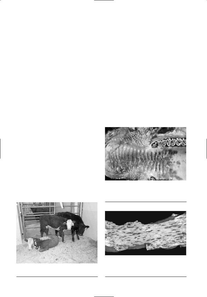
Figure 6.9. Postmortem image of the palate of calf suffering mucosal disease. Note mucosal ulceration and blunting of oral papillae. Similar lesions may be observed in cattle suffering severe acute BVDV infection.
Figure 6.8. Three age-matched beef calves from a herd suffering an outbreak of mucosal disease.
Figure 6.10. Postmortem image of esophageal ulcers and erosions in calf suffering mucosal disease. Similar lesions may be observed in cattle suffering severe acute BVDV infection.

Clinical Features |
113 |
Figure 6.11. Postmortem image of colon lesions in calf suffering mucosal disease. Similar lesions may be observed in cattle suffering severe acute BVDV infection.
CHRONIC MUCOSAL DISEASE
A small proportion of cattle with symptoms of acute MD do not die, but go on to develop a chronic form of the disease. These cattle typically present with persistent loose feces or intermittent diarrhea. Additional signs include ill thrift, mild-to-moderate anorexia, chronic recurrent bloat, interdigital erosions, and nonhealing erosive lesions of the skin. Ocular and nasal discharge may also be noted. Alopecia and areas of hyperkeratinization may develop, especially around the head and neck. Lameness may be manifest as chronic laminitis and abnormal hoof wall growth. Clinicopathologic findings often include anemia, neutropenia and thrombocytopenia.
VENEREAL INFECTIONS
Virus is present in the semen of bulls that are persistently infected with BVDV (Revell et al., 1988). Virus is also present in the semen of immunocompetent, transiently infected bulls undergoing acute infection (Kirkland et al., 1991). Furthermore, recent studies have demonstrated that transiently infected bulls can shed BVDV in semen for prolonged periods (months) after they have become non-viremic (Voges et al., 1998; Givens et al., 2003). Therefore, diagnostic testing of semen is recommended due to the potential for non-viremic bulls to shed BVDV in semen. A recent study demonstrated that the conventional testing method of virus isolation (VI) in semen was less sensitive compared to reverse tran- scription-nested polymerase chain reaction (RTnPCR).
Both acceptable and unacceptable semen quality has been reported from bulls that were TI or PI with BVDV. Although results of studies examining the effect of BVDV infection on conception rates have been conflicting, under certain circumstances
BVDV infection is likely associated with transient decreased conception rates (Baker, 1995).
REPRODUCTIVE CONSEQUENCES
Bovine viral diarrhea virus has been associated with several reproductive consequences, including reduced fertility and early embryonic death. All the major organs of the female reproductive system are permissive to BVDV, and the distribution of virus is similar between acutely infected and persistently infected cows (Fray et al., 2000). Although the precise mechanism responsible for reduced fertility is unknown, changes in ovarian function have been suggested. Ovarian hypoplasia has been reported in PI cows (Grooms et al., 1996), and diffuse interstitial ovaritis has been reported 61 days postinfection (Ssentongo et al., 1980). Grooms et al. (1998) demonstrated that the growth rate and diameter of dominant anovulatory and ovulatory follicles were significantly reduced following acute BVDV infection. Additionally, the numbers of subordinate follicles associated with the anovulatory and ovulatory follicle were also reduced following infection. Three possible mechanisms of how BVDV might compromise ovarian function have been suggested (Fray et al., 2000). First, BVDV might adversely affect pituitary gonadotroph function. Second, BVDV-suppressed plasma estrogen levels may adversely affect ovulation and estrus. Last, BVDV-induced general leukopenia may result in deficient ovarian leukocyte populations, because these cells are vital for normal follicular dynamics.
Kafi et al., (2002) demonstrated a detrimental effect of BVDV on in vitro fertilization and early embryo development; however, the mechanisms affecting these processes were not examined. Clearly, further studies are needed to define the effects of BVDV infection on the various hormones and cytokines that influence early reproductive events.
Infection of immunocompetent pregnant cattle can result in clinical manifestations in the dam similar to those described above—i.e., subclinical to severe, acute disease, or hemorrhagic syndrome. However, additional clinical outcomes in pregnant cattle are related to the potential transplacental transfer of virus to the fetus. Experimental studies have shown that BVDV crosses the placenta with near 100% efficiency; therefore, transplacental infection of the fetus should be anticipated (Duffell and Harkness, 1985). Importantly, the absence of clinical signs in the dam does not imply fetal protection or reduce the possibility of transplacental infection. The primary determinant of the outcome of fetal infection is clearly the gestational age of the

114 |
BVDV: Diagnosis, Management, and Control |
fetus, although other host and viral factors may also contribute to the final outcome.
Abortion results from BVDV infection during pregnancy, and the resultant fetal pathology has been recently reviewed (Fray et al., 2000). Infection of the dam during the preimplantation phase (<40 days) of pregnancy can result in a high incidence of embryonic or fetal mortality, whereas infection between 40 and 125 days gestation can result in fetal death, abortion, mummification, birth of PI calves, and to a lesser degree teratogeneisis. In the case of fetal death during the first third of gestation, fetal expulsion can vary and occur anywhere from days to months after infection (Kahrs, 2001). Significant rates of abortion (40%) have been reported when cattle are infected at 100 days gestation under experimental conditions (Done et al., 1980); however, lower rates are thought to prevail under field conditions. Fetal infections during midgestation (125–180 days) typically result in a high incidence of congenital abnormalities, which can approach 100% under experimental conditions (Baker, 1995). Fetal infection during the later stages of gestation does not typically result in abortion but is still possible (Bolin, 1990b). Overall, the incidence of abortion is low in immune herds, but can increase dramatically in nonimmune herds (Sanderson and Gnad, 2002). Furthermore, outbreaks of acute severe disease associated with BVDV type 2 have been associated with a higher incidence of abortion (Grooms et al., 2002).
Congenital defects result from transplacental infection of the fetus. It has long been known that BVDV can act as a teratogen (Mickelsen and Evermann, 1994). Transplacental infection of the fetus from approximately 100–180 days gestation can be manifest by several congenital defects. During this period, the fetus undergoes the final stages of organogenesis of the nervous system as well as immune system development. The mechanisms of viral-induced cellular damage are thought to include inhibition of cell growth and differentiation, or direct cell lysis (Castrucci et al., 1990). Some common congenital defects include CNS defects (cerebellar hypoplasia, hydrocephalus, hypomyelinogenesis), ocular defects (retinal atrophy and dysplasia, cataracts, microphthalmia), thymic hypoplasia, retarded growth, pulmonary hypoplasia, alopecia, hypotrichosis, brachygnathism, arthrogryposis, and other skeletal abnormalities (Baker, 1995).
IMMUNOSUPPRESSION
Acute BVDV infection results in the destruction of lymphoid tissue and immunosuppression (Chase et
al., 2004). Recent studies have examined lesions and tissue distribution of virus in calves experimentally infected with both highand low-virulence strains of BVDV type 2 (Liebler-Tenorio et al., 2002; LieblerTenorio et al., 2003a, 2003b). Widespread lymphoid depletion was noted in calves infected with either strain, and a strong correlation existed between the distribution of viral antigen and the lesions in lymphoid tissue. With the high-virulent BVDV strain, apoptosis and lymphocyte depletion was detected in all compartments where viral antigen was observed (Liebler-Tenorio et al., 2002). With the low-virulent strain, significant lymphoid depletion was rapidly followed by the clearance of viral antigen and subsequent recovery of lesions (Liebler-Tenorio et al., 2003a). Interestingly, viral antigen was not associated with lesions in non-lymphoid tissues (lung, liver, kidney, pancreas, testes, or heart), suggesting that the development of lesions are not solely a function of viral replication but are attributable to the host’s reaction to infection. Vaccination of cattle with modified live virus (MLV) vaccines has also been associated with temporary immunosuppression (Potgieter, 1995).
BVDV-induced immunosuppression may increase susceptibility of the host to other pathogens or exacerbate the pathogenicity of co-infecting organisms. Synergistic effects of BVDV infection have been described when animals are concurrently infected with
Mannheimia haemolytica, bovine herpesvirus type 1, or BRSV. BVDV infections have also been associated with concurrent infection with Salmonella spp., E. coli, bovine papular stomatitis virus, rotavirus, or coronavirus (Grooms, 1999).
The mechanism of BVDV immunosuppression appears to be multifactorial. Both lymphocytes and macrophages are targets of BVDV infection (Bruschke et al., 1998). Acute BVDV infection can result in transient leukopenia and depletion of lymphoid tissues (Bolin et al., 1985). Decreased B- lymphocytes, CD4+ and CD8+ T-lymphocytes, and neutrophils have been reported (Ellis et al., 1988). In vitro studies revealing potential causes of immunosuppression have been reviewed and include decreased mitogen stimulation of infected lymphocytes, decreased production of interferon, monocyte interleukin 1, interleukin 2, and tumor necrosis factor- , decreased monocyte chemotaxis, and impaired neutrophil-mediated, antibody-dependant, cell-mediated cytotoxicity (Grooms et al., 2002). Decreased bactericidal activity of neutrophils has also been described (Roth et al., 1981). Finally, BVDV-induced immunosuppression may indirectly

Clinical Features |
115 |
result from prostaglandin release by infected cells (Markham and Ramnaraine, 1985).
BOVINE RESPIRATORY DISEASE
Experimental studies have demonstrated that BVDV can establish infections in the respiratory tract of cattle and that differences in pneumopathogenicity exist between isolates (Potgieter et al., 1985). Despite this finding, the majority of evidence suggests that BVDV infection primarily has a negative impact upon the respiratory defenses, most likely through immunosuppressive effects (Callan and Garry, 2002). Indeed, synergistic effects have been documented between BVDV and M. haemolytica, bovine herpesvirus 1, BRSV, and Mycoplasma bovis (Potgeiter et al., 1984a; Potgeiter et al., 1984b; Pollreisz et al., 1997; Broderson and Kelling, 1998; Shahriar et al., 2002).
DURATION AND SEVERITY OF CLINICAL SIGNS
As stated earlier, infection of immunocompetent nonpregnant cattle can result in a wide spectrum of disease syndromes progressing from subclinical to severe. The incubation period for acute BVD is 5–7 days (Grooms et al., 2002). Signs of fever, lethargy, anorexia, oral lesions, oculonasal discharge, diarrhea, and decreased milk production in lactating cows follow. Viremia can last up to 15 days. The duration of clinical signs is therefore variable and depends on the duration of viremia, virulence of the infecting virus, the presence of secondary infections, and the normal regenerative capacity of affected tissues. In general, repair of lesions involving epithelial surfaces requires 1–2 weeks, and repair of lesions involving mucosal surfaces of the gastrointestinal tract require approximately 3–5 days. Results from experimental studies suggest that recovery is prolonged with more virulent strains of BVDV since the virus is more widespread in tissues and is more slowly cleared (Liebler-Tenorio et al., 2003b). In the absence of secondary bacterial infections, abatement of clinical signs would correlate to the normal regenerative capacity of the involved tissues.
Secondary infections influence both the duration and severity of clinical diseases associated with BVDV, especially the acute-transient infections. The fact that BVDV is immunosuppressive allows for secondary infections (synonyms include concurrent infections and polymicrobial infections) to have a profound effect on animal survival (Bolin, 2002). In some cases, such as the sheep-associated malignant
catarrhal fever (ovine herpesvirus-2) in bison, BVDV may actually serve as the secondary infection or as a dual infection (Evermann, unpublished data). There is laboratory and clinical evidence that nutritional deficiencies, such as selenium deficiency, predispose cattle to more severe BVDV acute infections and disease and concurrent bacterial infections, such as Hemophilus somnus.
Duration of clinical signs in cattle suffering acute MD is generally 3–10 days, whereby the eventual outcome is the death of the animal. Affected cattle that survive past this expected time frame are considered to develop chronic MD. Cattle with chronic MD typically have an unthrifty appearance with persistent loose stool or intermittent bouts of diarrhea, chronic bloat, decreased appetite, weight loss, interdigital and coronary erosions, and nonhealing skin lesions. Cattle suffering chronic MD rarely survive past 18 months of age and typically die due to debilitation and emaciation. Chronic MD is distinguished from chronically poor-doing PI calves because the latter are typically affected from birth.
RECOVERY FROM CLINICAL SIGNS
Cattle suffering acute BVDV infection without secondary infections generally recover without major complications. Time to recovery is related to the duration of viremia as well as the severity of lesions. In general, recovery can be observed within 2–4 weeks of the onset of signs. Recovery in cattle suffering secondary infections may be prolonged and is dependent on the severity of secondary infections. As stated previously, cattle suffering acute MD do not survive and death occurs approximately 3–10 days after of the onset of signs. Cattle suffering chronic MD typically do not survive beyond 18 months of age (Bolin, 1995).
OCCURRENCE IN OTHER RUMINANTS
Efforts to eradicate BVDV from cattle are hampered for several reasons, including short-term carrier cattle that have transient infections ranging from 2–8 weeks (Cherry et al., 1998), long-term PI carriers that continue to shed virus to herdmates for life (Bolin, 1995; Wittum et al., 2001), strain variation among BVDV isolates evading host immune defenses (Vilcek et al., 2001; Deregt and Loewen, 1995), and the wide host range of BVDV (Evermann et al., 1993; Nettleton and Entrican, 1995). The host range for BVDV and related pestiviruses (see Chapter 10) includes all ungulates belonging to the

116 |
BVDV: Diagnosis, Management, and Control |
order Artiodactyla, within which there are 11 species of swine, and up to 173 ruminant species (Van Campen, et al., 2001).
Clinical symptoms in wildlife, such as axis deer, roe deer, and moose, include fever, corneal opacity, and depression. The severity of the disease associated with BVDV appears to depend upon the viral strain, immune competence of the host animal, concurrent viral infection, and/or nutritional deficiencies, such as copper (Van Campen et al., 2001). Domestic animals that can be infected with BVDV include sheep, goats, and members of the camelidae family (camels, llamas, and alpacas) (Van Campen, et al., 2001; Goyal et al., 2002). Clinical symptoms include diarrhea in camels and llamas (Evermann, et al., 1993; Yousif, et al., 2004), abortion, and ill thrift in pregnant llamas (Belknap, et al., 2000), and congenital defects in camel calves (Yousif et al., 2004).
SUMMARY AND CONCLUSIONS
As clinicians and diagnosticians continue to observe and detect the multifaceted effects of BVDV, one must ask, “Is there such a thing as a true subclinical BVDV infection?” Recent epidemiological data would suggest that BVDV poses considerable economic constraints on a herd (Houe, 1999; Innocent et al., 1997; see Chapter 2). These include
•Immunosuppressive effects of the virus on calves postnatally
•Effects of transient infections upon pregnant cows and abortion
•Effects of transient infections upon reproductive age cattle and delayed rebreeding
•Congenital infections leading to calves with congenital defects and growth retardation, etc.
•Congenital infections resulting in PI calves
•Long-term survivability of PI heifers leading to future PI calves, mortality, and replacement costs
The impact that BVDV has on livestock production from both a subclinical and clinical perspective is of major concern (Houe, 1999; Kelling et al., 2000). A combination of rigorous testing for BVDV PI animals (see Chapter 12) and effective vaccination strategies (see Chapter 13) is imperative for the overall health of beef and dairy cattle (Smith and Grotelueschen, 2004).
REFERENCES
Ames TR: 1986, The causative agent of BVD: Its epidemiology and pathogenesis. Vet Med 81:848–869.
Archambault D, Beliveau C, Couture Y, et al.: 2000, Clinical response and immunomodulation following experimental challenge of calves with type 2 noncytopathogenic bovine viral diarrhea virus. Vet Res 31:215–227.
Baker JC: 1995, The clinical manifestations of bovine viral diarrhea infection. Vet Clin North Am Food Animal Pract 11:425–445.
Belknap EB, Collins JK, Larsen RS, et al.: 2000, Bovine viral diarrhea virus in new world camelids.
J Vet Diagn Invest 12:568–570.
Blowey RW, Weaver AD: 2003, Alimentary disorders. In: Color Atlas of Diseases and Disorders of Cattle,
2nd Ed. Eds. Blowey RW and Weaver AD, pp. 43–46, 123–124. Mosby-Elsevier Science, London.
Bolin SR: 1990a, The current understanding about the pathogenesis and clinical forms of BVD. Vet Med 85:1124–1132.
Bolin SR: 1990b, Bovine abortion caused by bovine viral diarrhea virus. In: Laboratory Diagnosis of Livestock Abortion, 3rd Ed. Ed. Kirkbridge CA, pp. 121–128. Iowa State University Press,
Ames, IA.
Bolin SR: 1995, The pathogenesis of mucosal disease.
Vet Clin North Am Food Anim Pract 11:489–500. Bolin SR: 2002, Bovine viral diarrhea virus in mixed
infections. In: Polymicrobial Diseases. Eds. Brogden KM, Guthmiller JM, pp. 33–50. ASM Press, Washington, D.C.
Bolin SR, McClurkin AW, Coria MF: 1985, Effects of bovine viral diarrhea virus on the percentages and absolute numbers of circulating B and T lymphocytes in cattle. Am J Vet Res 46:884–886.
Bolin SR, Ridpath JF: 1992, Differences in virulence between two noncytopathic bovine viral diarrhea viruses in calves. Am J Vet Res 53:2157–2163.
Bolin SR, Ridpath JR: 1996, The clinical significance of genetic variation among bovine viral diarrhea virus. Vet Med 91: 958–961.
Broderson BW, Kelling C: 1998, Effects of concurrent experimentally induced bovine respiratory syncytial virus and bovine viral diarrhea virus infection on respiratory tract and enteric diseases in calves. Am J Vet Res 59:1423–1450.
Brownlie J, Thomas I, Curwen A: 2000, Bovine virus diarrhoea virus—Strategic decisions for diagnosis and control. In Pract 22:176–187.
Bruschke CJM, Weerdmeester K, Van Dirschot JT, et al.: 1998, Distribution of bovine virus diarrhoea virus in tissues and white blood cells of cattle during acute infection. Vet Microbiol 64:23–32.
Callan RJ, Garry FB: 2002, Biosecurity and bovine respiratory disease. Vet Clin North Am Food Pract
18:57–77.