Материал: Атлас Ханц фениш

Внимание! Если размещение файла нарушает Ваши авторские права, то обязательно сообщите нам

332 Cranial nerves

1 VAGUS NERVE (X). N. vagus [X]. Tenth cranial nerve 1 (nerve of 4th and 5th pharyngeal arches). Together with the IX nerve, it exits the brain in the

2poserolateral sulcus and passes through the jugular foramen. Its supply region extends into the thoracic and abdominal cavities. A

32 Superior (jugular) ganglion. Ganglion superius. Small superior sensory ganglion of vagus situated

4in the jugular foramen. A

3Inferior (nodose) ganglion. Ganglion inferius. Large inferior, spindle-shaped ganglion of the

5vagus nerve. A

4Meningeal branch. Ramus meningeus. Recurrent

6branch from the superior ganglion supplying the dura of the posterior cranial fossa in the region of the transverse and occipital sinuses. A

75 Auricular branch. Ramus auricularis. Branch that arises from the superior ganglion of the vagus

8nerve, passes through the mastoid canal, exits through the tympanomastoid fissure and supplies

the posterior surface of the pinna and the posterio-

9inferior wall of the external acoustic meatus. A

6Communicating branch with IX nerve. Ramus

10communicans [cum. n. glossopharyngeo]. Anastomotic branch from the auricular ramus to the glossopharyngeal nerve. A

117 Pharyngeal branch. Ramus pharyngealis


(pharyngei). Branch radiating into the pharyngeal

12

plexus. A


8Pharyngeal plexus. Plexus pharyngealis. Nerve plexus below the middle pharyngeal constrictor

13formed by the glossopharyngeal and vagus nerves and the cervical sympathetic trunk. A

149 Superior cervical cardiac branches. Rami cardi-


aci cervicales superiores. Branches given off at

15

variably high cervical levels; they travel to the

deep part of the cardiac plexus. A

10 Superior laryngeal nerve. N. laryngealis superior.

16Nerve that arises from the inferior ganglion and passes downward medial to the internal carotid artery to supply the larynx. A

1711 External branch of superior laryngeal nerve

(external laryngeal nerve). Ramus externus.

18Nerve that branches to the inferior pharyngeal constrictor and then, covered by the infrahyoid musculature, passes to the cricothyroid muscle.

19A

12 Internal branch (internal laryngeal nerve).

20Ramus internus. Together with the superior laryngeal artery, it penetrates the thyrohyoid membrane and emerges below the mucosa of the

21piriform recess. It supplies the mucosae of the epiglottic valleculae, the epiglottis and the larynx as far down as the level of the vocal folds.

22A

13 Branch communicating with the recurrent laryngeal

23nerve. Ramus communicans [cum. n. laryngeali recurrenti]. A

24

14

Inferior cervical cardiac branches. Rami cardiaci


cervicales inferiores. On the right, they pass to


the deep part of the cardiac plexus; on the left,

25


they pass to the superficial part of the cardiac


plexus accompanied by the vagus nerve. A

15Recurrent laryngeal nerve. N. laryngealis recurrens. On the right, it loops around the subclavian artery whereas, on the left, it curves around the arch of the aorta before ascending in the groove between the trachea and esophagus. Its terminal branch penetrates the inferior pharyngeal constrictor and enters the larynx, where it supplies the mucosa up to the vocal folds and all intrinsic laryngeal muscles except the cricothyroid. It also provides a communicating branch to the internal laryngeal nerve. A

16Tracheal branches. Rami tracheales. A

17Esophageal branches. Rami oesophageales. A

17 a Rami pharyngeales. Pharyngeal branches to the inferior pharyngeal constrictor.

18Inferior laryngeal nerve. [[N. laryngeus inferior]]. Term sometimes used to denote the terminal branch of the recurrent laryngeal nerve, which supplies the intrinsic muscles of the larynx except the cricothyoid and projects a communicating branch to the internal laryngeal nerve. A

19[[R. communicans [cum ramo laryngeo interno]].

Communicating branch of the inferior (recurrent) laryngeal nerve to the internal laryngeal nerve. A

20Thoracic cardiac branches. Rami cardiaci thoracici. Rami to the thoracic inlet. A

21Bronchial branches. Rami bronchiales. Rami projected to the hilum of the lung below the recurrent laryngeal nerve. A

22Pulmonary plexus. Plexus pulmonalis. Nerve plexus located anterior and posterior to the hilum of the lung for innervation of bronchi, vessels and visceral pleura. A

23Esophageal plexus. Plexus oesophagealis. Nerve plexus around the esophagus formed directly by the two vagus nerves and superiorly also by the left recurrent laryngeal nerve. A

24Anterior vagal trunk. Truncus vagalis anterior. Weak anterior plexus emerging from the esophageal plexus and containing fibers from the both vagi. A

25Posterior vagal trunk. Truncus vagalis posterior. Better developed posterior nerve plexus arising from the esophageal plexus and containing fibers from both vagi. A

26Anterior gastric branches. Rami gastrici anteriores. Rami extending from the anterior vagal trunk to the anterior surface of the stomach. A

27Posterior gastric branches. Rami gastrici posteriores. Rami extending from the posterior vagal trunk to the posterior surface of the stomach. A

28Hepatic branches. Rami hepatici. Rami to the hilum of the liver. A

29Celiac branches. Rami coeliaci. Rami to the celiac plexus. A

30Renal branches. Rami renales. Rami to the renal plexus. A

31ACCESSORY NERVE (XI). N. accessorius [XI]. Eleventh cranial nerve. Its two roots unite in the skull and pass through the jugular foramen together with the IX and X nerves. B


Cranial nerves

333


32

Cranial roots (vagal part). Radices craniales

1


4


1


(pars vagalis). Fibers from the nucleus ambiguus


2


which leave the accessory nerve in the jugular


5


foramen and join the vagus nerve. B

3


6


2


33 Spinal roots (spinal part). Radices spinales (pars


spinalis). They arise from the base of the anterior


7


3


horn of the cervical spinal cord (C1−6) and form


a trunk which ascends into the subarachnoid

10


space of the skull where it unites temporarily


8


4


with fibers from the cranial roots. B


34 Accessory nerve trunk. Truncus nervi accessorii.

11


5


It is formed by the union of both roots. B


35

Internal branch. Ramus internus. Fiber tract as-

12


sociated with the vagus nerve and formed by the


13


6


united cranial roots of the accessory nerve. B

18


19


36

External branch. Ramus externus. United spinal

9


14


7


root fibers of the accessory nerve. They supply


16


the sternocleidomastoid and trapezius muscles.


20


B

15


8


37 Muscular branches. Rami musculares. Branches


17


supplying the sternocleidomastoid and trapezius


muscles. B


15


9


21


10


11


34 35

32

33

36

33

33

37

B Accessory nerve

22

23

25

24

29 27

26 28 28

30

30

A Vagus nerves with branches

12

13

14

15

16

17

18

19

20

21

22

23

24

25

Spinal nerves

334

1


1

HYPOGLOSSAL NERVE (XII). N. hypoglossus

15 Posterior (dorsal) branches. Rami posteriores.


[XII]. Twelfth cranial nerve. Formed by numer-


Posterior branches of the spinal nerve that


ous roots emerging from the brain between the


supply the nuchal muscles and the skin lateral
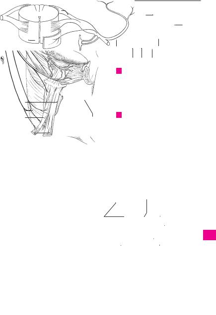
2


pyramid and olive. It passes through the hypo-


to the nuchal region and near the occiput. A


glossal canal and descends between the inter-

16

Medial branch of posterior ramus. Ramus

3


nal jugular vein and internal carotid artery. At


medialis. Branch with motor and sensory fibers


the level of the angle of the mandible it then


supplying the muscles and skin. A


proceeds anteriorly above the posterior margin

17

Lateral branch of posterior ramus. Ramus

4


of the floor of the mouth to enter the tongue. B


lateralis. Purely motor branch passing obliquely


2 Lingual branches. Rami linguales. Rami begin-


laterad into the muscles. A

5


ning lateral to the hyoglossus muscle and sup-

18

Suboccipital nerve. N. suboccipitalis. Poste-


plying the styloglossus, hyoglossus and genio-


rior branch of the first cervical spinal nerve. It


glossus muscles as well as the intrinsic muscles


exits between the vertebral artery and poste-


of the tongue. B


6


rior arch of the atlas and supplies the short


3

SPINAL NERVES. Nervi spinales. They are


muscles of the neck. D

7


formed by two roots and, in contrast to the

19

Greater occipital nerve. N. occipitalis major.


cranial nerves, they exit through the interverte-


Posterior branch of the second cervical spinal


bral foramina. A C


nerve. It emerges between the axis and ob-


4

Root filaments. Fila radicularia. Fine root fibers


8


liquus capitis inferior muscle, pierces the


emerging from the spinal cord within the ante-


trapezius and supplies the nuchal muscles and

9


rior and posterior roots of the individual spinal


skin of the occipital region. D


nerves. A


20

Third occipital nerve. N. occipitalis tertius.


5 Anterior (ventral) root. Radix anterior (mo-


Posterior branch of the third cervical spinal

10


toria). Motor root. A


nerve. It supplies the skin of the nuchal region


6 Posterior (dorsal) root. Radix posterior (sen-


close to the midline. D

11


soria). Sensory root. A


21 Anterior (ventral) branches. Rami anteriores.


7

Spinal (dorsal root) ganglion. Ganglion spinale


Anterior rami of cervical spinal nerves. They


(sensorium). Ganglion situated in the inter-


form the cervical and brachial plexuses. A

12


vertebral foramen, composed of pseudo-uni-

22 Cervical plexus. Plexus cervicalis. Nerve plexus


polar cells. It lies in the posterior root just in


formed by the anterior rami of spinal nerves

13


front of the site where it joins the anterior root. A


C1−4. They supply the skin and muscles of the


8

Spinal nerve trunk. Truncus nervi spinalis.


neck.


Segment between the union of the two roots

23 Nerve loop from C1−3. Ansa cervicalis [hypo-

14


and the first branch of the spinal nerve. A C


glossi]. A nerve loop in the neck (C1-C3) that


9

Anterior (ventral) branch. Ramus anterior.


supplies the infrahyoid muscles. B

15


Larger anterior branch of a spinal nerve. It com-

24

Anterior (ventral) root. Radix anterior. The


municates with adjacent anterior rami to form


anterior root, part of which supplies the genio-


large plexuses. In the thoracic region it be-


hyoid and thyrohyoid muscles via the hypo-

16


comes continuous with an intercostal nerve. A


glossal nerve. B


10

Posterior (dorsal) branch. Ramus posterior.

25

Posterior (dorsal) root. Radix posterior. Pos-

17


Weaker branch supplying the skin of the back


terior root. B


and autochthonous back muscles. A

26

Thyrohyoid branch of the ansa cervicalis.


18


11

Rami

communicantes.

Communicating


Ramus thyrohyoideus. Branch supplying the


branches connecting the spinal nerve and the


thyrohyoid muscle. B


sympathetic trunk. A


27

Lesser occipital nerve. N. occipitalis minor.


19


11 a

Gray communicating ramus. Ramus griseus.


Uppermost cutaneous branch of the cervical


Postganglionic part. A


plexus. It passes upward at the posterior mar-


11 b

White communicating ramus. Ramus albus.


gin of the sternocleidomastoid and, at the oc-

20


Preganglionic part. A


ciput, ramifies as a lateral communicating


12 Meningeal branch. Ramus meningeus. Deli-


nerve of the greater occipital nerve. D


21


cate, recurrent ramus. It passes in front of the

28 Great auricular nerve. N. auricularis magnus. It


spinal nerve to re-enter the vertebral canal


courses to the ear, thereby crossing the sterno-


through the intervertebral foramen and supply


cleidomastoid vertically somewhat above its

22


the meninges of the spinal cord, where it unites


middle. D


with other meningeal rami to form a plexus. It

29

Posterior (dorsal) branch. Ramus posterior. It

23


contains sensory and sympathetic fibers. A


supplies the skin of the posterior surface of the


13 Cauda equina. Collection of all spinal nerve


pinna and the adjacent area. D


roots extending from L1−2 caudally in addition

30

Anterior (ventral) ramus. Ramus anterior. It

24


to the filum terminale. C


supplies the skin of the anterior surface of the


14

CERVICAL NERVES. Nervi cervicales. Eight spi-


ear up to the angle of the mandible. D

25nal nerves emerging from the cervical spinal cord. B

Spinal nerves 335

3;8

1

14

26

25

24

23

3;8

13

4

16

6

17

5

4

7

10;15


12

11a 11b 9;21

ASpinal nerve with roots and branches

B Hypoglossal nerve

2and ansa cervicalis


18


19


29


30


27


20


28


C

Cauda equina

D

Nerves of the nape of the neck

1

2

3

4

5

6

7

8

9

10

11

12

13

14

15

16

17

18

19

20

21

22

23

24

25

336 Spinal nerves

1Transverse cervical (colli) nerve. N. transversus

1 colli. Arises from C3 and is the third nerve occupying the “nerve point” at the posterior margin

2of the middle third of the sternocleidomastoid

muscle where it turns anteriorly and passes beneath the platysma to supply the skin. It re-

3ceives motor fibers for the platysma from the cervical branch of the facial nerve. B

4

2

Superior branches. Rami superiores. They as-


cend to the suprahyoid region. B


3 Inferior branches. Rami inferiores. They de-

5scend to the infrahyoid region. B

4Supraclavicular nerves. Nn. supraclaviculares.

6Cutaneous branches from C3−4. They spread out fanlike to the shoulder and clavicular regions. B

5 Medial supraclavicular nerves. Nn. supra-

7claviculares mediales. They pass over the middle third of the clavicle and supply the skin of the

8neck in this region and the thorax as far as the sternal angle, as well as the sternoclavicular joint. B

9 6 Intermediate supraclavicular nerves. Nn. supraclaviculares intermedii. They descend beneath the platysma and over the middle third

10 of the clavicle to supply the skin up to the 4th rib. B

11 7 Lateral (posterior) supraclavicular nerves.

Nn. supraclaviculares laterales (posteriores). Posterior group of nerves supplying the skin over

12the acromion, deltoid muscle and the acromioclavicular joint. B

138 Phrenic nerve. N. phrenicus. It arises from C4

with additional rami from C3 and C5, extends on the scalenus anterior muscle, then passes

14through the middle mediastinum to the diaphragm. Some of its fibers extend further into the

15peritoneum. A C


9

Pericardial branch. Ramus pericardiacus.

16


Slender branch to the anterior surface of the peri-


cardium. A


10

Phrenicoabdominal branches. Rami phreni-

17


coabdominales. Fibers supplying the peritoneum


up to the gallbladder and pancreas. On the right


side they pass through the foramen for the vena

18


cava, on the left side further anteriorly through


the diaphragm near the left margin of the heart. A


19

11

Accessory phrenic nerves. Nn. phrenici acces-


sorii. Frequent additional roots of the phrenic

20


nerve from C5 and C6 via the nerve to the sub-


clavius. A C

12 BRACHIAL PLEXUS. Plexus brachialis. Nerve

21plexus formed by the anterior rami of spinal nerves C5−T1. Supplying the arm and part of the shoulder girdle, it passes between the scalenus

22anterior and medius extending as far as the head of the humerus. C

2312 a Nerve roots. Radices.

13 Trunks. Trunci. Three primary trunks make up the brachial plexus and each is usually formed

24from one or two anterior rami of spinal nerves.

14 Upper trunk. Truncus superior. Formed by the

25union of C5 and C6 spinal nerves, it generally arises lateral to the scalenus gap. C

15Middle trunk. Truncus medius. A continuation of C7 spinal nerve itself. C

16Lower trunk. Truncus inferior. Formed by the union of C8 and T1 spinal nerves; it lies within the scalenus gap posterior to the subclavian artery. C

17Anterior divisions. Divisiones anteriores. Portions of brachial plexus formed by the anterior branches of the three trunks. They supply the flexor muscles.

18Posterior divisions. Divisiones posteriores. Portions of brachial plexus formed by the posterior branchesofthethreetrunks.Theyunitetoformthe posterior cord and supply the extensor muscles.

18 a Cords. Fasciculi. Three nerve bundles formed by the union of branches (anterior and/or posterior) from the three trunks.

19SUPRACLAVICULAR PART. Pars supraclavicularis. Part of brachial plexus extending up to the superior margin of the clavicle. C

20Dorsal scapular nerve. N. dorsalis scapulae. Nerve that arises from C5 directly lateral to the intervertebral foramen, penetrates the scalenus medius and then courses below the levator scapulae and the two rhomboid muscles, which it innervates. C

21Long thoracic nerve. N. thoracicus longus. Nerve that arises from C5−7, penetrates the scalenus medius and travels on the serratus anterior, which it supplies. C

22Nerve to subclavius. N. subclavius. Slender nerve from the upper trunk with fibers from C4−6 for the subclavius muscle. It frequently sends a branch (11) to the phrenic nerve. C

23Suprascapular nerve. N. suprascapularis. Nerve that arises from C5−6, passes over the brachial plexus to the scapular notch and then goes under the superior transverse ligament of the scapula to innervate the supraand infraspinatus muscles. C

24INFRACLAVICULAR PART. Pars infraclaviculares. Portion of brachial plexus below the clavicle. It extends from the upper margin of the clavicle to the level where the cords divide into the individual nerves. C

25Lateral cord. Fasciculus lateralis. Located lateral to the axillary artery, it is formed by the union of the anterior divisions of the upper and middle trunks, thus from C5−7. C

26Medial cord. Fasciculus medialis. Cord located medial to the axillary artery. It is formed solely by the anterior division of the lower trunk, thus from C8−T1. C

27Posterior cord. Fasciculus posterior. Cord located posterior to the axillary artery; it is formed by the union of the posterior divisions of all three trunks, thus from C5−T1. C

28Medial pectoral nerve. N. pectoralis medialis. Nerve formed by fibers from the medial cord, thus from C8−T1. It supplies the pectoralis major and minor muscles. C

29Lateral pectoral nerve. N. pectoralis lateralis. Fibers from C5−7 that supply the two pectoral muscles. C