Материал: Bovine Viral Diarrhea Virus Diagnosis, Management, and Control
Vaccines |
221 |
vaccine-induced immune responses against bovine viral diarrhea virus type II in young calves. J Am Vet Med Assoc 219:351–356.
Endsley JJ, Quade MJ, Terhaar B, Roth JA: 2002, Bovine viral diarrhea virus type 1-and type 2- specific bovine T lymphocyte-subset responses following modified-live virus vaccination. Vet Therapeutics 3:364–372.
Flores EF, Ridpath JF, Weiblen R, et al.: 2002, Phylogenetic analysis of Brazilian bovine viral diarrhea virus type 2 (BVDV-2) isolates: eveidence for a subgenotype within BVDV-2. Virus Research 87:51–60.
Frey HR, Eicken K, Grummer B, Kenklies S, et al.: 2002, Foetal protection against bovine viral diarrhoea virus after two-step vaccination. J Vet Med B 49:489–493.
Fulton RW, Burge LJ: 2000, Bovine viral diarrhea virus types 1 and 2 antibody response in calves receiving modified live virus or inactivated vaccines. Vaccine 19:267–274.
Fulton RW, Confer AWW, Burge LJ, et al.: 1995, Antibody responses by cattle after vaccination with commercial viral vaccines containing bovine her- pesvirus-1, bovine viral diarrhea virus, parain- fluenza-3 virus, and bovine respiratory syncytial virus immunogens and subsequent revaccination at day 140. Vaccine 13:725–733.
Fulton RW, Cook BJ, Step DL, et al.: 2002b, Evaluation of health status of calves and the impact on feedlot performance: assessment of a retained ownership program for postweaning calves. Can J Vet Res 66:173–180.
Fulton RW, Purdy CW, Confer AW, et al: 2000a, Bovine viral diarrhea type 1 and 2 infections in feeder calves with respiratory disease: interactions with Pasteurella spp., parainfluenza-3 virus and bovine respiratory syncytial virus. Can J Vet Res 64:151–159.
Fulton RW, Ridpath JF, Confer AW, et al.: 2003a, Bovine viral diarrhoea virus antigenic diversity: impact on disease and vaccination programs.
Biologicals. In press.
Fulton RW, Ridpath JF, Saliki JT, et al.: 2002a, Bovine viral diarrhea virus (BVDV) 1b: Predominant subtype in calves in calves with respiratory disease. Can J Vet Res 66:181–190.
Fulton RW, Saliki JT, Burge LJ, et al: 1997, Neutralizing antibodies to type 1 and 2 bovine viral diarrhea viruses: detection by inhibition of viral cytopathology and infectivity by immunoperoxidase assay. Clin Diagn Immunol 4:380–383.
Fulton RW, Saliki JT, Burge LJ, Payton ME: 2003c, Humoral immune response and assessment of vaccine virus shedding in calves receiving modified
live virus vaccines containing bovine herpesvirus-1 and bovine viral diarrhoea virus 1a. J Vet Med B 50:31.
Fulton RW, Saliki JT, Confer AW, et al: 2000b, Bovine viral diarrhea virus cytopathic and noncytopathic biotypes and type 1 and 2 genotypes in diagnostic laboratory accessions: clinical and necropsy samples from cattle. J Vet Diagn Invest 12:33–38.
Fulton RW, Step DL, Ridpath JF, et al.: 2003b, Response of calves persistently infected with noncytopathic bovine viral diarrhea virus (BVDV) subtype 1b after vaccination with heterologous BVDV strains in modified live virus vaccines and
Mannheimia haemolytica bacterin-toxoid. Vaccine
21:2980–2985.
Giangaspero M, Harasawa, R: 1999, Ovine pestiviruses: Their taxonomic status revealed by palindromic nucleotide substitutions. Vet Microbiol 70:33–39.
Grooms DL, Brock KV, Ward LA: 1998, Detection of cytopathic bovine viral diarrhea virus in the ovaries of cattle following immunization with a modified live bovine viral diarrhea virus vaccine. J Vet Diagn Invest 10:130–134.
Grooms DL, Coe P: 2002, Neutralizing antibody responses in preconditioned calves following vaccination for respiratory viruses. Vet Therapeutics 3:119–127.
Harpin S, Hurley DJ, Mbikay M, et al.: 1999, Vaccination of cattle with a DNA plasmid encoding the bovine viral diarrhoea virus major glycoprotein E2. J Gen Virol 80:3137–3144.
Howard CJ, Clarke MC, Brownlie J: 1989, Protection against respiratory infection with bovine virus diarrhoea virus by passively acquired antibody. Vet Microbiol 19:195–203.
Howard CJ, Clarke MC, Sopp P, Brownlie J: 1994, Systemic vaccination with inactivated bovine virus diarrhoea virus protects against respiratory challenge. Vet Microbiol 42:171–179.
Jones L, Van Campen H, Xu ZC, et al.: 2001, Comparison of neutralizing antibodies to type 1a, 1b, and 2 bovine viral diarrhea virus from experimentally infected and vaccinated cattle. Bovine Pract 35:137–140.
Kirkpatrick J, Fulton RW, Burge LJ, et al.: 2001, Passively transferred immunity in newborn calves, rate of antibody decay, and effect on subsequent vaccination with modified live virus vaccine.
Bovine Pract 35:47–55.
Lambert G: 1973, Bovine viral diarrhea: Prophylaxis and postvaccinal reactions. J Am Vet Med Assoc 163:874–876.
Liess B, Orban S, Frey HR, et al.: 1984, Studies on transplacental transmissability of a bovine virus

222 |
BVDV: Diagnosis, Management, and Control |
diarrhoea (BVD) vaccine virus in cattle II. Inoculation of pregnant cows without detectable neutralizing antibodies to BVD virus 90–229 days before parturition (51st to 190th day of gestation). Zbl Vet Med B 31:669–681.
Makoschey B, Janssen MGJ, Vrijenhoek MP, et al.: 2001, An inactivated bovine virus diarrhoea virus (BVDV) type 1 vaccine affords protection against BVDV type 2. Vaccine 19:3261–3268.
McClurkin AW, Littledike ET, Cutlip RC, et al.: 1984, Production of cattle immunotolerant to bovine viral diarrhea virus. Can J Comp Med 48:156–161.
McKercher DG, Saito JK, Crenshaw GL, et al.: 1968, Complications in cattle following vaccination with a combined bovine viral diarrhea-infectious bovine rhinotracheitis vaccine. J Am Vet Med Assoc 152:1621–1624.
Nobiron I, Thompson I, Brownlie, J, Collins ME: 2003, DNA vaccination against bovine viral diarrhoea virus induces humoral and cellular responses in cattle with evidence for protection against viral challenge. Vaccine. In press.
Orban S, Liess B, Hafez SM, et al.: 1983, Studies on transplacental transmissibility of a bovine virus diarrhoea (BVDV) vaccine virus in cattle. I.
Inoculation of pregnant cows 15 to 90 days before parturition (190th to 265th day of gestation). Zbl Vet Med B 30:619–634.
Patel JR, Shilleto RW, Williams J, Alexander DCS: 2002, Prevention of transplacental infection of bovine foetus by bovine viral diarrhoea virus through vaccination. Arch Virol 147:2453–2463.
Pellerin C, vanden Hurk J, Lecomte J, Tijssen P: 1994, Identification of a new group of bovine viral diarrhea virus strains associated with severe outbreaks and high mortalities. Virology 203:260–268.
Perino LJ, Hunsaker BD: 1997, A review of bovine respiratory disease vaccine field efficacy. Bovine Pract 31:59–66.
Peter CP, Tyler DE, Ramsey FK: 1967, Characterization of a condition following vaccination with bovine virus diarrhea vaccine. J Am Vet Med Assoc 150:46–52.
Phillips RM, Heuschele WP, Todd JD: 1975, Evaluation of a bovine viral diarrhea vaccine produced in a porcine kidney cell line. J Am Vet Med Assoc 36:135–140.
Potgeiter LND: 1995, Immunology of bovine viral diarrhea virus. Vet Clin North Am: Food Anim Pract 11:501–520.
Ridpath JF, Bolin SR: 1995, Delayed onset postvaccinal mucosal disease as a result of genetic recombination between genotype 1 and genotype 2 BVDV. Virology 212:259–262.
Ridpath JF, Bolin SR: 1998, Differentiation of types 1a, 1b and 2 bovine viral diarrhea virus (BVDV) by PCR. Mol Cell Prob 12:101–106.
Ridpath JF, Bolin SR, Dubovi EJ: 1994, Segregation of bovine viral diarrhea virus into genotypes. Virology 205:66–74.
Ridpath JF, Neill JD, Endsley J, Roth JA: 2003, Effect of passive immunity on the development of a protective immune response against bovine viral diarrhea virus in calves. Am J Vet Res 64:65–69.
Rosner SF: 1968, Complications following vaccination of cattle against infectious bovine rhinotracheitis, bovine viral diarrhea-mucosal disease, and parainfluenza type 3. J Am Vet Med Assoc 152:898–902.
Roth JA, Kaeberle ML: 1983, Suppression of neutrophil and lymphocyte function induced by a vaccinal strain of bovine viral diarrhea virus with and without the administration of ACTH. Am J Vet Res 44:2366–2372.
Shimazaki T, Nakamura S, Taguchi K, Inoue Y, et al.: 2003, Efficacy of bovine viral diarrhea vaccine used in Japan against bovine viral diarrhea virus type 2 strain 890. J Vet Med Sci 65: 263–266.
Studer E, Bertoni G, Candrian U: 2002, Detection and characterization of pestivirus contaminations in human live viral vaccines. Biologicals 30:289–296.
Talens LT, Beckenhauer WH, Thurber ET, et al.: 1989, Efficacy of viral components of a nonabortigenic vaccine for prevention of respiratory and reproductive system diseases in cattle. J Am Vet Med Assoc 194:1273–1280.
van Campen H, Vorpahl P, Huzurbazar S, et al.: 2000, Evidence for bovine viral diarrhea virus type 2 (BVDV)-associated disease in beef herds vaccinated with a modified-live type 1 BVDV vaccine. J Vet Diagn Invest 12:263–265.
van Oirschot JT, Bruschke CJM, van Rijn PA: 1999, Vaccination of cattle against bovine viral diarrhoea.
Vet Microbiol 64:169–183.
van Rijn PA, van Gennip HGP, Leenderste CH, et al.: 1997, Subdivision of the Pestivirus genus based on envelope glycoprotein E2. Virology 237:337–348.

14 Management Systems and Control Programs
Robert L. Larson
INTRODUCTION
The management and control of bovine viral diarrhea virus (BVDV) infection in cattle herds must take into consideration two methods of virus transmission: postnatal horizontal transmission and gestational vertical transmission from a viremic dam to her fetus (Meyling et al., 1990). Postnatal horizontal transmission results in a transient infection that is usually mild or subclinical, but can result in severe disease if susceptible cattle are exposed to a virulent strain of the virus (Kelling et al., 2002; Hamers et al., 2000a). In addition, postnatal horizontal transmission can lead to vertical transmission of BVDV, because postnatal horizontal infection is the primary method by which a pregnant dam becomes viremic and subsequently infects her fetus.
PRINCIPAL RESERVOIRS OF BVDV
The primary source of BVDV in postnatal transmission is cattle persistently infected (PI) with BVDV. Persistently infected animals are much more efficient transmitters of BVDV than transiently infected animals because they shed large amounts of virus for a long period of time. Transiently infected animals experience a short period of viremia and shed virus in their body secretions and excretions from day 4–15 postinfection (Brownlie et al., 1987; Duffell and Harkness, 1985). In contrast, PI animals usually have a very high and persistent viremia, and BVDV is shed throughout life from virtually all secretions and excretions, including nasal discharge, saliva, semen, urine, tears, milk, and to a lesser extent, feces (Rae et al., 1987; Brock et al., 1991; Bezek et al., 1995; Brock et al., 1998). Horizontal transmission of BVDV to seronegative cattle has been shown to occur after only 1 hour of direct contact with a PI an-
imal (Traven et al., 1991). Over-the-fence contact with a PI animal from a neighboring herd can also introduce BVDV into a susceptible herd (Roeder and Harkness, 1986; Miller et al., 2002).
Although transiently infected cattle are far less efficient at transmitting the virus to susceptible incontact animals (Meyling and Jensen, 1988; Niskanen et al., 2000; Niskanen et al., 2002), the occurrence of seroconversion among assembled cattle without the presence of PI animals indicates that transmission from transiently infected animals does occur (Meyling et al., 1990). In a field study by Moerman et al. (1993) it was found that BVDV infections circulated within a herd over a period of 30 months in the absence of PI cattle and BVDV vaccination, but in the presence of transiently viremic cattle. Horizontal transmission of the virus from either persistently or transiently infected animals to susceptible cattle may be via inhalation or ingestion of virus-containing body fluids (Duffell and Harkness, 1985). Although aerosol transmission over short distances seems likely; the spread of infection is slow or absent when cattle are housed at long distances from PI animals (Wentink et al., 1991).
REPRODUCTIVE EFFECTS OF BVDV
Even mild or subclinical infections of pregnant and susceptible seronegative breeding females may result in conception failure, abortion, or vertical fetal infection. The immune status of the dam, the stage of gestation, and the viral biotype are important factors in determining the result of vertical infection. Transplacental infection occurs with high efficiency in pregnant, seronegative dams (Done, et al., 1980; McClurkin et al., 1984). However, naturally acquired immunity can prevent later fetal infection (Orban et
223
224 |
BVDV: Diagnosis, Management, and Control |
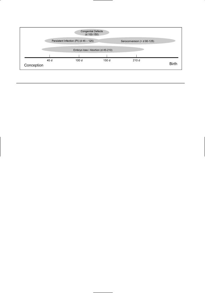
Figure 14.1. The effect of stage of gestation at the time of BVDV infection of susceptible pregnant cows on clinical outcome.
al., 1983). Infection of fetuses can lead to early embryonic death, abortion, congenital defects, stunting, and the birth of either PI or normal calves (Figure 14.1) (Baker, 1987). Fetal infection of susceptible dams with a cytopathic biotype of BVDV early in gestation will usually result in abortion. However, fetal infection with a noncytopathic biotype of BVDV will result either in abortion or, in a certain proportion of animals, the birth of a calf that is immunotolerant to and persistently infected with that particular noncytopathic strain of BVDV. Thus, PI cattle are the result of in utero exposure to the noncytopathic biotype of BVDV prior to the development of a competent fetal immune system (Casaro et al., 1971; McClurkin et al., 1984). Age at immune competence in the face of BVDV exposure is variable and has been reported to range from 90–125 days (Casaro et al., 1971; McClurkin et al., 1984; Roeder et al., 1986). If PI fetuses survive to term, they are continually viremic, but immunotolerant to the homologous BVDV (Duffell and Harkness, 1985; Roeder et al., 1986).
In addition to BVDV causing conception failure and abortion, reproductive efficiency can be decreased due to fatal congenital defects following fetal infection between 100 and 150 days of gestation (Duffell and Harkness, 1985). The teratogenic lesions associated with fetal infection with BVDV include microencephaly, cerebellar hypoplasia, hydranencephaly, hydrocephalus, hypomyelination of the spinal cord, cataracts, retinal degeneration, optic neuritis, microphthalmia, thymic aplasia, hypotrichosis, alopecia, brachygnathism, growth retardation, and pulmonary hypoplasia (Baker, 1987).
EXPOSURE OF HERDS TO PI CATTLE
Suckling calves are commonly in contact with the breeding herd and thus come in contact with dams in early stages of pregnancy. As a result, PI suckling calves may be a source of BVDV infection in breed-
ing herds, causing decreased pregnancy percentage, pregnancy loss, preweaning mortality and the induction of PI calves in the next generation (Wittum et al., 2001; Duffell and Harkness, 1985; McClurkin et al., 1984).
Although mortality of PI calves prior to weaning has been reported to be very high due to fatal congenital defects and secondary infections that cause enteritis, pneumonia, and arthritis (McClurkin et al., 1979; McClurkin et al., 1984), in some situations 17–50% of PI calves may reach breeding age (Barber et al., 1985; Binkhorst et al., 1983; Houe, 1993). Persistently infected females of breeding age are not only a source of horizontal transfer of BVDV, but also will produce a PI calf themselves, which if it survives, creates a familial clustering of PI animals (Houe et al., 1995; McLurkin et al., 1979).
The consequence of introducing a PI animal into a beef herd (confined breeding and calving seasons) depends on the timing of the introduction relative to the breeding season and the subsequent immunologic status of the herd during early gestation (Table 14.1). A likely scenario for a BVDV-exposed herd is to experience an initial peak of disease followed by low-level chronic reproductive losses in subsequent months and years. If a PI animal enters the herd either by birth or by purchase near the start of the breeding season, a high percentage of the herd may not be immunologically protected to the degree necessary to prevent viremia, conception failure, abortion, or fetal infection. If the PI animal is in contact with the breeding herd for a long enough period of time, the majority of the herd should become infected and seroconvert. Seropositive animals are less likely to have conception failures, abortions, or infected fetuses compared to seronegative animals. If no intervention is applied to the herd, the number of susceptible females the following year should be greatly decreased and the number of abortions and

Management Systems and Control Programs |
225 |
Table 14.1. Various outcomes of bovine viral diarrhea virus (BVDV) infection depending on transmission and host.
Timing and |
|
|
|
|
|
Route of |
|
|
|
|
|
Transmission |
Infected Animal |
|
|
Outcome |
|
|
|
|
|
||
Postnatal |
Seropositive nonpregnant animal |
• |
Mild or subclinical disease |
||
horizontal |
|
|
|
|
|
Susceptible nonpregnant animal |
• |
Mild or subclinical disease |
|||
transmission |
|||||
|
• |
Moderate to severe clinical disease with |
|||
|
|
||||
|
|
|
virulent strains |
||
|
|
|
|
||
|
Susceptible breeding animal |
• |
Conception failure |
||
|
|
• |
Conception success |
||
|
|
|
|
||
|
Susceptible pregnant animal |
• |
Fetal infection |
||
|
|
|
|
||
Gestational |
Fetus of persistently infected dam |
• |
Fetal infection |
||
vertical |
|
|
• |
Embryo loss/abortion |
|
transmission |
|
|
• |
Immunotolerant, persistently infected |
|
|
|
|
|
fetus/calf |
|
|
|
|
|
||
|
Fetus of susceptible dam (d 0 to d |
• |
Fetal infection |
||
|
90–125 gestation) — cytopathic biotype |
|
• |
Embryo loss/abortion |
|
|
|
|
|
||
|
Fetus of susceptible dam (d 0 to d |
• |
Fetal infection |
||
|
90–125 gestation) — noncytopathic |
|
• |
Embryo loss/abortion |
|
|
biotype |
|
• |
Immunotolerant, persistently infected |
|
|
|
|
|
fetus/calf |
|
|
|
|
• |
Fetal malformations (d 100–150): |
|
|
|
|
|
cerebellar hypoplasia, hypotrichosis, |
|
|
|
|
|
brachygnathism, depleted lymph |
|
|
|
|
|
nodes/tissue, etc. |
|
|
|
|
|
||
|
Fetus of susceptible pregnant animal with |
• |
Fetal infection |
||
|
immune-competent fetus (>90–125 d) |
|
• |
Abortion |
|
|
|
|
• |
Fetal malformations |
|
|
|
|
• |
Normal fetus/calf |
|
|
|
|
|
||
|
Fetus of seropositive pregnant dam |
• |
Fetal protection from infection |
||
|
(seropositive following natural infection) |
• |
Fetal infection ? |
||
|
|
|
|
||
|
Fetus of seropositive pregnant dam |
• |
Fetal protection from infection |
||
|
(seropositive following vaccination) |
• |
Fetal infection |
||
|
|
|
• |
Embryo loss/abortion ? |
|
|
|
|
• |
Immunotolerant, persistently infected |
|
|
|
|
|
fetus/calf |
|
|
|
|
• |
Normal fetus/calf |
|
|
|
|
|
|
|
infected fetuses (both PI and immunocompetent) should decrease. Thus, even in the absence of vaccination and culling, the number of PI animals and BVDV infections in a closed herd may be selflimiting over time (Houe, 1995). A model developed by Cherry et. al. (1998) indicates that in continuous calving situations, the proportion of PI animals in a dairy herd will reach an equilibrium of about
0.9–1.2% in closed herds with no BVDV control procedures (Cherry et al., 1998). This model does not hold true if the herd is not closed (replacement females and bulls are added). Prevalence of PI animals and the resulting problems would not be expected to reach equilibrium in BVDV-infected herds with no control measures that are purchasing heifers, particularly bred heifers.