Материал: part03

Page 266 |
DICOM PS3.3 2020a - Information Object Definitions |
A.32.4.3 VL Photographic Image IOD Content Constraints
A.32.4.3.1 Modality
The value of Modality (0008,0060) shall be XC.
A.32.4.3.2 Anatomic Region Sequence
For dermatology applications:
•the Baseline CID for Anatomic Region Sequence (0008,2218) is CID 4029 “Dermatology Anatomic Sites” •the Baseline CID for Anatomic Region Modifier Sequence (0008,2220) is CID 245 “Laterality with Median”
A.32.5 Video Endoscopic Image IOD
A.32.5.1 Video Endoscopic Image IOD Description
The Video Endoscopic Image IOD specifies the Attributes of Multi-frame Video Endoscopic Images.
A.32.5.2 Video Endoscopic Image IOD Entity-Relationship Model
This IOD uses the E-R Model in Section A.1.2, with only the Image IE below the Series IE. The Frame of Reference IE is not a com- ponent of this IOD.
Note
1.The video may include audio channel(s) for acquiring Patient voice or physiological sounds, healthcare professionals' commentary, or environmental sounds.
2.The Frame Pointers Module has not been included because the selection of relevant sub-sequence(s) is usually made in a second workflow step and stored into separate Key Object Selection Documents.
3.The Curve entity was previously include in the list of entities that are not used, but has been retired from DICOM. It is still not used in this IOD. See PS3.3-2004.
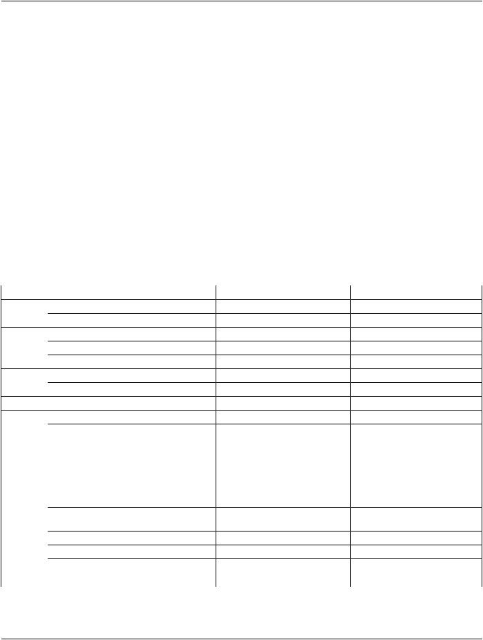
Table A.32.5-1. Video Endoscopic Image IOD Modules
IE |
Module |
Reference |
Usage |
Patient |
Patient |
C.7.1.1 |
M |
|
Clinical Trial Subject |
C.7.1.3 |
U |
Study |
General Study |
C.7.2.1 |
M |
|
Patient Study |
C.7.2.2 |
U |
|
Clinical Trial Study |
C.7.2.3 |
U |
Series |
General Series |
C.7.3.1 |
M |
|
Clinical Trial Series |
C.7.3.2 |
U |
EquipmentGeneral Equipment |
C.7.5.1 |
M |
|
Image |
General Image |
C.7.6.1 |
M |
|
General Reference |
C.12.4 |
U |
|
Cine |
C.7.6.5 |
M |
|
Multi-frame |
C.7.6.6 |
M |
|
Image Pixel |
C.7.6.3 |
M |
|
Acquisition Context |
C.7.6.14 |
M |
|
Device |
C.7.6.12 |
U |
- Standard -

|
DICOM PS3.3 2020a - Information Object Definitions |
Page 267 |
|
IE |
Module |
Reference |
Usage |
|
Specimen |
C.7.6.22 |
C - Required if the Imaging Subject |
|
|
|
is a Specimen |
|
VL Image |
C.8.12.1 |
M |
|
ICC Profile |
C.11.15 |
U |
|
SOP Common |
C.12.1 |
M |
|
Common Instance Reference |
C.12.2 |
U |
|
Frame Extraction |
C.12.3 |
C - Required if the SOP Instance |
|
|
|
was created in response to a |
|
|
|
Frame-Level retrieve request |
A.32.5.3 Video Endoscopic Image IOD Content Constraints
A.32.5.3.1 Modality
The value of Modality (0008,0060) shall be ES.
Note
The use of a single value for Modality recognizes the fact that the same acquisition equipment is often used for different purposes (e.g., laparoscopy and colonoscopy). This means that Modality is not useful to distinguish one type of endoscopy from another when browsing a collection of Studies. Therefore, the use of Procedure Code Sequence (0008,1032) and Anatomic Region Sequence (0008,2218) in the image instances and in the query response is recommended, though gath- ering sufficient information to populate these Attributes in an unscheduled workflow environment (i.e., in the absence of Modality Worklist) may require operator intervention.
A.32.5.3.2 Image Related Data Encoding
The Modality LUT Module, VOI LUT Module, Graphic Annotation Module and Overlay Plane Module shall not be present.
Note
The Curve Module (Retired) was previously include in the list of Modules that shall not be present, but has been retired from DICOM. It is still not permitted to be present. See PS3.3-2004.
A.32.5.3.3 Anatomic Region Sequence
The Defined Context Group for Anatomic Region Sequence (0008,2218) shall be CID 4040 “Endoscopy Anatomic Regions”.
A.32.6 Video Microscopic Image IOD
A.32.6.1 Video Microscopic Image IOD Description
The Video Microscopic Image IOD specifies the Attributes of Video Microscopic Images, including both imaging of specimens and direct microscopic imaging of the Patient (e.g., perioperative microscopy). Microscopic Images with Slide Coordinates shall not be encoded with this IOD.
A.32.6.2 Video Microscopic Image IOD Entity-Relationship Model
This IOD uses the E-R Model in Section A.1.2, with only the Image IE below the Series IE. The Frame of Reference IE is not a com- ponent of this IOD.
Note
1.The video may include audio channel for acquiring Patient voice or physiological sounds, healthcare professionals comment, or environment sounds.
- Standard -

Page 268 |
DICOM PS3.3 2020a - Information Object Definitions |
2.The Frame Pointers Module has not been included because the selection of relevant sub-sequence(s) is usually made in a second step and stored into separate Key Object Selection Documents.
3.The Curve entity was previously include in the list of entities that are not used, but has been retired from DICOM. It is still not used in this IOD. See PS3.3-2004.
4.TheSpecimenIdentificationModulewaspreviouslyincludedinthisIODbuthasbeenretired,anditsfunctionalityreplaced by the Specimen Module. See PS3.3-2008.
Table A.32.6-1. Video Microscopic Image IOD Modules
IE |
Module |
Reference |
Usage |
Patient |
Patient |
C.7.1.1 |
M |
|
Clinical Trial Subject |
C.7.1.3 |
U |
Study |
General Study |
C.7.2.1 |
M |
|
Patient Study |
C.7.2.2 |
U |
|
Clinical Trial Study |
C.7.2.3 |
U |
Series |
General Series |
C.7.3.1 |
M |
|
Clinical Trial Series |
C.7.3.2 |
U |
EquipmentGeneral Equipment |
C.7.5.1 |
M |
|
Image |
General Image |
C.7.6.1 |
M |
|
General Reference |
C.12.4 |
U |
|
Cine |
C.7.6.5 |
M |
|
Multi-frame |
C.7.6.6 |
M |
|
Image Pixel |
C.7.6.3 |
M |
|
Acquisition Context |
C.7.6.14 |
M |
|
Device |
C.7.6.12 |
U |
|
Specimen |
C.7.6.22 |
C - Required if the Imaging Subject |
|
|
|
is a Specimen |
|
VL Image |
C.8.12.1 |
M |
|
ICC Profile |
C.11.15 |
U |
|
SOP Common |
C.12.1 |
M |
|
Common Instance Reference |
C.12.2 |
U |
|
Frame Extraction |
C.12.3 |
C - Required if the SOP Instance |
|
|
|
was created in response to a |
|
|
|
Frame-Level retrieve request |
A.32.6.3 Video Microscopic Image IOD Content Constraints
A.32.6.3.1 Modality
The value of Modality (0008,0060) shall be GM.
A.32.6.3.2 Image Related Data Encoding
The Modality LUT Module, VOI LUT Module, Graphic Annotation Module and Overlay Plane Module shall not be present.
Note
The Curve Module (Retired) was previously include in the list of Modules that shall not be present, but has been retired from DICOM. It is still not permitted to be present. See PS3.3-2004.
- Standard -
DICOM PS3.3 2020a - Information Object Definitions |
Page 269 |
A.32.7 Video Photographic Image IOD
A.32.7.1 Video Photographic Image IOD Description
The Video Photographic Image IOD specifies the Attributes of VL Multi-frame photographic Images.
A.32.7.2 Video Photographic Image IOD Entity-Relationship Model
This IOD uses the E-R Model in Section A.1.2, with only the Image IE below the Series IE. The Frame of Reference IE is not a com- ponent of this IOD.
Note
1.The video may include audio channel for acquiring Patient voice or physiological sounds, healthcare professionals comment, or environment sounds.
2.The Frame Pointers Module has not been included because the selection of relevant sub-sequence(s) is usually made in a second step and stored into separate Key Object Selection Documents.
3.The Curve entity was previously include in the list of entities that are not used, but has been retired from DICOM. It is still not used in this IOD. See PS3.3-2004.
4.TheSpecimenIdentificationModulewaspreviouslyincludedinthisIODbuthasbeenretired,anditsfunctionalityreplaced by the Specimen Module. See PS3.3-2008.
Table A.32.7-1. Video Photographic Image IOD Modules
IE |
Module |
Reference |
Usage |
Patient |
Patient |
C.7.1.1 |
M |
|
Clinical Trial Subject |
C.7.1.3 |
U |
Study |
General Study |
C.7.2.1 |
M |
|
Patient Study |
C.7.2.2 |
U |
|
Clinical Trial Study |
C.7.2.3 |
U |
Series |
General Series |
C.7.3.1 |
M |
|
Clinical Trial Series |
C.7.3.2 |
U |
EquipmentGeneral Equipment |
C.7.5.1 |
M |
|
Image |
General Image |
C.7.6.1 |
M |
|
General Reference |
C.12.4 |
U |
|
Cine |
C.7.6.5 |
M |
|
Multi-frame |
C.7.6.6 |
M |
|
Image Pixel |
C.7.6.3 |
M |
|
Acquisition Context |
C.7.6.14 |
M |
|
Device |
C.7.6.12 |
U |
|
Specimen |
C.7.6.22 |
C - Required if the Imaging Subject |
|
|
|
is a Specimen |
|
VL Image |
C.8.12.1 |
M |
|
ICC Profile |
C.11.15 |
U |
|
SOP Common |
C.12.1 |
M |
|
Common Instance Reference |
C.12.2 |
U |
- Standard -

Page 270 |
DICOM PS3.3 2020a - Information Object Definitions |
|
|
IE |
Module |
Reference |
Usage |
|
Frame Extraction |
C.12.3 |
C - Required if the SOP Instance |
|
|
|
was created in response to a |
|
|
|
Frame-Level retrieve request |
A.32.7.3 Video Photographic Image IOD Content Constraints
A.32.7.3.1 Modality
The value of Modality (0008,0060) shall be XC.
A.32.7.3.2 Image Related Data Encoding
The Modality LUT Module, VOI LUT Module, Graphic Annotation Module and Overlay Plane Module shall not be present.
Note
The Curve Module (Retired) was previously include in the list of Modules that shall not be present, but has been retired from DICOM. It is still not permitted to be present. See PS3.3-2004.
A.32.8 VL Whole Slide Microscopy Image IOD
A.32.8.1 VL Whole Slide Microscopy Image IOD Description
TheVLWholeSlideMicroscopyImageIODspecifiestheAttributesofamulti-framevisiblelightwholeslidemicroscopyimageencoded as a tiled decomposition. Each frame encodes a single tile within a three-dimensional imaged volume at a uniform resolution.
Note
An entire set of tiles for an acquisition may be encoded in the frames of a single SOP Instance, in multiple SOP Instances ofasingleconcatenation,orinmultipleSOPInstancesinaSeries(withorwithoutconcatenations).E.g.,asingleSOPInstance may contain an entire low resolution image as a single tile (single frame), or a single SOP Instance may contain an entire high resolution, multi-focal depth, multi-spectral acquisition (multiple frames).
A.32.8.2 VL Whole Slide Microscopy Image IOD Entity-Relationship Model
This IOD uses the E-R Model in Section A.1.2, with only the Image IE below the Series IE.
A.32.8.3 VL Whole Slide Microscopy Image IOD Module Table
Table A.32.8-1 specifies the Modules of the VL Whole Slide Microscopy Image IOD.
Table A.32.8-1. VL Whole Slide Microscopy Image IOD Modules
IE |
Module |
Reference |
Usage |
Patient |
Patient |
C.7.1.1 |
M |
|
Clinical Trial Subject |
C.7.1.3 |
U |
Study |
General Study |
C.7.2.1 |
M |
|
Patient Study |
C.7.2.2 |
U |
|
Clinical Trial Study |
C.7.2.3 |
U |
Series |
General Series |
C.7.3.1 |
M |
|
Whole Slide Microscopy Series |
C.8.12.3 |
M |
|
Clinical Trial Series |
C.7.3.2 |
U |
Frame of |
Frame of Reference |
C.7.4.1 |
M |
Reference |
|
|
|
- Standard -