Page 1038 |
DICOM PS3.3 2020a - Information Object Definitions |
C.8.17.14.1.6 Bits Allocated, Bits Stored, and High Bit
These Attributes shall be determined based upon the Photometric Interpretation (0028,0004):
|
Photometric Interpretation |
Bits Allocated (0028,0100) |
Bits Stored (0028,0101) |
High Bit (0028,0102) |
|
(0028,0004) |
|
|
|
|
MONOCHROME2 |
8 |
8 |
7 |
|
PALETTE COLOR |
16 |
12 |
11 |
|
16 |
16 |
15 |
|
|
C.8.17.14.1.7RelationshipBetweenOphthalmicTomographyImageandOphthalmicOpticalCoherenceTomographyB-scan
Volume Analysis IODs
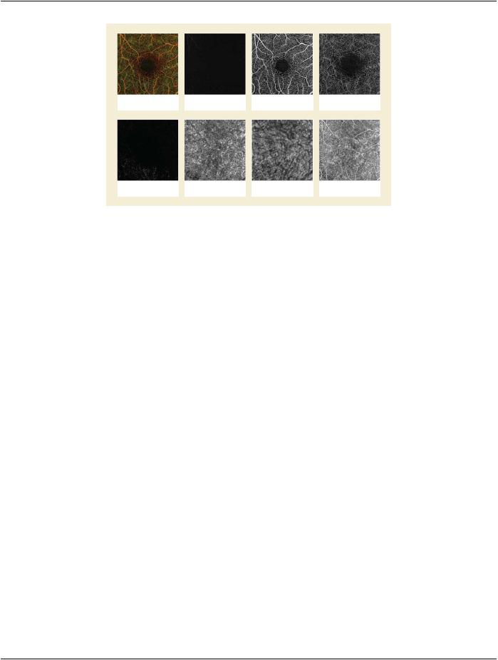
When generating an angiographic en face Image SOP Instance implementations need to understand the relationship between the Ophthalmic Tomography Image SOP Instance(s) and the Ophthalmic Optical Coherence Tomography B-scan Volume Analysis SOP Instance.
The Ophthalmic Optical Coherence Tomography B-scan Volume Analysis SOP Instance, which is a multi-frame SOP Instance, refer- ences one or more Ophthalmic Tomography Image SOP Instances using the Derivation Image Macro. The Derivation Image Macro defines Attributes at the Frame Level (i.e. each frame in the Ophthalmic Optical Coherence Tomography B-scan Volume Analysis SOP Instance, references an Ophthalmic Tomography Image SOP Instance and the Ophthalmic Tomography Image SOP Instance frame number that was used to generate the specific Ophthalmic Optical Coherence Tomography B-scan Volume Analysis frame).
Examples are shown in Section UUU.2 “Relationship Between Ophthalmic Tomography Image and Ophthalmic Optical Coherence Tomography B-scan Volume Analysis IODs” in PS3.17.
C.8.17.15 Ophthalmic Optical Coherence Tomography En Face Image Quality Rating
Module
Table C.8.17.15-1 specifies the Attributes for evaluating the quality of the derived en face image.
Table C.8.17.15-1. Ophthalmic Optical Coherence Tomography En Face Image Quality Rating Module
Attributes
Attribute Name |
Tag |
Type |
Attribute Description |
Ophthalmic En Face Image |
(0022,1628) |
1 |
Evaluation of the quality of the en face image. |
Quality Rating Sequence |
|
|
Only a single Item shall be included in this Sequence. |
|
|
|
>Include Table 10-26 “Numeric Value Macro Attributes” |
|
Defined CID 4243 shall be used for Concept Name Code |
|
|
|
Sequence (0040,A043) |
>Quality Threshold |
(0022,1630) |
1 |
Threshold for the quality value. If the Numeric Value |
|
|
|
(0040,A30A) of the Numeric Value Macro is equal or above |
|
|
|
the threshold, it is considered acceptable by the algorithm. |
The units of this Attribute shall be the same as defined in Measurement Units Code Sequence (0040,08EA) of the Numeric Value Macro.
 >Include Table 10-19 “Algorithm Identification Macro Attributes”
C.8.17.16 Ophthalmic Optical Coherence Tomography B-scan Volume Analysis Image
Module
Table C.8.17.16-1 specifies the Attributes that describe the Ophthalmic Optical Coherence Tomography B-scan Volume Analysis Image Module.
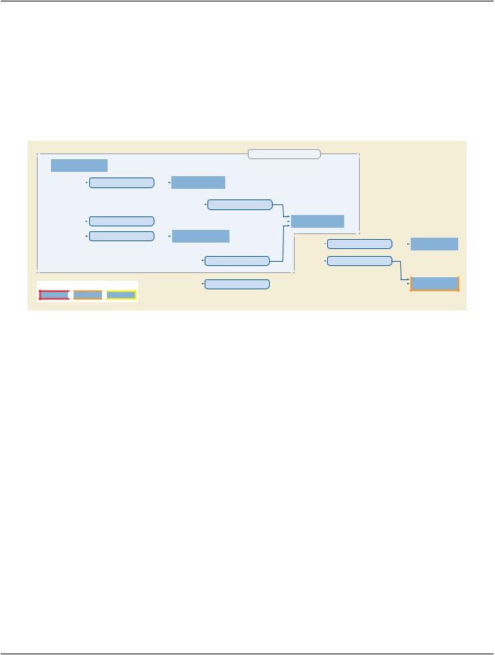

 Stage 2 Data
 Stage 3 Data