DICOM PS3.3 2020a - Information Object Definitions |
Page 1035 |
B
E
C
A
D
F
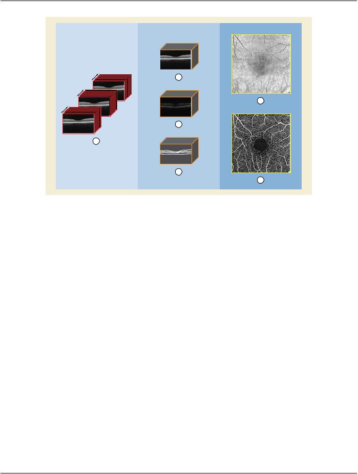
Figure C.8.17.14-1. Example of the Image Process Performed to Generate En Face Images
Figure Legend:
A.OCT proprietary B-scan data (possibly a DICOM Raw Data Instance)
B.Volumetric structural ophthalmic tomography image (Ophthalmic Tomography Image Instance)
C.OCT angiographic flow volume information (Ophthalmic Optical Coherence Tomography B-scan Volume Analysis Instance)
D.OCT surface mesh (Surface Segmentation Instance)
E.Structural en face image (Ophthalmic Optical Coherence Tomography En Face Image Instance)
F.En Face angiographic flow image (Ophthalmic Optical Coherence Tomography En Face Image Instance)
Stage 1:OCT technology is used to acquire a volumetric dataset from a retinal region of interest.This volumetric dataset (A) consists of multiple B-scans in a raster pattern, and multiple frames are acquired at each B-scan location. The B-scans are acquired in the manufacturer's proprietary format for analysis and storage. If this information is stored in DICOM, it can use the Raw Data Storage SOP Class.
Stage2:TheOCTproprietaryB-scandata(A)(orDICOMRawDataSOPInstance)isthenanalyzedtoderivethevolumetricstructural ophthalmic tomography image (B). From (B) one or more OCT surface meshes (D) are generated to delineate the anatomical boundaries. The difference in signal between the frames of each individual B-scan is analyzed to produce the OCT angiographic flow volume information (C).
Stage 3:Clinicians typically make their assessment based upon two types of OCT en face images. The structural OCT en face image
(E) is derived by using pixel information in (B) and two surface meshes (C). The vascular OCT en face image (F) may be derived using the volumetric structural ophthalmic tomography image (B) , the OCT surface mesh (D) and the OCT angiographic flow volume information (C).
EnfaceimagesaretypicallyderivedbytheacquisitionmodalitythatgeneratedtheOphthalmicTomographyImage,SurfaceSegment- ationandOphthalmicOpticalCoherenceTomographyB-scanVolumeAnalysisSOPInstancesorbyimageworkstationsthatreceived therespectiveOphthalmicImage,SurfaceSegmentationandOphthalmicOpticalTomographyB-scanVolumeAnalysisSOPInstances via DICOM Storage.

 >>Include Table 8.8-1 “Code Sequence Macro Attributes”
  BCID 4273 “Retinal Segmentation Surfaces”.