Материал: part03

Внимание! Если размещение файла нарушает Ваши авторские права, то обязательно сообщите нам

Page 1026​

DICOM PS3.3 2020a - Information Object Definitions​

OCT Frame 1

OCT Frame 2


1


N

1


L

M

Retinal Reference Image


(e,f)

(g,h)


(z,y)


(a,b)


(c,d)


Figure C.8.17.10-1. Relationship of Longitudinal (Nominally Perpendicular) OCT Frames to Reference​ Frame​

In the case of ophthalmic tomography (OPT) images with a transverse orientation, the OPT image corresponds to a sub-rectangle of​ the reference image. See Figure C.8.17.10-2. The OPT frame is nominally parallel to the reference image. Therefore, each corner of​ theOPTframecanalsocorrespondtoaparticularpixelonanassociatedimage.ItisassumedthattheedgesofOPTframeareexactly​ parallel to the edges of the reference image.​

Retinal Reference Image

(a,b)

(c,d)

OCT Frame 1

(a,b)

(c,d)

Figure C.8.17.10-2. Relationship of Transverse (Nominally Parallel) OCT Frame to Reference Frame​

- Standard -​

DICOM PS3.3 2020a - Information Object Definitions​

Page 1027​

C.8.17.11 Wide Field Ophthalmic Photography Stereographic Projection Module​

Table C.8.17.11-1 specifies the Attributes that describe the Wide Field Photography Stereographic Projection Module.​

Table C.8.17.11-1. Wide Field Ophthalmic Photography Stereographic Projection Module Attributes​

Attribute Name​

Tag​

Type​

Attribute Description​

Include Table 10-5 “General Anatomy Mandatory Macro​

The concept code for Anatomic Region Sequence (0008,2218) shall​

Attributes”​


be (81745001, SCT, "Eye"), and Defined CID 244 “Laterality” shall​


be used for Anatomic Region Modifier Sequence (0008,2220). Only​


asingleItemshallbeincludedinAnatomicRegionModifierSequence​


(0008,2220).​

Transformation Algorithm​

(0022,1513)​

1​

Software algorithm used for stereographic projection.​

Sequence​


Only a single Item shall be included in this Sequence.​


>IncludeTable10-19“AlgorithmIdentificationMacroAttributes”​

Ophthalmic Axial Length​

(0022,1019)​

1​

The axial length measurement used for the stereographic projection,​


in mm.​

Ophthalmic Axial Length​

(0022,1515)​

1​

The method used to obtain the Ophthalmic Axial Length.​

Method​


Enumerated values:​


MEASURED​ Measured axial length.​


ESTIMATED​ An estimated value based upon performing the​


examination (i.e., based upon surrogate markers of​


axial length).​


POPULATION​A length that represents a population norm (i.e., not​


based upon a measured axial length or surrogate​


markers of axial length).​

X Coordinates Center Pixel​

(0022,1528)​

1​

The horizontal angle covered on the sphere by the center pixel of the​

View Angle​


projected image, as measured from the center of the sphere, in​


degrees. See Section C.8.17.11.1.1 for further explanation.​

Y Coordinates Center Pixel​

(0022,1529)​

1​

The vertical angle covered on the sphere by the center pixel of the​

View Angle​


projected image, as measured from the center of the sphere, in​


degrees. See Section C.8.17.11.1.1 for further explanation.​

Ophthalmic FOV​

(0022,1517)​

3​

The field of view used to capture the ophthalmic image, in degrees.​


The field of view is the maximum image size displayed on the image​


plane, expressed as the angle subtended at the exit pupil of the eye​


by the maximum dimension 2r (where r equals the radius).​

C.8.17.11.1 Wide Field Ophthalmic Photography Stereographic Projection Attribute Descriptions​

C.8.17.11.1.1 Center Pixel View Angle​

TheCenterPixelViewAngle(0022,1528)comprisestworealnumbersXCENTERPIXELVIEWANGLEandYCENTERPIXELVIEWANGLE​ thatrepresentindegreestheanglealongthehorizontalaxisandtheverticalaxisrespectivelycoveredbythecenterpixelintheimage,​ where this angle is measured from the center of the sphere. These are used to convert pixel locations in the image to their corres-​ ponding locations on a sphere. x and y are pixel locations (may be sub pixels) in the image, x running from 0 to XPIXELS from the​ left-hand side of the image to the right-hand side, and y running from 0 to YPIXELS from top to bottom, and if λ denotes the azimuth​ or longitude on the sphere and ϕ the elevation or latitude on the sphere, both in degrees, then,​


-1 x'


1

λ = tan2

ρ

,


tan(c)


-1 y'sin c


ϕ = sin


ρ


- Standard -​

Page 1028​

DICOM PS3.3 2020a - Information Object Definitions​

where​

tan2-1 used to define λ represents the two-argument inverse tangent function:​


-1 y


tan


x


tan-1

y

+ 180


x


tan-1

y

- 180

tan2-1

x

(y, x) =

180


+

2


180


- 2


undefined

x > 0

y ≥ 0, x < 0 y < 0, x < 0 y > 0, x = 0 y < 0, x = 0 y = 0, x = 0

and​

x' =

x - XPIXELS XCENTERPIXELVIEWANGLE


2


y' =

- y + YPIXELS YCENTERPIXELVIEWANGLE


2


and​


ρ =


x'2 + y'2


c = 2tan

-1 ρ π


2 180


The above equations assume the use of degrees throughout. Radians can be used, if XCENTERPIXELVIEWANGLE and​ YCENTERPIXELVIEWANGLE are given in radians, if all trigonometric functions are switched to their radian-equivalent, and if the​ factor π/180 is removed from the definition of c.​

C.8.17.12 Wide Field Ophthalmic Photography 3D Coordinates Module​

Table C.8.17.12-1 specifies the Attributes that describe the Wide Field Ophthalmic Photography 3D Coordinates Module.​

Table C.8.17.12-1. Wide Field Ophthalmic Photography 3D Coordinates Module Attributes​

Attribute Name​

Tag​

Type​

Attribute Description​

Include Table 10-5 “General Anatomy Mandatory Macro​

TheconceptcodeforAnatomicRegionSequence(0008,2218)shall​

Attributes”​


be (81745001, SCT, "Eye"), and Defined CID 244 “Laterality” shall​


beusedforAnatomicRegionModifierSequence(0008,2220).Only​


a single Item shall be included in Anatomic Region Modifier​


Sequence (0008,2220).​

TransformationMethodCode​

(0022,1512)​

1​

Method used to map the 2D Pixel Image data in this SOP Instance​

Sequence​


to the 3D Cartesian coordinates in the Dimensional to Two Three​


Dimensional Map Sequence (0022,1518).​


Only a single Item shall be included in this Sequence.​


See Section C.8.17.12.1.1 for further explanation.​

>Include Table 8.8-1 “Code Sequence Macro Attributes”​

DCID 4245 “Wide Field Ophthalmic Photography Transformation​


Method”​

Transformation Algorithm​

(0022,1513)​

1​

Software algorithm that performed the mapping.​

Sequence​


Only a single Item shall be included in this Sequence.​


- Standard -​


DICOM PS3.3 2020a - Information Object Definitions​

Page 1029​

Attribute Name​

Tag​

Type​

Attribute Description​


>Include Table 10-19 “Algorithm Identification Macro Attributes”​


Ophthalmic Axial Length​

(0022,1019)​

1​

The axial length measurement used when performing the 2D pixel​


imagemapping into 3D Cartesian coordinates, in mm.​


Ophthalmic Axial Length​

(0022,1515)​

1​

The method used to obtain the Ophthalmic Axial Length.​

Method​


Enumerated values:​


MEASURED​ Measured axial length.​


ESTIMATED​ An estimated value based upon performing the​


examination (i.e., based upon surrogate markers​


of axial length).​


POPULATION​Alengththatrepresentsapopulationnorm(i.e.,not​


based upon a measured axial length or surrogate​


markers of axial length).​


Ophthalmic FOV​

(0022,1517)​

3​

The field of view used to capture the ophthalmic image, in degrees.​


Thefieldofviewisthemaximumimagesizedisplayedontheimage​


plane, expressed as the angle subtended at the exit pupil of the​


eye by the maximum dimension 2r (where r equals the radius).​

Two Dimensional to Three​

(0022,1518)​

1​

A sparsely sampled map of 2D image pixels (with sub pixel​

Dimensional Map Sequence​


resolution) to 3D coordinates.​


EachframeshallbereferencedonceandonlyonceinthisSequence​


in Referenced Frame Numbers (0040,A136).​


One or more Items shall be included in this Sequence.​

>ReferencedFrameNumber​

(0008,1160)​

1​

References one or more frames within this SOP Instance to which​


this Sequence Item applies. The first frame shall be denoted as​


frame number one.​


>Number of Map Points​

(0022,1530)​

1​

The number of points in the map. Shall include one or more points.​

>Two Dimensional to Three​

(0022,1531)​

1​

See Section C.8.17.12.1.2 for further explanation.​


Dimensional Map Data​


C.8.17.12.1 Wide Field Ophthalmic Photography 3D Coordinates Attribute Descriptions​

C.8.17.12.1.1 Transformation Method Code Sequence​

IfTransformationMethodCodeSequence(0022,1512)is(111791,DCM,"Sphericalprojection")allthecoordinatesintheTwoDimen-​ sionaltoThreeDimensionalMapData(0022,1531)shalllieonaspherewithadiameterthatshallbeequaltoOphthalmicAxialLength​ (0022,1019).​

If Transformation Method Code Sequence (0022,1512) is (111792, DCM, "Surface contour mapping") the coordinates in the Two​ Dimensional to Three Dimensional Map Data (0022,1531) are based upon the contour of the eye, therefore it cannot be assumed to​ be a spherical surface.​

C.8.17.12.1.2 Two Dimensional to Three Dimensional Map Data​

Two Dimensional to Three Dimensional Map Data (0022,1531) is used to convey a sparsely sampled map of 2D image pixels (with​ sub pixel resolution) to 3D coordinates.​

The origin of the 3D points shall be the Ophthalmic Coordinate System which is based upon the corneal vertex (i.e., the x, y and z​ coordinates of 0.0, 0.0, 0.0, in mm). See Section C.8.30.3.1.4.​

All data points are encoded as a floating point 5-tuple where the values are:​

1st value = 2D horizontal location (a sub pixel location between 0 and number of columns)​

2nd value = 2D vertical location (a sub pixel location between 0 and number of rows)​

- Standard -​

Page 1030​

DICOM PS3.3 2020a - Information Object Definitions​

3rd value = x 3D-coordinate​

4th value = y 3D-coordinate​

5th value = z 3D-coordinate​

The ordering is 2D horizontal location1, 2D vertical location1, 3Dx1, 3Dy1, 3Dz1, … 2D horizontal locationn, 2D vertical locationn, 3Dxn,​ 3Dyn, 3Dzn.​

C.8.17.13 Wide Field Ophthalmic Photography Quality Rating Module​

Table C.8.17.13-1 specifies the Attributes that evaluate the quality of the projection or mapping used for a wide field ophthalmic​ photography image.​

Table C.8.17.13-1. Wide Field Ophthalmic Photography Quality Rating Module Attributes​

Attribute Name​

Tag​

Type​

Attribute Description​

Wide Field Ophthalmic​

(0022,1525)​

1​

Type of metric and metric value used to evaluate the quality of the​

Photography Quality Rating​


projection or mapping used for the wide field ophthalmic​

Sequence​


photography image for this SOP Instance.​


Only a single Item shall be included in this Sequence.​

>Include Table 10-26 “Numeric Value Macro Attributes”​

DCID 4243 “Ophthalmic Quality Metric Type” shall be used for​


Concept Name Code Sequence (0040,A043).​

>Wide Field Ophthalmic​

(0022,1526)​

1​

Quality threshold value and software algorithm used to provide the​

PhotographyQualityThreshold​


wide field ophthalmic photography projection or mapping quality​

Sequence​


rating for this SOP Instance.​


Only a single Item shall be included in this Sequence.​

>>Wide Field Ophthalmic​

(0022,1527)​

1​

Quality rating threshold value for acceptable wide field ophthalmic​

PhotographyThresholdQuality​


photography projection or mapping.​

Rating​


Note​


The units of this Attribute is the same as defined in​ Measurement Units Code Sequence (0040,08EA) of the​ Wide Field Ophthalmic Photography Quality Rating​ Sequence (0022,1525). The threshold value is not the​ same as the Attribute Numeric Value (0049,A30A) of the​ Wide Field Ophthalmic Photography Quality Rating​ Sequence (0022,1525). Therefore, it conveys the least​ stringent value that is acceptable, not the actual rating for​ this SOP Instance.​

>Include Table 10-19 “Algorithm Identification Macro Attributes”​

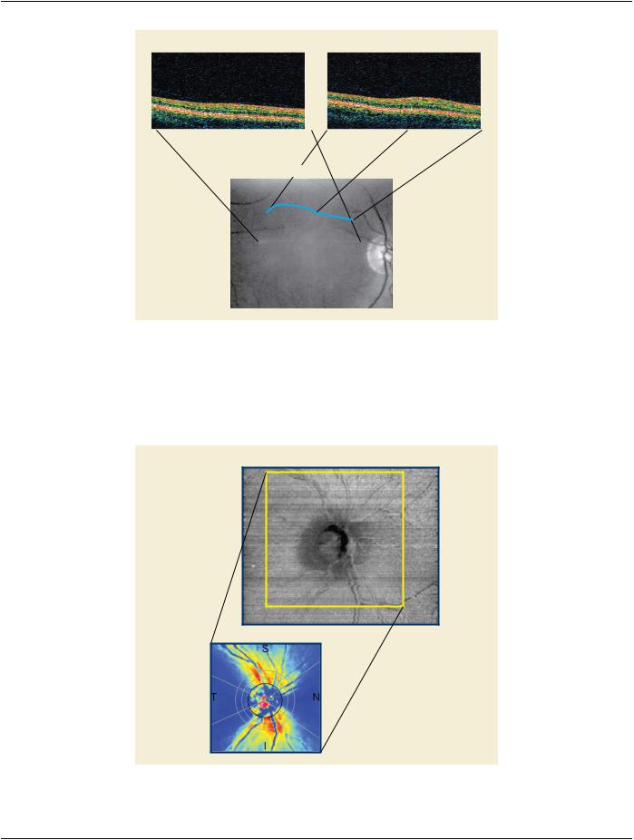
C.8.17.14 Ophthalmic Optical Coherence Tomography En Face Image Module​

Table C.8.17.14-1 specifies the Attributes that describe the Ophthalmic Optical Coherence Tomography En Face Image Module.​

Table​

Table C.8.17.14-1. Ophthalmic Optical Coherence Tomography En Face Image Module Attributes​

Attribute Name​

Tag​

Type​

Attribute Description​

Image Type​

(0008,0008)​

1​

Image identification characteristics.​


See Section C.8.17.14.1.5 for specialization.​

Instance Number​

(0020,0013)​

1​

A number that identifies this SOP Instance.​

- Standard -​