Материал: ЭКГ 2 инглиш
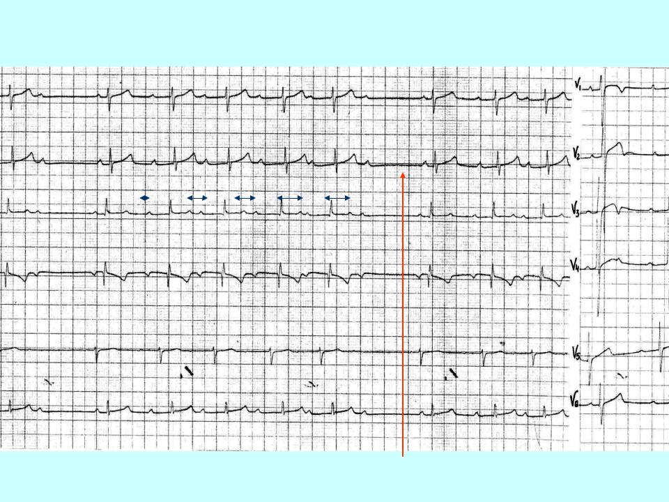
Mobitz type I:
I |
|
|
|
|
|
|
V1 |
|
|
|
|
|
|
|
|
II |
p |
p |
p |
p |
p |
p |
p |
V2
III
V3
AVR
V4
AVL
V5
AVF
V6
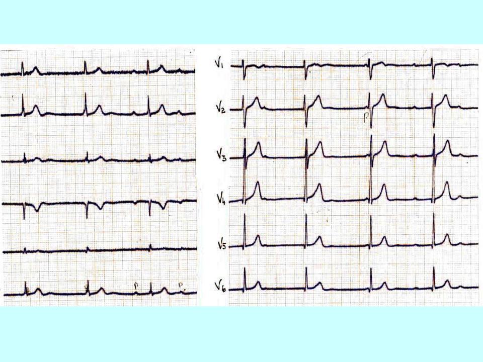
3rd degree AV block (complete AV-block):
I |
V1 |
p |
p |
p |
p |
p |
II |
V2 |
III |
V3 |
AVR |
V4 |
AVL |
V5 |
AVF |
V6 |
|
Независимое сокращение предсердий и желудочков

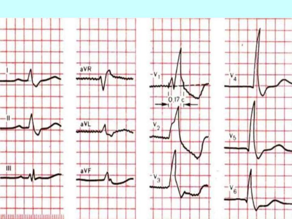
Right bundle branch block:
Main ECG signs of incomplete right bundle branch block:
1.Cleavage of the QRS complex in lead V1 like rSr or rSR '.
2.Broadened (up to o, 11-o, 12s) or normal duration of the QRS complex 3.Increase in the activation time of the right ventricle in lead V1 more than o, o3 from.
4.Absence of typical widening and deepening of the S wave in V6 and I standard leads.
Main ECG signs of complete right bundle branch block:
1.A split, M-shaped QRS complex of the rSR ', rsR', RSR 'or RsR '(and R'> R) in V1 V2 sometimes II and aVF leads.
2.Broadened (up to about, 12 s and more) QRS complex, as well as an increase in time internal deviation (activation of the right ventricle) in V1 V2 leads more about, about7 - 0.08 s.
3- Discordant downward displacement of the R (S) -T segment and the T wave (asymmetric biphasic or negative) in relation to the main tooth of the complex QRS in V1 sometimes V2 III and aVF leads.
4.Wide (more about, 04 s), deep and often serrated S wave in V6, V5, I, aVL and sometimes II leads.
Right bundle branch block:
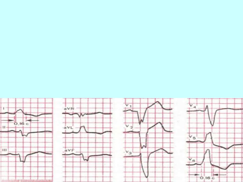
Left bundle branch block :
Main ECG signs of complete blockade of both branches of the left bundle branch Gisa:
1.Wide (more than about 12 s), varied in shape, often split QRS complex, usually represented by one R wave in V, Vs, I, a VL leads.
2.Increasing the time of the internal deviation (activation of the left
ventricle) more about, about8 s in V5, Vo leads.
3.Discordant downward displacement of the R (S) -T segment and the T wave (asymmetric biphasic or negative) in relation to the main tooth of the complex QRS in V5, Vs, I and aVL leads.
4.Widened S wave (or QS) in V, 1 V2 leads.