Материал: Internal_diseases_propedeutics._Part_II._Diagnostics_of_cardiovascular_diseases
ventricle, LA — left atrium, RV — right ventricle, RA — right atrium, RSPV — right superior pulmonary vein.
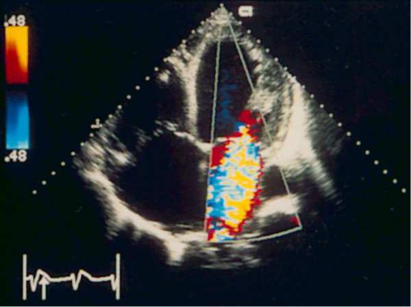
Fig.3 The position of the apical four-chamber hearts. Color Doppler study systolic. Severe mitral insufficiency. The jet has a large diameter at the level of the cusps of the mitral valve, the flow reaches the opposite wall of the left atrium occupies almost all the left atrium and enters the pulmonary veins — these trait s characterize severe mitral regurgitation.
96
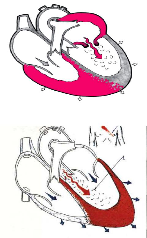
Fig.4. Mitral stenosis
Fig.5.Aortic incompetence
97
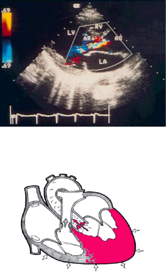
Fig.6. Aortic regurgitation, the severity — from small to moderat e. Color Doppler study of the position of the parasternal long axis of left ventricle. Motley regulatorului flow begins at the level of closin g of the flaps of the aortic valve. LV — left ventricle, LA — left atrium, RV — right ventricle, Ao — ascending aorta, AR — aortic regurgitation.
Fig.7.Aortic stenosis
98
References:
1.Избранные вопросы пропедевтики внутренних болезней: издание для студентов и практикующих врачей. Часть 1/ В.А. Семенов, В.в. гноевых, Е.А.
Черкашина, А.Ю. Смирнова; под. Ред. Проф. В.В. Гноевых.- Ульяновск, 2014.- 208с.
2.Internal diseases propedeutics / Ivashkin V.T., Okhlobystin A.V. - М. : ГЭОТАР-
Медиа, 2014.- 176 с.;
3.Пропедевтика внутренних болезней. Учебно-методическое пособие. Часть VI. Introduction to internal diseases. Manual. Part VI. / Ослопов В.Н., Садыкова А.Р.,
Карамышева И.В. – Казань: КГМУ, 2006. - 75 с.
99
100